

High-Spatial-Resolution Multishot Multiplexed Sensitivity-encoding Diffusion-weighted Imaging for Improved Quality of Breast Images and Differentiation of Breast Lesions: A Feasibility Study
- PMID: 33778712
- PMCID: PMC7983772
- DOI: 10.1148/rycan.2020190076
High-Spatial-Resolution Multishot Multiplexed Sensitivity-encoding Diffusion-weighted Imaging for Improved Quality of Breast Images and Differentiation of Breast Lesions: A Feasibility Study
Abstract
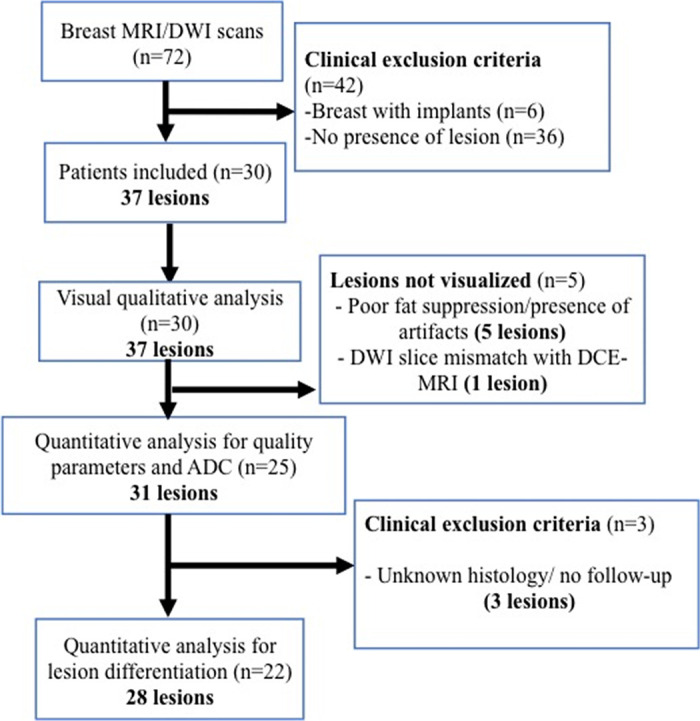
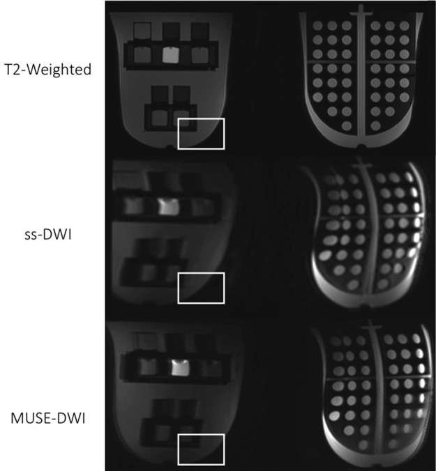
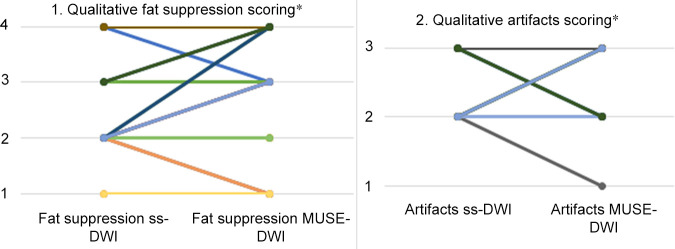
Multishot multiplexed sensitivity-encoding diffusion-weighted imaging is a feasible and easily implementable routine breast MRI protocol that yields high-quality diffusion-weighted breast images.Purpose: To compare multiplexed sensitivity-encoding (MUSE) diffusion-weighted imaging (DWI) and single-shot DWI for lesion visibility and differentiation of malignant and benign lesions within the breast.Materials and Methods: In this prospective institutional review board-approved study, both MUSE DWI and single-shot DWI sequences were first optimized in breast phantoms and then performed in a group of patients. Thirty women (mean age, 51.1 years ± 10.1 [standard deviation]; age range, 27-70 years) with 37 lesions were included in this study and underwent scanning using both techniques. Visual qualitative analysis of diffusion-weighted images was accomplished by two independent readers; images were assessed for lesion visibility, adequate fat suppression, and the presence of artifacts. Quantitative analysis was performed by calculating apparent diffusion coefficient (ADC) values and image quality parameters (signal-to-noise ratio [SNR] for lesions and fibroglandular tissue; contrast-to-noise ratio) by manually drawing regions of interest within the phantoms and breast tumor tissue. Interreader variability was determined using the Cohen κ coefficient, and quantitative differences between MUSE DWI and single-shot DWI were assessed using the Mann-Whitney U test; significance was defined at P < .05.Results: MUSE DWI yielded significantly improved image quality compared with single-shot DWI in phantoms (SNR, P = .001) and participants (lesion SNR, P = .009; fibroglandular tissue SNR, P = .05; contrast-to-noise ratio, P = .008). MUSE DWI ADC values showed a significant difference between malignant and benign lesions (P < .001). No significant differences were found between MUSE DWI and single-shot DWI in the mean, maximum, and minimum ADC values (P = .96, P = .28, and P = .49, respectively). Visual qualitative analysis resulted in better lesion visibility for MUSE DWI over single-shot DWI (κ = 0.70).Conclusion: MUSE DWI is a promising high-spatial-resolution technique that may enhance breast MRI protocols without the need for contrast material administration in breast screening.Keywords: Breast, MR-Diffusion Weighted Imaging, OncologySupplemental material is available for this article.© RSNA, 2020.
2020 by the Radiological Society of North America, Inc.
Conflict of interest statement
Disclosures of Conflicts of Interest: I.D.N. disclosed no relevant relationships. R.L.G. disclosed no relevant relationships. E.A.M. disclosed no relevant relationships. T.L. disclosed no relevant relationships. M.M.F. Activities related to the present article: disclosed no relevant relationships. Activities not related to the present article: is and employee of and holds stock in GE Healthcare. Other relationships: disclosed no relevant relationships. A.G. Activities related to the present article: disclosed no relevant relationships. Activities not related to the present article: is an employee of GE Healthcare. Other relationships: disclosed no relevant relationships. K.P. editorial board member for Radiology: Imaging Cancer. S.B.T. disclosed no relevant relationships.
Figures
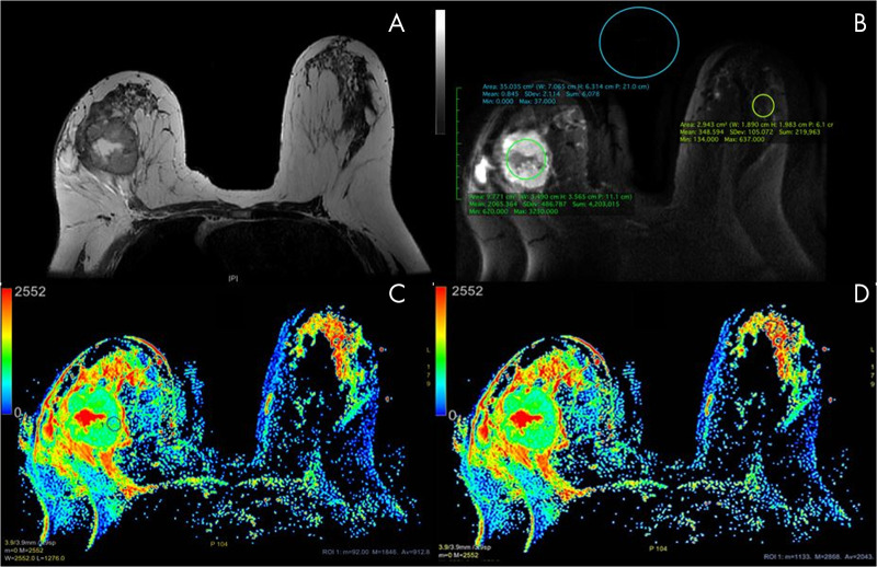
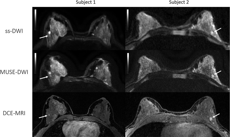
![Axial images from two patients with biopsy-proven invasive ductal carcinoma. Left: Patient 1, a 36-year-old woman with a 9-mm mass in the upper outer quadrant of the right breast (arrow). Right: Patient 2, a 57-year-old woman with a 36-mm necrotic mass in the right upper breast (arrow). Both readers assigned category 1 (better overall image quality with multishot multiplexed sensitivity-encoding diffusion-weighted imaging [MUSE-DWI] than with single-shot DWI [ss-DWI]) for overall image quality since MUSE DWI (b value, 800 sec/mm2) showed better lesion delineation compared with single-shot DWI (b value, 800 sec/mm2). Note also that artifact seen with single-shot DWI is partially corrected at MUSE DWI in patient 1.](https://cdn.ncbi.nlm.nih.gov/pmc/blobs/4a32/7983772/8bc64ca6290c/rycan.2020190076.fig6.jpg)
References
-
- Jezzard P, Balaban RS. Correction for geometric distortion in echo planar images from B0 field variations. Magn Reson Med 1995;34(1):65–73. - PubMed
-
- van Pul C, Roos FG, Derksen OS, et al. A comparison study of multishot vs. single-shot DWI-EPI in the neonatal brain: reduced effects of ghosting compared to adults. Magn Reson Imaging 2004;22(9):1169–1180. - PubMed
