Comparative Study
. 2021 Jun;95(6):2081-2107.
doi: 10.1007/s00204-021-03031-1. Epub 2021 Mar 29.

Functional alterations by a subgroup of neonicotinoid pesticides in human dopaminergic neurons

Affiliations
Comparative Study

Functional alterations by a subgroup of neonicotinoid pesticides in human dopaminergic neurons

Dominik Loser et al. Arch Toxicol. 2021 Jun.

Abstract

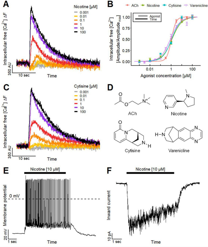
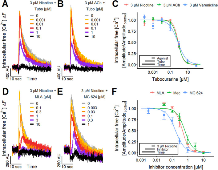
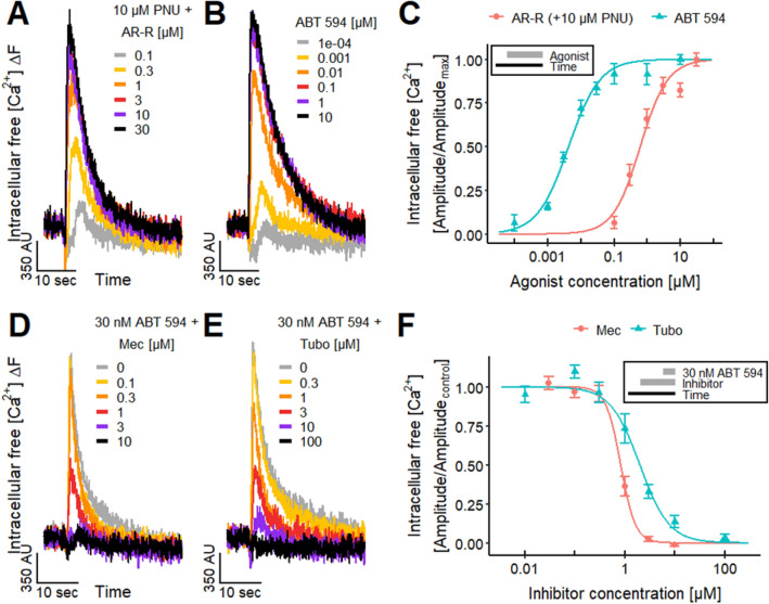
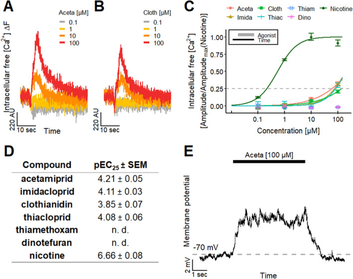
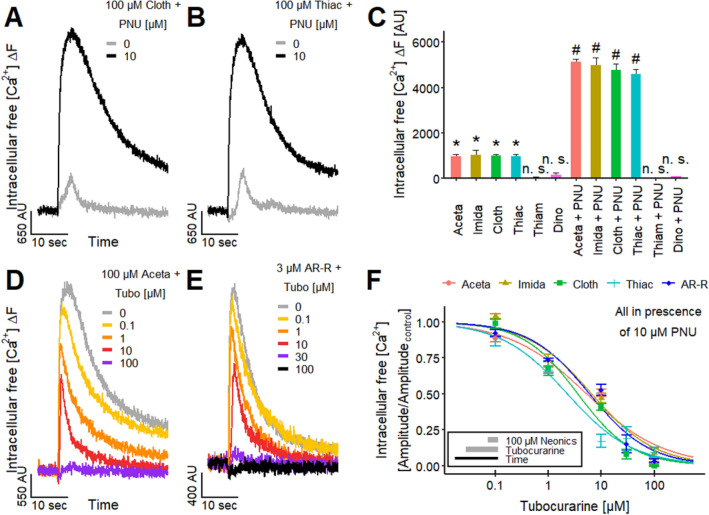
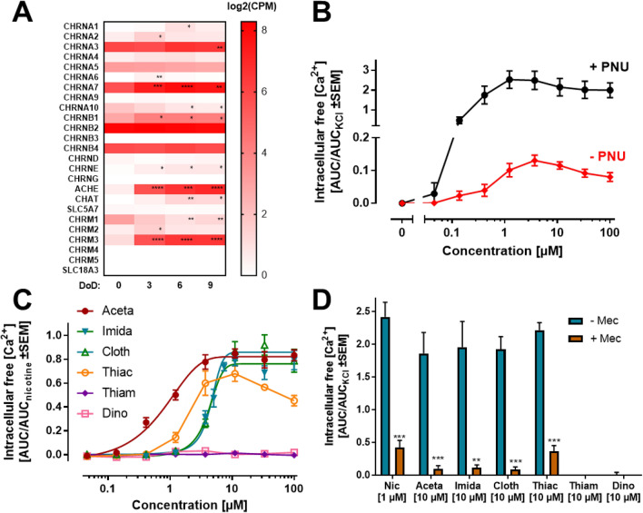
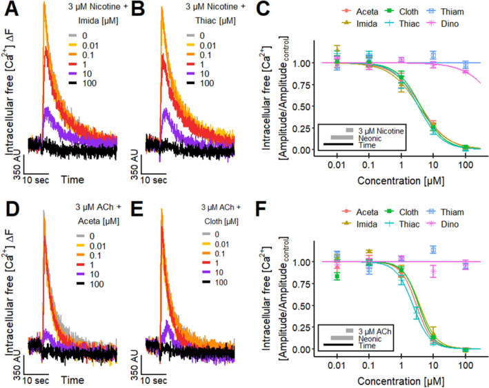
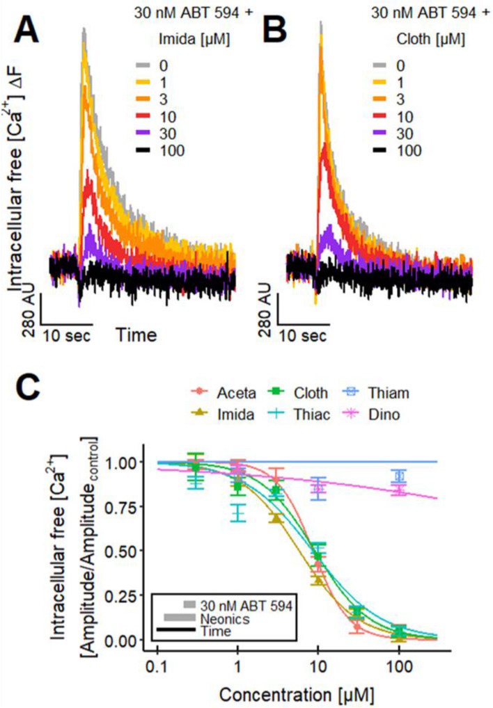
Neonicotinoid pesticides, originally developed to target the insect nervous system, have been reported to interact with human receptors and to activate rodent neurons. Therefore, we evaluated in how far these compounds may trigger signaling in human neurons, and thus, affect the human adult or developing nervous system. We used SH-SY5Y neuroblastoma cells as established model of nicotinic acetylcholine receptor (nAChR) signaling. In parallel, we profiled dopaminergic neurons, generated from LUHMES neuronal precursor cells, as novel system to study nAChR activation in human post-mitotic neurons. Changes of the free intracellular Ca2+ concentration ([Ca2+]i) were used as readout, and key findings were confirmed by patch clamp recordings. Nicotine triggered typical neuronal signaling responses that were blocked by antagonists, such as tubocurarine and mecamylamine. Pharmacological approaches suggested a functional expression of α7 and non-α7 nAChRs on LUHMES cells. In this novel test system, the neonicotinoids acetamiprid, imidacloprid, clothianidin and thiacloprid, but not thiamethoxam and dinotefuran, triggered [Ca2+]i signaling at 10-100 µM. Strong synergy of the active neonicotinoids (at low micromolar concentrations) with the α7 nAChR-positive allosteric modulator PNU-120596 was observed in LUHMES and SH-SY5Y cells, and specific antagonists fully inhibited such signaling. To provide a third line of evidence for neonicotinoid signaling via nAChR, we studied cross-desensitization: pretreatment of LUHMES and SH-SY5Y cells with active neonicotinoids (at 1-10 µM) blunted the signaling response of nicotine. The pesticides (at 3-30 µM) also blunted the response to the non-α7 agonist ABT 594 in LUHMES cells. These data show that human neuronal cells are functionally affected by low micromolar concentrations of several neonicotinoids. An effect of such signals on nervous system development is a toxicological concern.

Keywords: Desensitization; Live-cell calcium imaging; Molecular docking; Neurotoxicity; Nicotine.


Conflict of interest statement

The authors declare no conflict of interest.

Figures

Fig. 1
Fig. 1
Identification of functional nicotinic acetylcholine receptors (nAChRs) on LUHMES neurons. a Traces of Ca2+-imaging recordings show the concentration-dependent effects of the nAChR agonist nicotine on LUHMES neurons. b Concentration–response curves for the effects of nAChR agonists acetylcholine (ACh), nicotine, cytisine and varenicline with pEC50 values of 5.98 ± 0.03, 5.93 ± 0.05, 5.95 ± 0.05 and 6.08 ± 0.04, respectively. Note the treatment scheme (upper left corner), illustrating the experimental design. Detailed data on n numbers are found in table S4. c Ca2+-imaging signals evoked by the addition of cytisine. d Chemical structures of the tested nAChR agonists. e, f Manual patch clamp recordings of the responses of LUHMES neurons evoked by the application of 10 µM nicotine for 5 s. e Firing of multiple action potentials with a long-lasting depolarization of the membrane potential (n = 28) recorded in current-clamp. f Slowly inactivating inward current (n = 12) measured in voltage-clamp
Fig. 2
Fig. 2
Characterization of the nAChRs. a, b Ca2+-imaging signals of the effects of the pre-applied non-selective nAChR antagonist tubocurarine (Tubo) on the responses of LUHMES neurons triggered by a 3 µM nicotine and b 3 µM ACh. c Inhibitory effect of Tubo on the signals evoked by the acute exposure to 3 µM of nicotine, ACh and varenicline. The resulting pIC50 values were 6.08 ± 0.04 for nicotine, 6.13 ± 0.04 for ACh and 6.13 ± 0.09 for varenicline. d, e Traces of the effects of d MLA and e MG 624 on the Ca2+-imaging signals stimulated by 3 µM nicotine. f Concentration–response curves for the effects of MLA, mecamylamine (Mec) and MG 624 on the responses triggered by the acute exposure to 3 µM nicotine. The pIC50 values were 6.33 ± 0.04 for MLA, 6.17 ± 0.05 for Mec and 6.80 ± 0.07 for MG 624. Note the treatment schemes (lower left corner), illustrating the experimental design. Detailed data on n numbers are found in table S4
Fig. 3
Fig. 3
Differential agonist responses on nAChRs. a, b Signals of Ca2+-imaging triggered by the selective α7 nAChR agonist a AR-R 17779 (AR-R), after the pretreatment with 10 µM PNU-120596 (PNU), a positive allosteric modulator of α7 nAChR, and the selective non-α7 nAChR agonist b ABT 594. c Agonistic effect of AR-R in presence of 10 µM PNU and ABT 594 yielded pEC50 values of 6.20 ± 0.05 and 8.36 ± 0.05, respectively. d, e Ca2+-imaging traces of the effects of d Mec and e Tubo, which were preincubated for 4.5 min, on the response of the LUHMES neurons evoked by 30 nM ABT 594. f The concentration–response curves of the effects of Mec and Tubo on the response evoked by 30 nM ABT 594 resulted in pIC50 values of 6.08 ± 0.03 and 5.70 ± 0.05, respectively. Note the treatment schemes, illustrating the experimental design. Detailed data on n numbers are found in table S4
Fig. 4
Fig. 4
Effect of neonicotinoids on LUHMES neurons. a, b Traces of Ca2+-imaging show the effects of the neonicotinoids a Aceta and b Cloth on LUHMES neurons. c Concentration-dependent effect of the neonicotinoids Aceta, Imida, Cloth, Thiac, Thiam and Dino and the positive control nicotine. Amplitudes were normalized to the maximal amplitude evoked by nicotine. Note the treatment scheme (upper left corner), illustrating the experimental design. d Table with corresponding pEC25 values for the tested neonicotinoids and nicotine. Detailed data on n numbers are found in table S6. e Manual patch clamp recording of a long-lasting depolarization of the membrane potential during the application of 100 µM Aceta for 5 s (n = 4). The Aceta-induced depolarization from a holding potential of − 70 mV was not sufficient to evoke action potential firing
Fig. 5
Fig. 5
Effects of neonicotinoids on Ca2+-signaling on the level of individual neurons. a LUHMES neurons during control and during the application of 10 µM nicotine and 100 µM Aceta. Pictures of single-cell Ca2+-imaging recordings were taken with the Cell Observer (Carl Zeiss Microscopy). Images are shown in false color to enhance the interpretability. b Traces of single-cell Ca2+-imaging recordings of six cells (marked in a) with and without response to the application of 100 µM Aceta. Threshold for response detection is defined as mean + 3 × SD of negative control recordings. c Percentage of cells that responded to the application of nicotine, Aceta and Imida in single-cell Ca2+-imaging recordings. Changes are significant (*p < 0.05, t test) for 10 µM Aceta and Imida. Using more stringent ANOVA with Dunnett’s post hoc test, there was a significant difference for 100 µM, but only a trend (p > 0.05 for 10 µM). This range of effect significance agrees well with calculations of the Imida benchmark concentration (BMC10 = 11.2 µM) and its upper 95% confidence limit (BMCU10 = 26 µM). d Fraction of cells reacting to Cloth, Thiac, Thiam and Dino at a concentration of 100 µM. Note the enlarged y-axis. c, d Statistical significance was determined against negative control recordings (*, significant by ANOVA; n. s., not significant). Detailed data on n numbers and percentages of responsive cells are found in table S7 and S8, respectively
Fig. 6
Fig. 6
Effect of neonicotinoids on α7 nAChR. a, b Ca2+-imaging traces of the effects of the neonicotinoids a Cloth and b Thiac during control and in the presence of 10 µM PNU, which was preincubated for 4.5 min. c Effects of the neonicotinoids Aceta, Imida, Cloth, Thiac, Thiam and Dino (100 µM) on the Ca2+-imaging signal of LUHMES neurons in absence and presence of 10 µM PNU. Statistical significance was determined between the recordings of the neonicotinoids without PNU and negative control recordings (*, significant; n. s., not significant) and between the recordings of each neonicotinoid without and with PNU (#, significant; n. s., not significant). d, e Traces of Ca2+-imaging showing the effect of pre-applied tubocurarine (Tubo) on the signals evoked by d 100 µM Aceta and e 3 µM AR-R, both in the presence of pre-applied 10 µM PNU. f The concentration–response curves illustrate the effects of Tubo on the response of the LUHMES neurons to the acute exposure to the neonicotinoids Aceta, Imida, Cloth and Thiac (100 µM) and the α7 agonist AR-R (3 µM). All recordings were performed in the presence of 10 µM PNU, which was preincubated for 4.5 min. The resulting pIC50 values were 5.28 ± 0.10, 5.18 ± 0.09, 5.45 ± 0.08, 5.64 ± 0.08 and 5.22 ± 0.09 for Aceta, Imida, Cloth, Thiac and AR-R, respectively. Note the treatment scheme (lower left corner), illustrating the experimental design. Detailed data on n numbers are found in table S6
Fig. 7
Fig. 7
Nicotine signaling in SH-SY5Y cells. a The expression of genes coding for nAChR subunits was determined by whole-transcriptome RNA-sequencing during differentiation of SH-SY5Y cells. The raw counts were normalized to counts per million total counts (CPM) and log2-transformed. Significance of changes between day of differentiation (DoD) zero and DoD3-9 was evaluated by ANOVA with Dunnett’s multiple comparison test. *p < 0.05, **p < 0.01, ***p < 0.001, ****p < 0.0001. b The SH-SY5Y cells were differentiated for 72 h, and used on DoD3 for Ca2+-imaging experiments. The increase of the [Ca2+]i triggered by nicotine was measured in the presence or absence of 10 µM PNU. The responses were evaluated as the area under the curve (AUC) of the increased fluorescence of the calcium-sensitive dye Fura-2 for 0–150 s after compound addition. An example of an original recording is shown in figure S5C. The responses were normalized to the [Ca2+]i response of SH-SY5Y cells after depolarization with 30 mM KCl (AUCKCl) (n = 4–6). c The [Ca2+]i response of SH-SY5Y cells triggered by Aceta, Imida, Cloth, Thiac, Thiam and Dino was measured in the presence of 10 µM PNU. The AUC of the response (0–150 s) was normalized to the AUC of the response evoked by the treatment of the cells with 11 µM nicotine (AUCnicotine) (n = 3). The estimated pEC50 values were 6.10 ± 0.07, 5.38 ± 0.03, 5.33 ± 0.01 and 5.73 ± 0.01 for Aceta, Imida, Cloth and Thaic, respectively. d The [Ca2+]i responses of SH-SY5Y cells triggered by nicotine, Aceta, Imida, Cloth, Thiac, Thiam and Dino were measured in the presence of 10 µM PNU, and the presence or absence of Mec (125 µM). The AUC of the responses was normalized to the AUCKCl (n = 3–5). Significance was evaluated by multiple t tests. *p < 0.05, **p < 0.01, ***p < 0.001
Fig. 8
Fig. 8
Effect of neonicotinoids on responses evoked by nicotine and ACh. a, b Ca2+-imaging traces displaying the responses of LUHMES neurons to the acute exposure to 3 µM nicotine in the presence of the neonicotinoids a Imida and b Thiac, which were preincubated for 4.5 min. c Concentration-dependent effects of the pre-applied neonicotinoids Aceta, Imida, Cloth, Thiac, Thiam and Dino on the nicotine-evoked responses. The resulting pIC50 values were 5.40 ± 0.08, 5.47 ± 0.10, 5.41 ± 0.07 and 5.48 ± 0.08 for Aceta, Imida, Cloth and Thiac, respectively. Thiam and Dino did not show an effect. d, e Ca2+-imaging traces showing the effects of the neonicotinoids d Imida and e Thiac, which were preincubated for 4.5 min, on the signals evoked by the application of 3 µM ACh. f Effects of the pre-applied neonicotinoids Aceta, Imida, Cloth, Thiac, Thiam and Dino on the response of LUHMES neurons triggered by 3 µM ACh. The pIC50 values were 5.53 ± 0.09, 5.43 ± 0.08, 5.46 ± 0.07 and 5.64 ± 0.04 for Aceta, Imida, Cloth and Thiac, respectively. Thiam and Dino had no effect. Note the treatment schemes (lower left corner), illustrating the experimental design. Detailed data on n numbers are found in table S6
Fig. 9
Fig. 9
Effect of neonicotinoids on responses evoked by ABT 594. a, b Ca2+-imaging traces of the effects of the neonicotinoids a Imida and b Cloth, which were preincubated for 4.5 min, on the responses stimulated by the acute exposure to 30 nM ABT 594. c The concentration-dependent effects of the pre-applied neonicotinoids Aceta, Imida, Cloth, Thiac, Thiam and Dino on the ABT 594-induced response resulted in pIC50 values of 5.07 ± 0.03, 5.24 ± 0.03, 5.05 ± 0.05 and 5.08 ± 0.06 for Aceta, Imida, Cloth and Thiac, respectively. No pIC50 values could be determined for Thiam and Dino. Note the treatment scheme (lower left corner), illustrating the experimental design. Detailed data on n numbers are found in table S6

Similar articles

Cited by

References

    1. Abou-Donia MB, Goldstein LB, Bullman S, et al. Imidacloprid induces neurobehavioral deficits and increases expression of glial fibrillary acidic protein in the motor cortex and hippocampus in offspring rats following in utero exposure. J Toxicol Environ Health A. 2008;71:119–130. doi: 10.1080/15287390701613140. - DOI - PubMed
    1. Agrawal L, Vimal SK, Shiga T. Role of serotonin 4 receptor in the growth of hippocampal neurons during the embryonic development in mice. Neuropharmacology. 2019;158:107712. doi: 10.1016/j.neuropharm.2019.107712. - DOI - PubMed
    1. Alijevic O, McHugh D, Rufener L, et al. An electrophysiological characterization of naturally occurring tobacco alkaloids and their action on human α4β2 and α7 nicotinic acetylcholine receptors. Phytochemistry. 2020;170:112187. doi: 10.1016/j.phytochem.2019.112187. - DOI - PubMed
    1. Alkondon M, Pereira EFR, Eisenberg HM, Albuquerque EX. Choline and selective antagonists identify two subtypes of nicotinic acetylcholine receptors that modulate GABA release from CA1 interneurons in rat hippocampal slices. J Neurosci. 1999;19:2693–2705. doi: 10.1523/JNEUROSCI.19-07-02693.1999. - DOI - PMC - PubMed
    1. Arias HR, Feuerbach D, Targowska-Duda K, et al. Pharmacological and molecular studies on the interaction of varenicline with different nicotinic acetylcholine receptor subtypes. Potential mechanism underlying partial agonism at human α4β2 and α3β4 subtypes. Biochim Biophys Acta BBA Biomembr. 2015;1848:731–741. doi: 10.1016/j.bbamem.2014.11.003. - DOI - PubMed

Publication types

MeSH terms