Review
. 2021 Apr 7;22(4):e51078.
doi: 10.15252/embr.202051078. Epub 2021 Mar 28.

Ubiquitin-dependent regulation of transcription in development and disease

Affiliations
Review

Ubiquitin-dependent regulation of transcription in development and disease

Kevin G Mark et al. EMBO Rep. .

Abstract

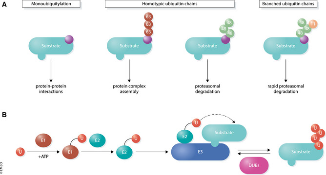
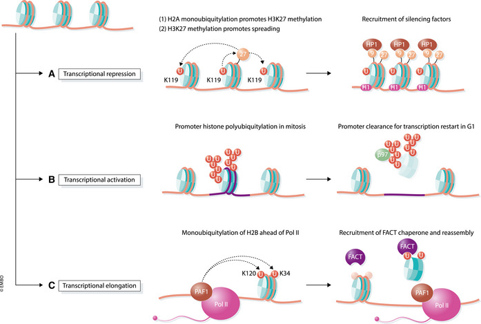
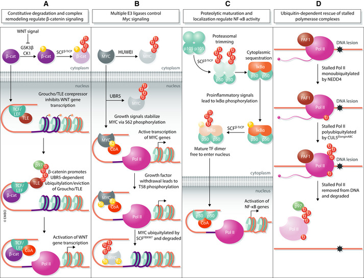
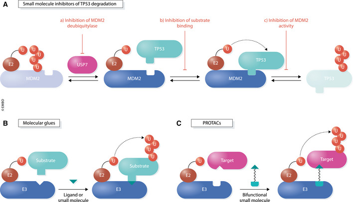
Transcription is an elaborate process that is required to establish and maintain the identity of the more than two hundred cell types of a metazoan organism. Strict regulation of gene expression is therefore vital for tissue formation and homeostasis. An accumulating body of work found that ubiquitylation of histones, transcription factors, or RNA polymerase II is crucial for ensuring that transcription occurs at the right time and place during development. Here, we will review principles of ubiquitin-dependent control of gene expression and discuss how breakdown of these regulatory circuits leads to a wide array of human diseases.

Keywords: RNA polymerase II; histone modification; transcription; ubiquitin.


Conflict of interest statement

M.R. is a co‐founder and consultant to Nurix and scientific advisory board member to Monte Rosa.

Figures

Figure 1
Figure 1. The ubiquitin modification system
(A) Various structures and functions of different types of ubiquitin conjugates. Monoubiquitylation (left) involves transfer of a single ubiquitin to a substrate. E3 ligases can also connect several ubiquitin molecules together using the C‐terminus of one subunit and one of seven lysine (K) residues (K6, K11, K27, K29, K33, K48, K63) or the N‐terminal methionine (M1) on the other. Different ubiquitin topologies adopt distinct structural conformations which direct a wide array of substrate outcomes. (B) Ubiquitylation occurs through an enzymatic cascade. E1 enzymes use ATP to form a high‐energy thioester bond to ubiquitin through an active site cysteine residue. Charged E1s transfer ubiquitin to one of ~ 40 E2 ubiquitin‐conjugating enzymes, which then bind to one of ~ 600 E3 ubiquitin ligases that facilitate the transfer of ubiquitin onto a specific substrate. Approximately 100 DUBs remove ubiquitin from substrates to reverse the ubiquitylation process.
Figure 2
Figure 2. Transcriptional effects of histone ubiquitylation
(A) Monoubiquitylation of histone H2A at K119 by PRC1 promotes transcriptional silencing. This modification recruits PRC2 to trimethylate histone H3 at K27, a mark which in turn recruits PRC1 to ubiquitylate additional H2A histones, resulting in spreading. H3K27me3 also leads to H3K9 trimethylation, which together with ubiquitylated H2A, recruit factors that compact chromatin and silence transcription. (B) Histone polyubiquitylation promotes transcriptional reactivation following cell division. During mitosis, the APC/C E3 ligase is recruited to specific promoters to polyubiquitylate histones, leading to their extraction by p97/VCP and proteasomal degradation. This action licenses these promoters for rapid transcriptional reactivation when cells enter G1. (C) Monoubiquitylation of histone H2B at K120 and K34 mediates transcriptional elongation. As part of the PAF1 elongation complex, the E3 ligases RNF20/RNF40 and MSL1/MSL2 travel with RNA Pol II and ubiquitylate H2B on nucleosomes obstructing RNA Pol II’s path. H2B ubiquitylation promotes nucleosome remodeling, through the FACT histone chaperone, to allow polymerase passage. Ubiquitin is then removed by H2B DUBs to reestablish chromatin behind RNA Pol II.
Figure 3
Figure 3. Ubiquitin‐dependent regulation of transcriptional machinery
(A) In the absence of WNT signal, the β‐catenin transcription factor (β‐cat) is constitutively degraded by a destruction complex consisting of kinases GSK3β and CK1 as well as the ubiquitin ligase SCFβTrCP. The presence of WNT ligand stabilizes β‐catenin by inhibiting its phosphorylation, thus allowing it to translocate into the nucleus. At TCF/LEF transcriptional complexes, β‐catenin facilitates the exchange of co‐repressors for co‐activators (CoA) through UBR5‐mediated ubiquitylation and p97/VCP‐dependent extraction of the repressive Groucho/TLE subunit. (B) MYC levels are kept low in the cytoplasm and nucleus by the E3 ligases HUWE1 and UBR5, respectively. Upstream growth signals stabilize and activate MYC through phosphorylation of the S62 residue. This primes MYC for subsequent phosphorylation at nearby T58, which occurs upon growth factor withdrawal. T58‐phosphorylated MYC is recognized by the SCFFBXW7 E3 ligase and degraded to prevent subsequent re‐initiation of transcription. (C) The proinflammatory transcription factor NF‐κB is synthesized as a 105‐kDa precursor (p105) that is ubiquitylated by the SCFβTrCP E3 ligase and partially processed by the proteasome to yield a mature 50‐kDa form (p50). Mature NF‐κB dimers are sequestered in the cytoplasm by an inhibitor, IκBα, until cellular signals lead to degradation of IκBα and liberation of mature NF‐κB. Only then can mature NF‐κB enter the nucleus and initiate transcription of target genes. (D) RNA Pol II complexes that have stalled, such as at a DNA lesion, are first monoubiquitylated by the E3 ligase NEDD4. A second E3, CUL5ElonginABC, adds K48‐linked polyubiquitin chains to monoubiquitylated RNA Pol II leading to p97/VCP‐dependent removal of the stalled complex from DNA.
Figure 4
Figure 4. Regulation of transcription by small molecules targeting ubiquitin components
(A) Mechanisms of small molecules that prevent degradation of the transcription factor and tumor suppressor, TP53, by its E3 ubiquitin ligase, MDM2. (a) Inhibitors of the USP7 deubiquitylase lead to increased autoubiquitylation and destabilization of MDM2. (b) Small molecules, such as the Nutlins, block TP53 degradation by disrupting the substrate‐ligase interaction surface. (c) Compounds that block the E3 ligase activity of MDM2, but not substrate binding, also result in accumulation of TP53. (B) Molecular glues are small molecules that alter the surface of proteins, namely an E3 ligase and substrate, to promote their association. These compounds often display low binding affinities for each interactor but simultaneously bind both components to enhance their interaction. (C) PROTACs (proteolysis‐targeting chimeras) are heterobifunctional molecules that consist of two small molecules tethered by a linker. One of the small molecules binds a target protein while the other binds an E3 ligase. In this manner, the cell’s endogenous ubiquitin–proteasome system can be used to rapidly and selectively eliminate any given protein target‐of‐interest.

References

    1. Adhikary S, Marinoni F, Hock A, Hulleman E, Popov N, Beier R, Bernard S, Quarto M, Capra M, Goettig S et al (2005) The ubiquitin ligase HectH9 regulates transcriptional activation by Myc and is essential for tumor cell proliferation. Cell 123: 409–421 - PubMed
    1. Adorno M, Sikandar S, Mitra SS, Kuo A, Nicolis Di Robilant B, Haro‐Acosta V, Ouadah Y, Quarta M, Rodriguez J, Qian D et al (2013) Usp16 contributes to somatic stem‐cell defects in Down's syndrome. Nature 501: 380–384 - PMC - PubMed
    1. Ahmad KF, Melnick A, Lax S, Bouchard D, Liu J, Kiang CL, Mayer S, Takahashi S, Licht JD, Prive GG (2003) Mechanism of SMRT corepressor recruitment by the BCL6 BTB domain. Mol Cell 12: 1551–1564 - PubMed
    1. Anindya R, Aygun O, Svejstrup JQ (2007) Damage‐induced ubiquitylation of human RNA polymerase II by the ubiquitin ligase Nedd4, but not Cockayne syndrome proteins or BRCA1. Mol Cell 28: 386–397 - PubMed
    1. Anindya R, Mari PO, Kristensen U, Kool H, Giglia‐Mari G, Mullenders LH, Fousteri M, Vermeulen W, Egly JM, Svejstrup JQ (2010) A ubiquitin‐binding domain in Cockayne syndrome B required for transcription‐coupled nucleotide excision repair. Mol Cell 38: 637–648 - PMC - PubMed

Publication types