. 2021 Aug;31(8):1272-1281.
doi: 10.1089/thy.2020.0931. Epub 2021 Jun 4.

The Iodide Transport Defect-Causing Y348D Mutation in the Na+/I- Symporter Renders the Protein Intrinsically Inactive and Impairs Its Targeting to the Plasma Membrane

Affiliations

The Iodide Transport Defect-Causing Y348D Mutation in the Na+/I- Symporter Renders the Protein Intrinsically Inactive and Impairs Its Targeting to the Plasma Membrane

Andrea Reyna-Neyra et al. Thyroid. 2021 Aug.

Abstract

Background: The sodium/iodide (Na+/I-) symporter (NIS) mediates active transport of I- into the thyroid gland. Mutations in the SLC5A5 gene, which encodes NIS, cause I- transport defects (ITDs)-which, if left untreated, lead to congenital hypothyroidism and consequent cognitive and developmental deficiencies. The ITD-causing NIS mutation Y348D, located in transmembrane segment (TMS) 9, was reported in three Sudanese patients. Methods: We generated cDNAs coding for Y348D NIS and mutants with other hydrophilic and hydrophobic amino acid substitutions at position 348 and transfected them into cells. The activity of the resulting mutants was quantitated by radioiodide transport assays. NIS glycosylation was investigated by Western blotting after endoglycosidase H (Endo H) and PNGase-F glycosidase treatment. Subcellular localization of the mutant proteins was ascertained by flow cytometry analysis, cell surface biotinylation, and immunofluorescence. The intrinsic activity of Y348D was studied by measuring radioiodide transport in membrane vesicles prepared from Y348D-NIS-expressing cells. Our NIS homology models and molecular dynamics simulations were used to identify residues that interact with Y348 and investigate possible interactions between Y348 and the membrane. The sequences of several Slc5 family transporters were aligned, and a phylogenetic tree was generated in ClustalX. Results: Cells expressing Y348D NIS transport no I-. Furthermore, Y348D NIS is only partially glycosylated, is retained intracellularly, and is intrinsically inactive. Hydrophilic residues other than Asp at position 348 also yield NIS proteins that fail to be targeted to the plasma membrane (PM), whereas hydrophobic residues at this position, which we show do not interact with the membrane, rescue PM targeting and function. Conclusions: Y348D NIS does not reach the PM and is intrinsically inactive. Hydrophobic amino acid substitutions at position 348, however, preserve NIS activity. Our findings are consistent with our homology model's prediction that Y348 should face the side opposite the TMS9 residues that coordinate Na+ and participate in Na+ transport, and with the notion that Y348 interacts only with hydrophobic residues. Hydrophilic or charged residues at position 348 have deleterious effects on NIS PM targeting and activity, whereas a hydrophobic residue at this position rescues NIS activity.

Keywords: Y348D NIS; intracellular retention of NIS; iodide transport defect-causing NIS mutations; molecular dynamics simulations; sodium/iodide symporter.


Conflict of interest statement

No competing financial interests exist.

Figures

FIG. 1.
FIG. 1.
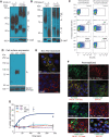
The Y348D amino acid substitution, which is in TMS9, yields a NIS protein that does not transport I. (A) Experimentally tested secondary structure model of NIS showing 13 TMSs (colored cylinders) and 3 glycosylation sites (branches). ITD-causing NIS mutations, including Y348D, are shown next to the affected positions. Mutations that yield proteins that are both intrinsically inactive and retained intracellularly are shown in orange rectangles, nonfunctional proteins in green rectangles, and intrinsically active but intracellularly retained proteins in blue rectangles. An extracellularly facing HA tag was engineered at the N-terminus. (B) NIS homology model [cf. (23)] (inwardly open conformation). (C) New NIS homology model (outwardly open conformation), generated using as a template the Na+-coupled sialic acid symporter, which has 25% sequence identity and 46% sequence similarity to NIS. The location of the Y348 residue (yellow) in TMS9 (orange) is shown in both B and C. (D, E) Y348 is on the side in TMS9 opposite that of S353 and T354, which coordinate Na+ (21). (F) Electrostatic surface of WT and Y348D NIS. Positive and negative potentials are shown in blue and red, respectively, and the position of Y348 is indicated by a black arrow. (G) Steady-state I transport assay (30 min) in MDCK II cells transfected with control (pcDNA3.1), human WT, or Y348D NIS HA-cDNA in the presence or absence of ClO4 (gray bars) at 10 μM I/140 mM Na+. ClO4, perchlorate; HA, hemagglutinin; I, iodide; ITD, I transport defect; MDCK-II, Madin-Darby canine kidney; NIS, sodium/iodide symporter; TMSs, transmembrane segments; WT, wild type.
FIG. 2.
FIG. 2.
Y348D NIS fails to mature fully and is retained intracellularly. Immunoblot analysis of membrane proteins (10 μg) extracted from MDCK II cells transfected with WT or Y348D NIS cDNA and treated with (+) or without (−) Endo H (A) or PNGase-F (B). NIS protein forms were detected using an affinity-purified anti-hNIS antibody against residues 618–633 of the carboxy terminus of hNIS. Letters to the right of the blots indicate differently glycosylated states of NIS with different relative electrophoretic mobilities (A: ∼90 kDa: mature NIS protein; B, B′: ∼60 and ∼120 kDa: core glycosylated NIS and its dimer, respectively; C, C′: ∼50 and ∼100 kDa: deglycosylated NIS and its dimer, respectively). E-cadherin was used as a loading control. (C) Flow cytometry analysis (FACS) using an anti-HA antibody to detect total NIS expression (permeabilized cells) or exclusively at the cell surface (nonpermeabilized cells) in MDCK II cells. (D) Cell surface biotinylation reveals fully glycosylated WT NIS (A) but not Y348D NIS at the PM of MDCK II cells. NIS was detected by immunoblot using the same anti-NIS antibody as in panels (A, B). The alpha subunit of the Na+/K+ ATPase was used as a loading control. (E) Immunofluorescence reveals WT but not Y348D NIS at the PM in nonpermeabilized cells, indicated by colocalization with the Na+/K+ ATPase (merge). (F) Immunofluorescence under permeabilized conditions shows WT NIS at the PM and expressed intracellularly. Y348D NIS is retained intracellularly. Nonpermeabilized and permeabilized WT and Y348D NIS-transfected MDCK II cells were immunostained with an anti-HA, an anti-Na+/K+-ATPase, and an anti-GM130 antibody, followed by anti-rabbit Alexa 488 and anti-mouse Alexa 594-conjugated antibodies. The overlay of the two images is shown (merge). Scale bar = 10 μm. (G) I transport assays in MVs from control, WT, and Y348D NIS-transduced COS-7 cells carried out at 20 μM I in the presence or absence of 100 mM Na+ for the periods of time indicated (100 μg of protein for each time point). Navy blue (WT, +Na+), pink (WT, –Na+), cyan (Y348D, +Na+), red (Y348D, –Na+), yellow (control). Error bars indicate SE for triplicate determination. COS-7, African green monkey kidney fibroblast-like cell line 7; Endo H, endoglycosidase H; FC, flow cytometry; GM130, Golgi matrix protein 130; MVs, membrane vesicles; PM, plasma membrane; SE, standard error; hNIS, human NIS.
FIG. 3.
FIG. 3.
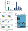
Hydrophilic amino acids at position 348 render NIS inactive. MDCK-II cells were transfected with control (pcDNA3.1) WT, Y348H, or Y348Q NIS cDNAs. (A) Steady-state I transport assay (30 min) carried out in the absence (blue bars) or presence (gray bars) of ClO4 at 10 μM or 100 μM I/140 mM Na+. Error bars indicate SE for four independent experiments. (B) Flow cytometry analysis (FACS) using an anti-HA antibody to detect NIS expression in MDCK-II cells at the cell surface (nonpermeabilized) or anywhere within the cell (permeabilized). (C) Immunoblot analysis of membrane proteins (10 μg) obtained from MDCK-II cells transfected with WT, Y348H, or Y348Q NIS, treated with (+) or without (−) Endo H. Letters to the right indicate the relative electrophoretic mobility of the NIS molecules, which depends on the glycosylation of the proteins, as in Figure 2.
FIG. 4.
FIG. 4.
Hydrophobic residues at position 348 yield active NIS proteins. (A) Localization of Y348D in TMS9 and residues in TMS1 (L24), TMS5 (L156, S170, and I174), and TMS9 (L344, A345, and L352) that may interact with Y348D. (B) Steady-state I transport assay (30 min) in control, WT, Y348A, Y348L, and Y348F NIS-transfected MDCK II cells in the absence (blue bars) or presence (gray bars) of ClO4 at 10 μM or 100 μM I and 140 mM Na+. (C) Flow cytometry analysis of total expression and cell surface expression of NIS mutants, carried out as in the experiment whose results are shown in Figure 3B. (D) Immunoblot analysis of membrane proteins (10 μg) obtained from MDCK-II cells transfected with Y348A, Y348L, or Y348F NIS and treated with (+) or without (−) Endo H. Letters to the right indicate the relative electrophoretic mobility of the NIS molecules, which depends on the glycosylation of the proteins (cf. Fig. 2).

References

    1. Zimmermann MB, Boelaert K. 2015Iodine deficiency and thyroid disorders. Lancet Diabetes Endocrinol 3:286–295 - PubMed
    1. The Iodine Global Network 2020Global Scorecard of Iodine Nutrition in 2020 in the General Population Based on School-Age Children (SAC). IGN, Ottawa, Canada
    1. Carrasco N1993Iodide transport in the thyroid gland. Biochim Biophys Acta 1154:65–82 - PubMed
    1. Dai G, Levy O, Carrasco N. 1996Cloning and characterization of the thyroid iodide transporter. Nature 379:458–460 - PubMed
    1. Levy O, De la Vieja A, Ginter CS, Riedel C, Dai G, Carrasco N. 1998. N-linked glycosylation of the thyroid Na+/I- symporter (NIS). Implications for its secondary structure model. J Biol Chem 273:22657–22663 - PubMed

Publication types

LinkOut - more resources