. 2021 Mar 12:(169):10.3791/62385.
doi: 10.3791/62385.

Ganglioside Extraction, Purification and Profiling

Affiliations

Ganglioside Extraction, Purification and Profiling

Mitchell J Porter et al. J Vis Exp. .

Abstract

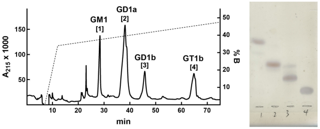
Gangliosides are glycosphingolipids that contain one or more sialic acid residues. They are found on all vertebrate cells and tissues but are especially abundant in the brain. Expressed primarily on the outer leaflet of the plasma membranes of cells, they modulate the activities of cell surface proteins via lateral association, act as receptors in cell-cell interactions and are targets for pathogens and toxins. Genetic dysregulation of ganglioside biosynthesis in humans results in severe congenital nervous system disorders. Because of their amphipathic nature, extraction, purification, and analysis of gangliosides require techniques that have been optimized by many investigators in the 80 years since their discovery. Here, we describe bench-level methods for the extraction, purification, and preliminary qualitative and quantitative analyses of major gangliosides from tissues and cells that can be completed in a few hours. We also describe methods for larger scale isolation and purification of major ganglioside species from brain. Together, these methods provide analytical and preparative scale access to this class of bioactive molecules.


Conflict of interest statement

Disclosures

The authors claim no competing interests.

Figures

Figure 1:
Figure 1:. Major brain gangliosides and their biosynthetic precursors.
Structures are shown using Symbol Nomenclature for Glycans.
Figure 2:
Figure 2:. Ganglioside TLC equipment and set up.
A twin trough chamber is filled to ≈ 0.5 cm on both sides with running solvent. The plate is placed against one side with the origin end immersed in the running buffer. The chamber is covered with an acrylic box to avoid air currents. Panel A, side view prior to plate insertion. The solvent level is visible a few mm above the chamber bottom; Panel B, front view during development. The solvent front is visible at about 40% of the way up the plate.
Figure 3:
Figure 3:. TLC plate of resolved mixed ganglioside.
TLC plate of resolved mixed ganglioside standards (left lane) and purified mixed bovine grey matter gangliosides (right lane) after resorcinol staining and heating with glass cover plate clipped in place. Standard gangliosides (top to bottom) are GM3, GM2, GM1, GD3, GD1a, GD1b, GT1b and GQ1b. After cooling, the plate can be imaged and/or the cover plate taped in place for storage.
Figure 4:
Figure 4:. TLC of mouse brain gangliosides.
TLC of mouse brain gangliosides from wild-type (WT) and St3gal single and double-null mice purified as in Protocol 1. This figure has been modified from Sturgill et al, 2012.
Figure 5:
Figure 5:. MS of permethylated wild-type and St3gal2/3-double-null mouse brain gangliosides purified as in section 1.
Note that GD1a and GD1b resolve by TLC (Figure 4) but have the same mass so are not distinguishable by onedimensional MS. This figure has been modified from Sturgill et al, 2012.
Figure 6:
Figure 6:. Representative HPLC of bovine brain gangliosides.
The elution gradient (%B, dotted line) is overlaid on the absorbance (A215, solid line) for the first 75 min of the cycle. Peaks (A215) were collected (numbers in brackets) and subjected to TLC as in Protocol 3. Lane numbers refer to the peak numbers on the chromatogram.
Figure 7:
Figure 7:. TLC plate of resolved mixed ganglioside.
TLC plate of resolved mixed gangliosides standards (lane 1) along with post-saponification partitioned gangliosides (lane 2) and released fatty acids (lane 3). Lipids, including gangliosides, are detected with p-anisaldehyde stain. Standard gangliosides (top to bottom) are GM3, GM2, GM1, GD3, GD1a, GD1b, and GT1b.

References

    1. Schnaar RL The Biology of Gangliosides. Advances in Carbohydrate Chemistry and Biochemistry. 76, 113–148 (2019). - PubMed
    1. DeMarco ML, Woods RJ Atomic-resolution conformational analysis of the GM3 ganglioside in a lipid bilayer and its implications for ganglioside-protein recognition at membrane surfaces. Glycobiology. 19 (4), 344–355 (2009). - PMC - PubMed
    1. Schnaar RL Gangliosides of the vertebrate nervous system. Journal of Molecular Biology. 428, 3325–3336 (2016). - PMC - PubMed
    1. Klenk E Über die Ganglioside, eine neue Gruppe von zuckerhaltigen Gehirnlipoiden [About gangliosides, a new group of sugar-containing brain lipids], Hoppe-Seyler’s Zeitschrift für Physiologische Chemie. 273, 76–86 (1942).
    1. Uemura S, Go S, Shishido F, Inokuchi J Expression machinery of GM4: the excess amounts of GM3/GM4S synthase (ST3GAL5) are necessary for GM4 synthesis in mammalian cells. Glycoconjugate Journal. 31 (2), 101–108 (2014). - PubMed

Publication types

LinkOut - more resources