Multicenter Study
. 2021 Mar 1;4(3):e213538.
doi: 10.1001/jamanetworkopen.2021.3538.

Utility of Fetal Cardiovascular Magnetic Resonance for Prenatal Diagnosis of Complex Congenital Heart Defects

Affiliations
Multicenter Study

Utility of Fetal Cardiovascular Magnetic Resonance for Prenatal Diagnosis of Complex Congenital Heart Defects

Daniel Ryd et al. JAMA Netw Open. .

Erratum in

  • Errors in Figure Titles and Article Information.
    [No authors listed] [No authors listed] JAMA Netw Open. 2021 Apr 1;4(4):e2111261. doi: 10.1001/jamanetworkopen.2021.11261. JAMA Netw Open. 2021. PMID: 33890998 Free PMC article. No abstract available.
  • Author Name Change.
    [No authors listed] [No authors listed] JAMA Netw Open. 2022 Mar 1;5(3):e225825. doi: 10.1001/jamanetworkopen.2022.5825. JAMA Netw Open. 2022. PMID: 35289868 Free PMC article. No abstract available.

Abstract

Importance: Prenatal diagnosis of complex congenital heart defects reduces mortality and morbidity in affected infants. However, fetal echocardiography can be limited by poor acoustic windows, and there is a need for improved diagnostic methods.

Objective: To assess the clinical utility of fetal cardiovascular magnetic resonance imaging in cases in which fetal echocardiography could not visualize all relevant anatomy.

Design, setting, and participants: This cohort study was conducted between January 20, 2017, and June 29, 2020, at Skåne University Hospital (Lund, Sweden), a tertiary center for pediatric cardiology and thoracic surgery. Participants were fetuses referred for fetal cardiovascular magnetic resonance examination by a pediatric cardiologist after an inconclusive echocardiograph.

Exposures: Fetal cardiovascular magnetic resonance examination requested by the patient's pediatric cardiologist.

Main outcomes and measures: Any change in patient management because of diagnostic information gained from fetal cardiovascular magnetic resonance imaging.

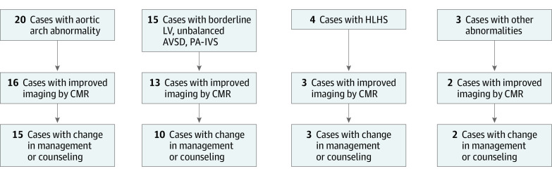
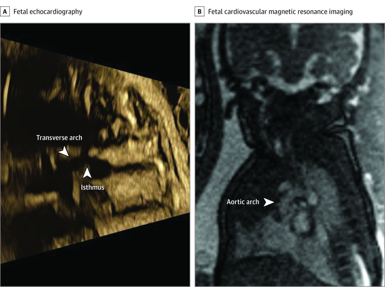
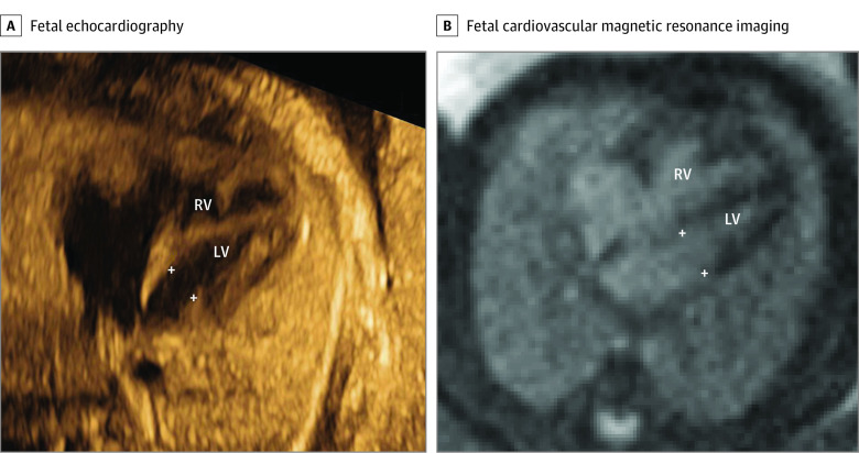
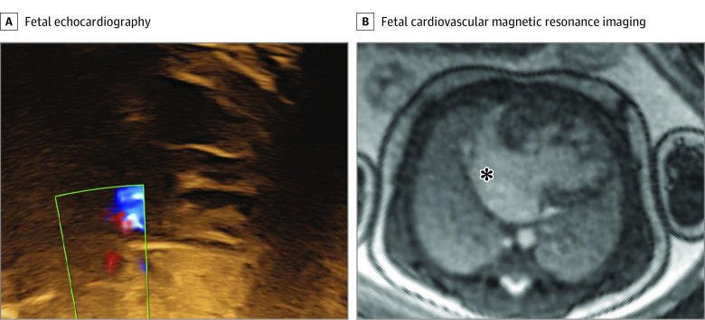
Results: A total of 31 fetuses underwent cardiovascular magnetic resonance examination at a median gestational age of 36 weeks (range, 31-39 weeks). Overall, fetal cardiovascular magnetic resonance imaging had clinical utility, affecting patient management and/or parental counseling in 26 cases (84%). For aortic arch anatomy including signs of coarctation (20 fetuses), fetal cardiovascular magnetic resonance imaging added diagnostic information in 16 cases (80%). For assessment of univentricular vs biventricular outcome in borderline left ventricle, unbalanced atrioventricular septal defect, and pulmonary atresia with intact ventricular septum (15 fetuses), fetal cardiovascular magnetic resonance imaging visualized intracardiac anatomy and ventricular function, allowing assessment of outcome in 13 cases (87%). In 4 fetuses with hypoplastic left heart syndrome, fetal cardiovascular magnetic resonance imaging helped delivery planning in 3 cases (75%). Finally, fetal cardiovascular magnetic resonance imaging provided valuable information for parental counseling in 21 cases (68%).

Conclusions and relevance: In this cohort study, fetal cardiovascular magnetic resonance imaging added clinically useful information to what was available from echocardiography. These findings suggest that fetal CMR has the potential to affect clinical decision-making in challenging cases of congenital heart defects with inconclusive data from echocardiography. Fetal cardiovascular magnetic resonance imaging showed an association with clinical decision-making, including mode of delivery and early postnatal care, as well as with parental counseling.


Conflict of interest statement

Conflict of Interest Disclosures: Dr Arheden reported being an owner of Imacor Ab outside the submitted work. No other disclosures were reported.

Figures

Figure 1.
Figure 1.. Overview of the Study Population and Utility of Fetal Cardiovascular Magnetic Resonance (CMR)
A flowchart of the included main groups of suspected pathology, in how many cases fetal CMR led to improved imaging and information, and change in management or counseling. AVSD indicates atrioventricular septal defect; LV, left ventricle; HLHS, hypoplastic left heart syndrome; PA-IVS, pulmonary atresia with intact ventricular septum.
Figure 2.
Figure 2.. Evaluation of Aortic Arch Anatomy in a Fetus With Suspected Aortic Arch Hypoplasia and/or Coarctation
Fetal echocardiography (A) showed suspected aortic arch hypoplasia and/or coarctation. The arrows indicate a narrow transverse aortic arch and aortic isthmus. However, fetal cardiovascular magnetic resonance imaging showed a normal-sized aortic arch with no signs of coarctation (B). The CMR findings were confirmed by postnatal echocardiography (not shown). Contrast and brightness in the CMR image have been optimized for printing (case 8; eTable in the Supplement).
Figure 3.
Figure 3.. Measurement of Atrioventricular Valve Annulus for Assessment of Univentricular vs Biventricular Outcome
A 4-chamber image by fetal echocardiography (A) showed an underdeveloped mitral valve annulus (5.6 mm; z score, −3.2) and a non–apex-forming left ventricle; however, the case was suspected to be underestimated or misdiagnosed due to suboptimal image projection. A 4-chamber image by fetal cardiovascular magnetic resonance (B) showed a normal-sized mitral valve annulus (9.4 mm; z score −0.3), 2 papillary muscles, and a narrow, almost apex-forming, left ventricle (LV) with normal function, suggesting biventricular outcome, as confirmed postpartum. White crosses indicate the mitral annulus. Contrast and brightness in the CMR image have been optimized for printing (case 13) (eTable in the Supplement). RV indicates right ventricle.
Figure 4.
Figure 4.. Evaluation of Atrial Restriction in a Fetus With Hypoplastic Left Heart Syndrome
Fetal echocardiography could not visualize the atrial cavity or Doppler flow across the atrial septum due to poor acoustic windows (A). Fetal cardiovascular magnetic resonance showed a large interatrial communication, indicated by the asterisk, and no nutmeg pattern (B). Therefore, the risk of restrictive atrial septum was considered low (albeit a membrane could not be ruled out), and the fetus was planned for a vaginal delivery without cardiac catheterization laboratory on standby. Contrast and brightness in the CMR image have been optimized for printing (case 5) (eTable in the Supplement).
Figure 5.
Figure 5.. Basic Assessment in a Fetus With Risk Factors for Cardiac Malformation and a Very Poor Acoustic Window
The fetal heart and vessels were not visible at all during fetal echocardiography, due to mother having obesity (A). Fetal cardiovascular magnetic resonance imaging (MRI) showed normal-sized ventricles with normal systolic function and at least 1 normal pulmonary vein (PV) without obvious direct or indirect signs of aberrant pulmonary veins (B), superior venae cavae (SVC) and inferior venae cavae (IVC) and normal ascending aorta (Ao) (C), and normal main pulmonary artery (MPA) (D). This information ruled out major congenital heart disease, and delivery was planned at the hospital closest to the patient’s home. A nonurgent postnatal echocardiography examination was performed before discharge, which showed a normal cardiovascular anatomy. Contrast and brightness in the CMR images have been optimized for printing (case 30) (eTable in the Supplement).

Comment in

Similar articles

Cited by

References

    1. van der Linde D, Konings EEM, Slager MA, et al. . Birth prevalence of congenital heart disease worldwide: a systematic review and meta-analysis. J Am Coll Cardiol. 2011;58(21):2241-2247. doi:10.1016/j.jacc.2011.08.025 - DOI - PubMed
    1. Wen SW, Liu S, Joseph KS, Rouleau J, Allen A. Patterns of infant mortality caused by major congenital anomalies. Teratology. 2000;61(5):342-346. doi:10.1002/(SICI)1096-9926(200005)61:5<342::AID-TERA5>3.0.CO;2-7 - DOI - PubMed
    1. Bakker MK, Bergman JEH, Krikov S, et al. . Prenatal diagnosis and prevalence of critical congenital heart defects: an international retrospective cohort study. BMJ Open. 2019;9(7):e028139. doi:10.1136/bmjopen-2018-028139 - DOI - PMC - PubMed
    1. Levey A, Glickstein JS, Kleinman CS, et al. . The impact of prenatal diagnosis of complex congenital heart disease on neonatal outcomes. Pediatr Cardiol. 2010;31(5):587-597. doi:10.1007/s00246-010-9648-2 - DOI - PMC - PubMed
    1. Satomi G, Yasukochi S, Shimizu T, Takigiku K, Ishii T. Has fetal echocardiography improved the prognosis of congenital heart disease? comparison of patients with hypoplastic left heart syndrome with and without prenatal diagnosis. Pediatr Int. 1999;41(6):728-732. doi:10.1046/j.1442-200x.1999.01154.x - DOI - PubMed

Publication types