. 2021 Mar 29;11(1):51.
doi: 10.1186/s13613-021-00837-1.

Clinical performance of lung ultrasound in predicting ARDS morphology

Affiliations

Clinical performance of lung ultrasound in predicting ARDS morphology

Andrea Costamagna et al. Ann Intensive Care. .

Abstract

Background: To assess diagnostic performance of lung ultrasound (LUS) in identifying ARDS morphology (focal vs non-focal), compared with the gold standard computed tomography.

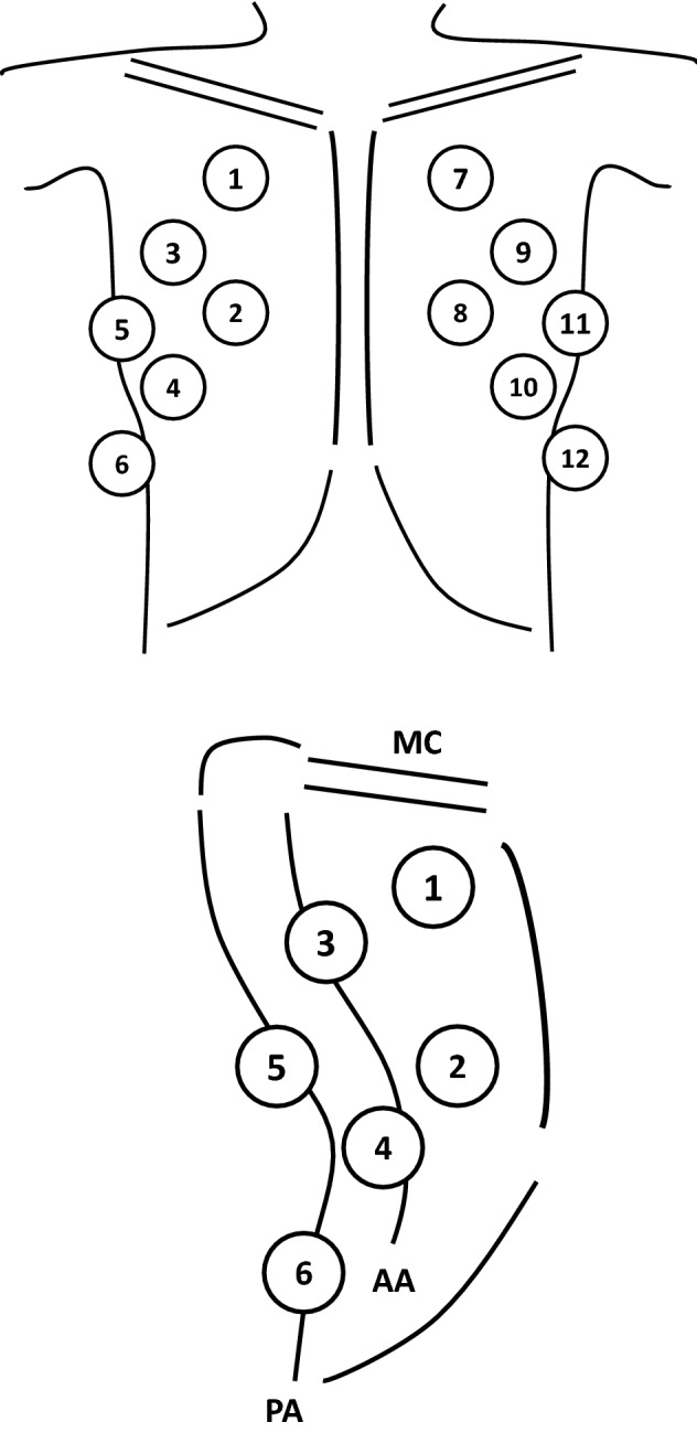
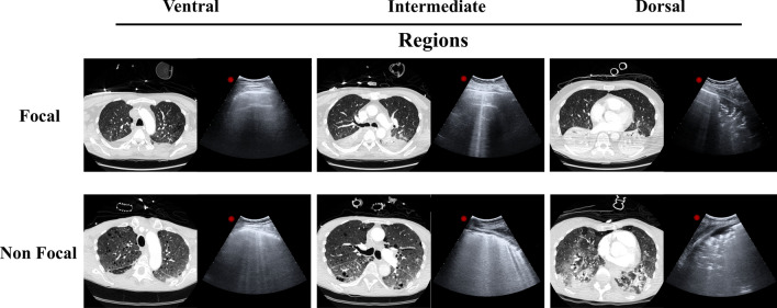
Methods: Mechanically ventilated ARDS patients undergoing lung computed tomography and ultrasound were enrolled. Twelve fields, were evaluated. LUS score was graded from 0 (normal) to 3 (consolidation) according to B-lines extent. Total and regional LUS score as the sum of the four ventral (LUSV), intermediate (LUSI) or dorsal (LUSD) fields, were calculated. Based on lung CT, ARDS morphology was defined as (1) focal (loss of aeration with lobar distribution); (2) non-focal (widespread loss of aeration or segmental loss of aeration distribution associated with uneven lung attenuation areas), and diagnostic accuracy of LUS in discriminating ARDS morphology was determined by AU-ROC in training and validation set of patients.

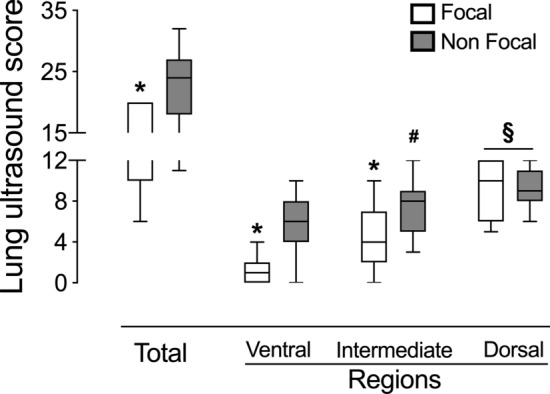
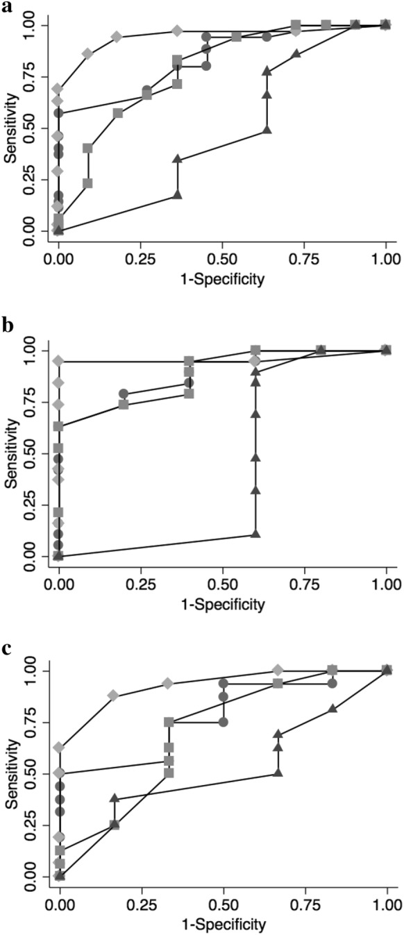
Results: Forty-seven patients with ARDS (25 training set and 22 validation set) were enrolled. LUSTOT, LUSV and LUSI but not LUSD score were significantly lower in focal than in non-focal ARDS morphologies (p < .01). The AU-ROC curve of LUSTOT, LUSV, LUSI and LUSD for identification of non-focal ARDS morphology were 0.890, 0.958, 0.884 and 0.421, respectively. LUSV value ≥ 3 had the best predictive value (sensitivity = 0.95, specificity = 1.00) in identifying non-focal ARDS morphology. In the validation set, an LUSV score ≥ 3 confirmed to be highly predictive of non-focal ARDS morphology, with a sensitivity and a specificity of 94% and 100%.

Conclusions: LUS had a valuable performance in distinguishing ARDS morphology.

Keywords: ARDS; ARDS morphology; Bedside tests; Lung ultrasound; Point of care diagnostic tests; Respiratory monitoring.


Conflict of interest statement

The authors report the absence of conflicts of interest related to the submitted work.

Figures

Fig. 1
Fig. 1
Representative image of ultrasound anatomical landmarks
Fig. 2
Fig. 2
Representative lung TC images and their corresponding LUS images in ventral, intermediate and dorsal lung regions in focal and non-focal ARDS morphologies
Fig. 3
Fig. 3
Total and regional LUS score in focal and non-focal ARDS morphologies in the overall population. *p < 0.01 focal vs non-focal ARDS morphologies; #p < 0.01 Ventral vs Intermediate lung regions in non-focal ARDS morphology; §p < 0.01 Ventral and §p < 0.05 Intermediate vs Dorsal lung regions in focal and non-focal ARDS morphology
Fig. 4
Fig. 4
Combined Receiver Operating Characteristic (ROC) curves of overall (panel A), training set (panel B) and validation set (panel C) for total (circles) and regional ventral (rhombus), intermediate (squares) and dorsal (triangles) LUS score in identifying non-focal ARDS morphologies. AUCROC (95% CI) of LUSV for non-focal ARDS was 0.948 (0.888–1.000), 0.958 (0.881–1.000) and 0.932 (0.832–1.000) in overall, training set and validation set, respectively

Similar articles

Cited by

References

    1. Thompson BT, Chambers RC, Liu KD. Acute respiratory distress syndrome. N Engl J Med. 2017;377(6):562–572. doi: 10.1056/NEJMra1608077. - DOI - PubMed
    1. Constantin JM, Grasso S, Chanques G, Aufort S, Futier E, Sebbane M, et al. Lung morphology predicts response to recruitment maneuver in patients with acute respiratory distress syndrome. Crit Care Med. 2010;38(4):1108–1117. doi: 10.1097/CCM.0b013e3181d451ec. - DOI - PubMed
    1. Gattinoni L, Caironi P, Cressoni M, Chiumello D, Ranieri VM, Quintel M, et al. Lung recruitment in patients with the acute respiratory distress syndrome. N Engl J Med. 2006;354(17):1775–1786. doi: 10.1056/NEJMoa052052. - DOI - PubMed
    1. Lichtenstein DA, Meziere GA. Relevance of lung ultrasound in the diagnosis of acute respiratory failure: the BLUE protocol. Chest. 2008;134(1):117–125. doi: 10.1378/chest.07-2800. - DOI - PMC - PubMed
    1. Bouhemad B, Liu ZH, Arbelot C, Zhang M, Ferarri F, Le-Guen M, et al. Ultrasound assessment of antibiotic-induced pulmonary reaeration in ventilator-associated pneumonia. Crit Care Med. 2010;38(1):84–92. doi: 10.1097/CCM.0b013e3181b08cdb. - DOI - PubMed