. 2021 Mar 29;15(3):e0009300.
doi: 10.1371/journal.pntd.0009300. eCollection 2021 Mar.

Early control of viral load by favipiravir promotes survival to Ebola virus challenge and prevents cytokine storm in non-human primates

Affiliations

Early control of viral load by favipiravir promotes survival to Ebola virus challenge and prevents cytokine storm in non-human primates

Stéphanie Reynard et al. PLoS Negl Trop Dis. .

Abstract

Ebola virus has been responsible for two major epidemics over the last several years and there has been a strong effort to find potential treatments that can improve the disease outcome. Antiviral favipiravir was thus tested on non-human primates infected with Ebola virus. Half of the treated animals survived the Ebola virus challenge, whereas the infection was fully lethal for the untreated ones. Moreover, the treated animals that did not survive died later than the controls. We evaluated the hematological, virological, biochemical, and immunological parameters of the animals and performed proteomic analysis at various timepoints of the disease. The viral load strongly correlated with dysregulation of the biological functions involved in pathogenesis, notably the inflammatory response, hemostatic functions, and response to stress. Thus, the management of viral replication in Ebola virus disease is of crucial importance in preventing the immunopathogenic disorders and septic-like shock syndrome generally observed in Ebola virus-infected patients.


Conflict of interest statement

The authors have declared that no competing interests exist.

Figures

Fig 1
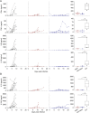
Fig 1. Virological, hematological, and biochemical parameters.
Individual values from control (black, n = 10), treated and fatally-infected (red, n = 4), and treated and surviving (blue, n = 5) animals were measured during the course of the experiment and are presented. A. Viral loads and viral titers were determined by RT-PCR and plaque assay, respectively, to determine the absolute values of genome copies and infectious particles. B. Lymphocyte and platelet counts were determined using a hematological analyzer and expressed as Giga/L (109/L). C. The prototypic hepatic enzymes ALP, AST, and ALT were determined, along with blood urea nitrogen, to evaluate liver and kidney function during EVD.
Fig 2
Fig 2. Inflammatory and anti-inflammatory mediators.
A. Inflammatory soluble mediators were measured at each sampling point during the course of the experiment by Luminex assay or ELISA for IFNα. The animals tested and the representation of the values are the same as in Fig 1. The whisker plots present the dispersion of values at day 7. P-values are indicated for pairwise comparisons (*p < 0.05; **p < 0.01). Ctrl corresponds to the control NHPs, fatal and non-fatal correspond to treated NHPs, depending on the outcome. B. Anti-inflammatory soluble mediators were analyzed and are presented as in A.
Fig 3
Fig 3. Chemokine expression.
Chemokines were measured by Luminex assay and the results are presented as in Fig 2.
Fig 4
Fig 4. T-cell response.
A. Cytokines involved in the T-cell response are presented as in Fig 2. The activation of T lymphocytes was evaluated by flow cytometry. CD69 expression was calculated for CD4 and CD8 T-cell populations. Results are presented as for the Luminex assay. B. Soluble cytotoxic molecules were assayed and are presented as in Fig 2.
Fig 5
Fig 5. Correlation between viremia and cytokine expression.
A. PCA, including results from all parameters analyzed and for which measurable values were obtained, is presented. Control animals (black), treated animals with a fatal infection (red), and treated animals who survived (blue) are presented individually. The trend for the evolution of the protein level is presented by an arrow. B. Dot plots presenting the correlation between the viral load and protein level. Protein values from each animal are plotted for each timepoint. Control animals are indicated by black dots, fatally-infected and treated animals by red dots, and survivors by blue dots. A Pearson correlation coefficient and the associated p value was calculated and is presented in each plot. C. Proteomic analysis was performed on plasma samples from days 0, 3, and 10. Proteins involved in the inflammatory pathway and significantly regulated between group and timepoint are represented (ATRN: Attractin, CD5L: CD5 molecule like, IL1RAP: IL1 receptor accessory protein, KLKB1: Kallikrein B1 and KNG1: Kininogen 1). The evolution of the protein level was plotted depending on the level of viremia (left: high viremia, right: low viremia). The colors of the dots and lines are as in B. P values were calculated depending on timepoint and group (low or high viremia) and are indicated (*p < 0.05; **p < 0.01; ***p < 0.001). The p value stated at day 0 of the high-viremia group (left) indicates that the protein level actually evolves depending on the viremia and/or the time point. For the graph of low viremia group (right), the p value at day 0 indicates the difference on day 0 between the low- and high-viremia groups. For days 3 and 10, the p values indicate the significance of the change relative to day 0 inside the group.
Fig 6
Fig 6. Dysregulation of pathways involved in homeostasis.
A. The coagulation pathway was studied based on the results of the proteomic analysis. The proteins significantly regulated in this pathway are represented as in Fig 5C (F5: Coagulation factor V, HRG: Histidine Rich Glycoprotein, F13B: Coagulation factor XIII B chain, SERPINC1: Serpin family C member 1, KLKB1: Kallikrein B1, SERPIND1: Serpin family D member 1, KNG1: Kininogen 1, PROC: Anticoagulant protein C, PROZ: Protein Z and FGG: Fibrinogen gamma chain). B. The complement pathway was also analyzed and the results are presented as in Fig 5C (C1QB: Complement C1q B chain, C1QC: Complement C1q C chain, MBL2: Mannose Binding Lectin 2, C7: Complement C7, MASP2: MBL-assocoated serine protease 2, C1QA: Complement C1q A chain, CR1: Complement Receptor Type 1, MASP1: MBL-associated serine protease 1, CR2: Complement Receptor type 2 and C1RL: Complement C1r subcomponent like). C. Heat-shock proteins that were significantly regulated in the stress-response pathway are represented as in Fig 5C (HSPA4: Heat Shock 70kDa Protein 4 and HSP90AB1: Heat Shock Protein 90kDa Protein 1 beta).

Similar articles

Cited by

References

    1. Nguyen THT, Guedj J, Anglaret X, Laouenan C, Madelain V, Taburet A-M, et al.. Favipiravir pharmacokinetics in Ebola-Infected patients of the JIKI trial reveals concentrations lower than targeted. PLoS Negl Trop Dis. 2017. February;11(2):e0005389. 10.1371/journal.pntd.0005389 - DOI - PMC - PubMed
    1. Sissoko D, Laouenan C, Folkesson E, M’Lebing A-B, Beavogui A-H, Baize S, et al.. Experimental Treatment with Favipiravir for Ebola Virus Disease (the JIKI Trial): A Historically Controlled, Single-Arm Proof-of-Concept Trial in Guinea. Lipsitch M, editor. PLOS Med [Internet]. 2016. March 1 [cited 2020 Jan 28];13(3):e1001967. Available from: https://dx.plos.org/10.1371/journal.pmed.1001967 - DOI - PMC - PubMed
    1. St Claire MC, Ragland DR, Bollinger L, Jahrling PB. Animal Models of Ebolavirus Infection. Comp Med. 2017. June;67(3):253–62. - PMC - PubMed
    1. Bixler SL, Bocan TM, Wells J, Wetzel KS, Van Tongeren SA, Dong L, et al.. Efficacy of favipiravir (T-705) in nonhuman primates infected with Ebola virus or Marburg virus. Antiviral Res [Internet]. 2018. March;151:97–104. Available from: https://linkinghub.elsevier.com/retrieve/pii/S0166354217307374 10.1016/j.antiviral.2017.12.021 - DOI - PubMed
    1. Guedj J, Piorkowski G, Jacquot F, Madelain V, Nguyen THT, Rodallec A, et al.. Antiviral efficacy of favipiravir against Ebola virus: A translational study in cynomolgus macaques. Boyles T, editor. PLOS Med [Internet]. 2018. March 27;15(3):e1002535. Available from: 10.1371/journal.pmed.1002535 - DOI - PMC - PubMed

MeSH terms