. 2021;13(4):211-224.
doi: 10.1159/000514546. Epub 2021 Mar 29.

MASP-1 and MASP-3 Bind Directly to Aspergillus fumigatus and Promote Complement Activation and Phagocytosis

Affiliations

MASP-1 and MASP-3 Bind Directly to Aspergillus fumigatus and Promote Complement Activation and Phagocytosis

Anne Rosbjerg et al. J Innate Immun. 2021.

Abstract

Activation of the complement system is mediated by the interaction between pathogens and pattern recognition molecules (PRMs); mannose-binding lectin (MBL), ficolins, and collectin-10/-11 from the lectin pathway and C1q from the classical pathway. Lectin pathway activation specifically depends on proteases named MBL-associated serine proteases (MASPs) that are found in complexes with PRMs. In this study, we hypothesize that MASPs can recognize selected pathogens independently of PRMs. Using different clinical strains of opportunistic fungi, we have observed that MASPs directly recognize certain fungal pathogens in a way that can facilitate complement activation. Among these were Aspergillus fumigatus - a dangerous pathogen, especially for immunocompromised patients. In flow cytometry and fluorescence microscopy, we found that MASP-1 and -3 bound to all A. fumigatus growth stages (conidia, germ tubes, and hyphae), whereas rMASP-2 and the nonproteolytic rMAP-1 did not. Bound rMASPs could recruit rMBL and rficolin-3 to A. fumigatus conidia in a nonclassical manner and activate complement via rMASP-2. In experiments using recombinant and purified components, rMASP-1 increased the neutrophilic phagocytosis of conidia. In serum where known complement activation pathways were blocked, phagocytosis could be mediated by rMASP-3. We have encountered an unknown pathway for complement activation and found that MASP-1 and MASP-3 have dual functions as enzymes and as PRMs.

Keywords: Aspergillosis; Fungi; Lectin pathway; Mannose-binding lectin-associated serine protease; Pattern recognition molecules.


Conflict of interest statement

The authors have no conflict of interest to declare.

Figures

Fig. 1
Fig. 1
Recombinant and purified MASP-1 and −3 bind to a. fumigatus. a Histogram showing binding of the following MASP variants to A. fumigatus strain 6871 resting conidia: rMASP-1, −2, and −3, rMAP-1, rMASP-1/-3 heavy chain, and purified MASP-1/-3 from serum. b, c The purification was evaluated in Western blots detecting MASP-1 and −3 (b) and MASP-3 (c). Lane 1: rMASP-1. Lane 2: rMASP-3. Lane 3: purified MASP-1/-3 mix. The shown histogram is representative of 3 independent experiments (see also online suppl. Fig. 1 for further evaluation of the serum-purified MASPs). MASP, mannose-binding lectin-associated serine protease; MFI, mean fluorescence intensity.
Fig. 2
Fig. 2
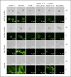
MASP-1 and MASP-3 bind to 4 growth stages of A. fumigatus. Resting conidia, swollen conidia, germ tubes, and hyphae from A. fumigatus strain Af293 (rows) were incubated with different MASP variants (columns). We applied a primary mAb (8B3) that recognizes a common epitope on all the included MASP variants. Binding was detected in fluorescence microscopy using an Alexa flour 488-conjugated secondary pAb. Both BF and F images are shown. Images were obtained on a Zeiss Axio Observer using a Plan-Apochromat. ×63/1.40 Oil DIC M27. Exposure time and image editing (brightness/background) were done equally between the samples and the negative controls (no MASPs). Shown images are representative of 2 independent experiments. MASP, mannose-binding lectin-associated serine protease; BF, bright-field; F, fluorescent.
Fig. 3
Fig. 3
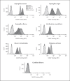
MASP binding to different opportunistic fungi. Histograms showing binding of rMASP-1, rMASP-3, and rMAP-1 to conidia from various opportunistic fungi; A. terreus, A. niger, A. flavus, L. corymbifera, R. arrhizus, M. circinelloides, and C. albicans. Binding was measured with anti-MASP-1/-3/MAP-1 mAb 8B3 using flow cytometry. Histograms are representative of 2 independent experiments. MASP, mannose-binding lectin-associated serine protease; MFI, mean fluorescence intensity.
Fig. 4
Fig. 4
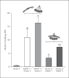
MASPs recruit PRMs to the fungal surface. A. fumigatus strain 6871 resting conidia were incubated with rMASP-1, rMASP-3, and rMAP-1 to test the recruitment of rficolin-3 (a), rMBL + MBL inhibitor (b), and rMBL + mannose (c). Results represent the means of 3 independent experiments ±SD. *p ≤ 0.05, **p ≤ 0.01, 1-way ANOVA, Bonferroni's correction. MASP, mannose-binding lectin-associated serine protease; PRMs, pattern recognition molecules; MBL, mannose-binding lectin; MFI, mean fluorescence intensity.
Fig. 5
Fig. 5
Preformed complexes of rficolin-3 and rMASPs bind to A. fumigatus. Preformed complexes of rficolin-3 and rMASP-1 or rMASP-3 were incubated with A. fumigatus strain 6871 resting conidia and afterward binding of rficolin-3 was measured using flow cytometry. Stepwise incubations as shown in Fig. 4 were also included. Results represent the means of 3 independent experiments ±SD. *p ≤ 0.05, **p ≤ 0.01, 1-way ANOVA, Bonferroni's correction. MASP, mannose-binding lectin-associated serine protease; MFI, mean fluorescence intensity.
Fig. 6
Fig. 6
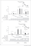
MASP-recruited PRM/MASP-2 complexes activate complement. A. fumigatus strain 6871 resting conidia were incubated with rMASP-1 or rMASP-3 to recruit PRM/MASP-2 complexes before the addition of purified C2, C4, and C3 and deposition of C4b (a), C3b (b) on the cell surface was measured by flow cytometry. When using rMBL/rMASP-2 complexes, both situation 1 and 2 in the top right drawing is possible. For rficolin-3/rMASP-2 complexes, only rMASP-recruitment, situation 2, is possible as ficolin-3 does not bind independently. Results represent the means of 3 independent experiments ±SD. *p ≤ 0.05, **p ≤ 0.01, 1-way ANOVA, Bonferroni's correction. MASP, mannose-binding lectin-associated serine protease; PRM, pattern recognition molecule; MBL, mannose-binding lectin; MFI, mean fluorescence intensity.
Fig. 10
Fig. 10
Illustration of the mechanism behind MASP recruitment of PRMs and subsequent complement activation. MASP-1 and MASP-3 can bind directly to the opportunistic pathogenic fungus A. fumigatus in a manner possibly implicating the CCP2 and protease domains of the MASPs. In theory, MASPs can bind with either 1 or 2 “arms” of the dimer − here the latter is depicted. The bound MASPs possibly recruit PRMs and thereby facilitate complement activation leading to C3b deposition on A. fumigatus. MASP, mannose-binding lectin-associated serine protease; PRM, pattern recognition molecule.
Fig. 7
Fig. 7
MASP-1 facilitates phagocytosis of A. fumigatus in a pure system. A. fumigatus strain 6871 FITC-conjugated resting conidia were incubated with rMASP-1 or rMASP-3 to recruit rPRM/rMASP-2 complexes. Next, purified C4, C2, and C3 were added and subsequently neutrophils isolated from human blood. Phagocytosis was measured by flow cytometry and based on that the phagocytic index was calculated as the percentage of phagocytizing neutrophils × MFI. Situation 1 and 2 in the top right drawing represent the action of rMBL/rMASP-2 complexes, whereas only situation 2 occurs for rficolin-3/rMASP-2. Graphs show the phagocytic index after rMASP-recruitment of rMBL/rMASP-2 complexes or rficolin-3/rMASP-2 complexes. Results represent the means of 3 independent experiments ±SD. *p ≤ 0.05, **p ≤ 0.01, 1-way ANOVA, Bonferroni's correction. MASP, mannose-binding lectin-associated serine protease; MFI, mean fluorescence intensity; PRMs, pattern recognition molecules; MBL, mannose-binding lectin.
Fig. 8
Fig. 8
A. fumigatus-bound MASPs activate complement in serum. A. fumigatus strain 6871 resting conidia were incubated with rMASP-1 or rMASP-3 and then added to MBL-defect serum ± C1q inhibitory mAb. Deposition of C4b, C3b was detected by flow cytometry. Results represent the means of 3 independent experiments ±SD. *p ≤ 0.05, 1-way ANOVA, Bonferroni's correction. MASP, mannose-binding lectin-associated serine protease; MBL, mannose-binding lectin.
Fig. 9
Fig. 9
MASP-3 facilitates phagocytosis of A. fumigatus in a serum system. A. fumigatus strain 6871 FITC-conjugated resting conidia were incubated with rMASP-1 or rMASP-3 before adding MBL-defect serum ± C1q inhibitory mAb and subsequently neutrophils isolated from human plasma. Phagocytosis was measured by flow cytometry and based on that the phagocytic index was calculated as the percentage of phagocytizing neutrophils × MFI. Results represent the means of 3 independent experiments ±SD. *p ≤ 0.05, 1-way ANOVA, Bonferroni's correction. MASP, mannose-binding lectin-associated serine protease; MFI, mean fluorescence intensity; MBL, mannose-binding lectin.

Similar articles

Cited by

References

    1. Garred P, Genster N, Pilely K, Bayarri-Olmos R, Rosbjerg A, Ma YJ, et al. A journey through the lectin pathway of complement-MBL and beyond. Immunol Rev. 2016;274((1)):74–97. - PubMed
    1. Wallis R. Interactions between mannose-binding lectin and MASPs during complement activation by the lectin pathway. Immunobiology. 2007;212((4–5)):289–99. - PMC - PubMed
    1. Bohlson SS, Garred P, Kemper C, Tenner AJ. Complement nomenclature-deconvoluted. Front Immunol. 2019;10:1–6. - PMC - PubMed
    1. Matsushita M, Thiel S, Jensenius JC, Terai I, Fujita T. Proteolytic activities of two types of mannose-binding lectin-associated serine protease. J Immunol. 2000;165((5)):2637–42. - PubMed
    1. Dobó J, Szakács D, Oroszlán G, Kortvely E, Kiss B, Boros E, et al. MASP-3 is the exclusive pro-factor D activator in resting blood: the lectin and the alternative complement pathways are fundamentally linked. Sci Rep. 2016;6:31877. - PMC - PubMed

Publication types

MeSH terms

Substances