. 2021 May;593(7857):142-146.
doi: 10.1038/s41586-021-03471-w. Epub 2021 Mar 29.

Escape of SARS-CoV-2 501Y.V2 from neutralization by convalescent plasma

Collaborators, Affiliations

Escape of SARS-CoV-2 501Y.V2 from neutralization by convalescent plasma

Sandile Cele et al. Nature. 2021 May.

Abstract

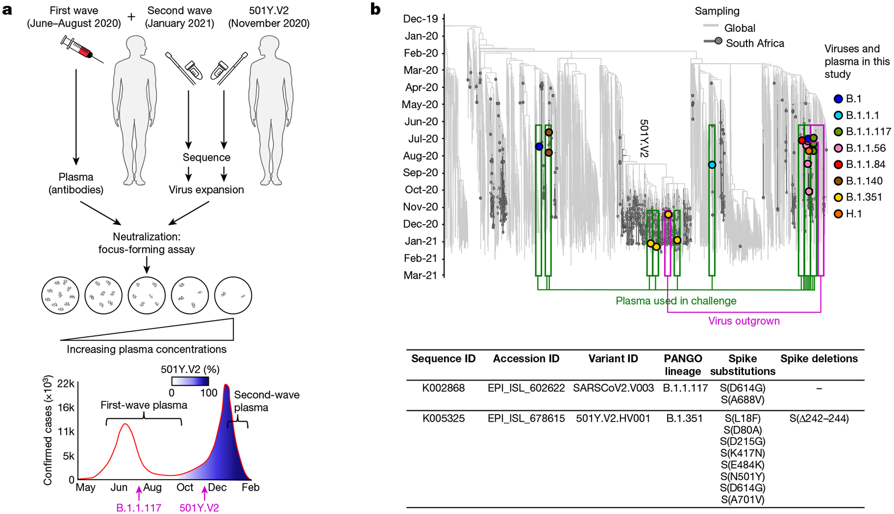
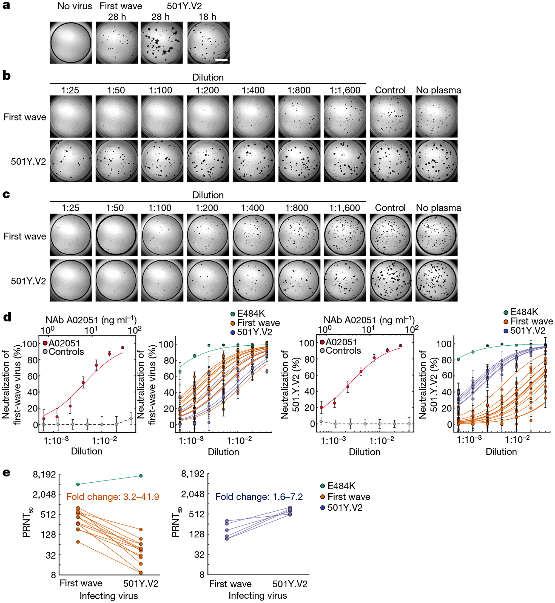
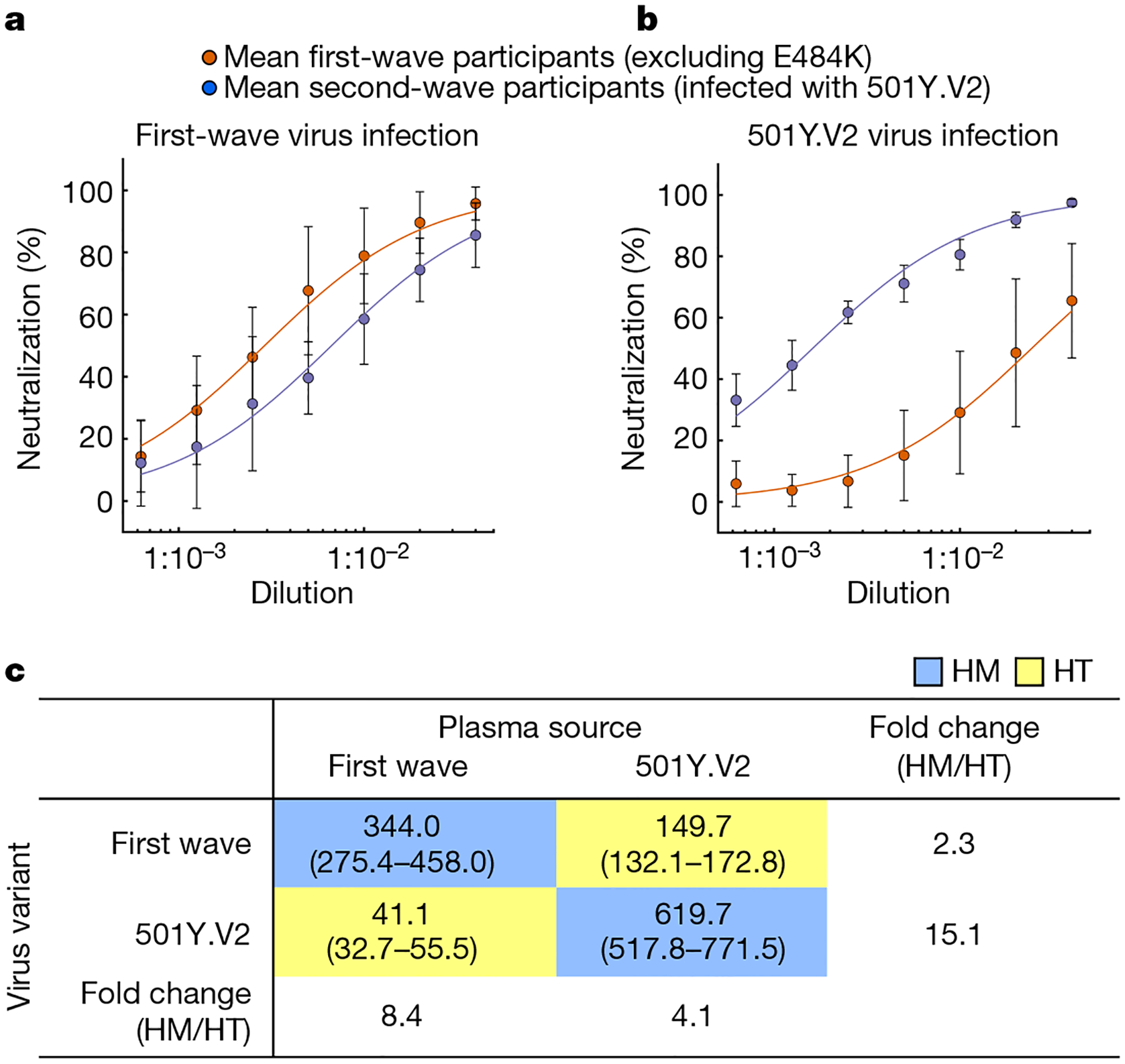
SARS-CoV-2 variants of concern (VOC) have arisen independently at multiple locations1,2 and may reduce the efficacy of current vaccines that target the spike glycoprotein of SARS-CoV-23. Here, using a live-virus neutralization assay, we compared the neutralization of a non-VOC variant with the 501Y.V2 VOC (also known as B.1.351) using plasma collected from adults who were hospitalized with COVID-19 during the two waves of infection in South Africa, the second wave of which was dominated by infections with the 501Y.V2 variant. Sequencing demonstrated that infections of plasma donors from the first wave were with viruses that did not contain the mutations associated with 501Y.V2, except for one infection that contained the E484K substitution in the receptor-binding domain. The 501Y.V2 virus variant was effectively neutralized by plasma from individuals who were infected during the second wave. The first-wave virus variant was effectively neutralized by plasma from first-wave infections. However, the 501Y.V2 variant was poorly cross-neutralized by plasma from individuals with first-wave infections; the efficacy was reduced by 15.1-fold relative to neutralization of 501Y.V2 by plasma from individuals infected in the second wave. By contrast, cross-neutralization of first-wave virus variants using plasma from individuals with second-wave infections was more effective, showing only a 2.3-fold decrease relative to neutralization of first-wave virus variants by plasma from individuals infected in the first wave. Although we tested only one plasma sample from an individual infected with a SARS-CoV-2 variant with only the E484K substitution, this plasma sample potently neutralized both variants. The observed effective neutralization of first-wave virus by plasma from individuals infected with 501Y.V2 provides preliminary evidence that vaccines based on VOC sequences could retain activity against other circulating SARS-CoV-2 lineages.


Conflict of interest statement

Competing interests The authors declare no competing interests.

Figures

Extended Data Fig. 1 |
Extended Data Fig. 1 |. Neutralization of first-wave and 501Y.V2 variants by convalescent plasma from first-wave infections using equal infection incubation times.
a, A representative focus-forming assay using plasma from participant 039-13-0015. b, c, Plasma neutralization of first-wave virus (b) and 501Y.V2 variants (501Y.V2.HV001 and 501Y.V2.HVdF002) (c). Coloured circles represent mean ± s.e.m. from 8 independent neutralization experiments using plasma from n = 6 participants convalescing from an infection with first-wave variants in the first peak of the pandemic in South Africa. Correspondingly coloured lines are fits of the sigmoidal equation with ID50 as the fitted parameter. Data from both 501Y.V2 variants were combined to obtain a more accurate fit as neutralization of 501Y.V2 virus infection was low in the range of plasma concentrations used. The matched infections with first-wave virus that were done in parallel with each 501Y.V2 variant were also combined. One experiment was removed in the process of quality control owing to plate edge effects, which were subsequently corrected by adding sterile water between wells. Black points represent a pool of plasma from three uninfected control individuals. The transmission index (Tx) is the number of foci in the presence of the plasma dilution normalized to the number of foci in the absence of plasma. d, Plasma ID50 values and ratios for first-wave and 501Y.V2 variants. Knockout (KO) was scored as ID50 > 1. ND, not defined. e, Plasma neutralization of all first-wave and all 501Y.V2 variants combined. Data are mean ± s.e.m. across all plasma donors (n = 6) from 8 independent neutralization experiments.
Extended Data Fig. 2 |
Extended Data Fig. 2 |. Neutralization of first-wave and 501Y.V2 variants by convalescent plasma: representative experiments of the first set of participant plasma tested.
Top, neutralization of first-wave virus. Bottom, neutralization of 501Y.V2. Rows are plasma dilutions, ranging from 1:25 to 1:1,600. The last three columns comprise plasma from a pool of uninfected participants, the no-plasma control and no-virus control, respectively. The first column is the neutralizing antibody A2051, with antibody concentrations in ng ml−1 (magenta). First-wave plasma donors are marked with a red line.
Extended Data Fig. 3 |
Extended Data Fig. 3 |. Neutralization of first-wave and 501Y.V2 variants by convalescent plasma: representative experiments of the second set of participant plasma tested.
Top, neutralization of first-wave virus. Bottom, neutralization of 501Y.V2. Rows are plasma dilutions, ranging from 1:25 to 1:1,600. The last three columns comprise plasma from a pool of uninfected participants, the no-plasma control and no-virus control, respectively. The first column is the neutralizing antibody A2051, with antibody concentrations in ng ml−1 (magenta). First-wave plasma donors are marked with a red line; second-wave plasma donors are marked with a blue line; and the plasma donor who was infected with SARS-CoV-2 with the E484K substitution only is marked with a green line.
Extended Data Fig. 4 |
Extended Data Fig. 4 |. Neutralization of first-wave and 501Y.V2 variants by convalescent plasma: representative experiments of the third set of participant plasma tested.
Top, neutralization of first-wave virus. Bottom, neutralization of 501Y.V2. Rows are plasma dilutions, ranging from 1:25 to 1:1,600. The last three columns comprise plasma from a pool of uninfected participants, the no-plasma control and no-virus control, respectively. The first column is the neutralizing antibody A2051, with antibody concentrations in ng ml−1 (magenta). First-wave plasma donors are marked with a red line and second-wave plasma donors are marked with a blue line.
Extended Data Fig. 5 |
Extended Data Fig. 5 |. Fit of combined data for each plasma dilution to a normal distribution.
The function normplot in MATLAB v.2019b was used to assess the fit of the data (blue crosses) to a normal distribution (solid red line). For each plot, one data point is the Tx result for one experiment for one participant at the specified dilution. The number of total experiments per viral variant was n = 42 for first-wave plasma and n = 21 for second-wave plasma. Lack of pronounced curvature of the data in the range of the solid line indicates that the data are a reasonably good fit to a normal distribution. See https://www.mathworks.com/help/stats/normplot.html for additional information.
Fig. 1 |
Fig. 1 |. Study design and sequences of SARS-CoV-2 variants.
a, We obtained convalescent plasma and sequenced the matching infecting virus from individuals with COVID-19 during the first and second waves of SARS-CoV-2 infections in South Africa. A variant that lacked the mutations in the RBD and NTD of 501Y.V2 was expanded from one participant infected in the first wave of infections in South Africa, and 501Y.V2 was expanded from a participant at the beginning of the second wave. Live-virus neutralization was assessed using a focus-forming assay. Conditions were: neutralization of non-VOC virus by plasma elicited against first-wave, non-VOC virus, neutralization of 501Y.V2 virus by plasma elicited against 501Y.V2 virus, neutralization of 501Y.V2 virus by plasma elicited against first-wave non-VOC virus, and neutralization of non-VOC virus by plasma elicited against 501Y.V2. b, Top, phylogenetic relationships and mutations in the virus sequences. Variants that elicited the antibody immunity in the plasma samples are highlighted in green boxes. Variants that were expanded are highlighted in magenta boxes. The y axis denotes the time of sampling. Bottom, substitutions and deletions that are present in the spike protein of the SARS-CoV-2 expanded variants used in the live-virus neutralization assay. See Supplementary Table 1 for a complete list of mutations in the viral genomes of variants that elicited plasma immunity and the expanded variants.
Fig. 2 |
Fig. 2 |. Neutralization of first-wave and 501Y.V2 variants by convalescent plasma elicited by first-wave and 501Y.V2 infections.
a, Focus formation by first-wave and 501Y.V2 virus variants. To obtain similar focus sizes, the incubation time with 501Y.V2 was reduced to 18 h. Scale bar, 2 mm. b, c, A representative focus-forming assay using plasma from participant 039-13-0015, who was infected with a first-wave variant (b), and participant 039-02-0033, who was infected with 501Y.V2 (c). Columns are plasma dilutions—which range from 1:25 to 1:1,600—a plasma pool from three uninfected individuals (control) and a no-plasma control (no plasma). d, Quantified neutralization per participant for the first-wave virus variants (left two plots) and 501Y.V2 (right two plots). Red points are neutralization by the A02051 neutralizing antibody (NAb), grey points show neutralization by the plasma pool from uninfected individuals (control), green points indicate neutralization by plasma from the participant who was infected with the S(E484K)-mutant virus, orange points are neutralization by plasma from participants who were infected by first-wave variants, and blue points are neutralization by plasma from participants who were infected with 501Y.V2. Data are mean and s.e.m. of 3–4 independent experiments per plasma sample of participants convalescing from infection with the first-wave (n = 14) or 501Y.V2 (n = 6) virus variants or 10 independent experiments for A02051 and uninfected plasma controls. Solid lines of the corresponding colour are fitted values using a sigmoidal equation. From left to right, the plots show the following analyses. First plot, neutralization of the first-wave virus by the neutralizing antibody A02051 (PRNT50 = 6.5 ng ml−1; 95% confidence intervals, 3.9–9.1 ng ml−1) and control plasma. Second plot, neutralization of first-wave virus by plasma from participants convalescing from infection with first-wave or 501Y.V2 viruses. Third plot, neutralization of the 501Y.V2 variant by the neutralizing antibody A02051 (PRNT50 = 3.5 ng ml−1 (2.9–4.1 ng ml−1)) and control plasma. Fourth plot, neutralization of the 501Y.V2 variant by plasma from participants convalescing from infection with first-wave or 501Y.V2 viruses. e, Decrease in PRNT50 in cross-neutralization. Left, neutralization of first-wave or 501Y.V2 virus variants by first-wave plasma. Right, neutralization of 501Y.V2 or first-wave virus variants by second-wave plasma. The fold change was calculated as PRNT50 of the homologous virus/PRNT50 of the heterologous virus and ranged from 3.2 to 41.9 for first-wave plasma, and from 1.6 to 7.2 for second-wave plasma. The fold change in PRNT50 elicited by the S(E484K)-mutant virus was excluded.
Fig. 3 |
Fig. 3 |. Cross-neutralization of first infection wave and 501Y.V2 virus across all participants.
Neutralization of the virus from the first wave (a) or 501Y.V2 (b). Sigmoidal fits were performed to the means of plasma neutralization results from the first wave (red points) and second wave (blue points) across all participants excluding a participant with plasma immunity elicited by SARS-CoV-2 with only the E484K substitution. Data are mean ± s.d. of n = 13 plasma donors infected with a first-wave virus and n = 6 plasma donors infected with a second-wave virus. c, Plasma PRNT50 as a function of plasma source (columns) and infecting viral variant (rows). Blue rectangles highlight homologous (HM) neutralization for which virus and infection wave are matched; yellow rectangles highlight heterologous, cross-neutralization (HT) for which virus and plasma are from different infection waves.

Similar articles

Cited by

References

    1. Tegally H et al. Detection of a SARS-CoV-2 variant of concern in South Africa. Nature 10.1038/s41586-021-03402-9 (2021). - DOI - PubMed
    1. Faria NR et al. Genomics and epidemiology of a novel SARS-CoV-2 lineage in Manaus, Brazil. Preprint at 10.1101/2021.02.26.21252554 (2021). - DOI - PMC - PubMed
    1. Madhi SA et al. Efficacy of the ChAdOx1 nCoV-19 COVID-19 vaccine against the B.1.351 variant. N. Engl. J. Med 10.1056/NEJMoa2102214 (2021). - DOI - PMC - PubMed
    1. Piccoli L et al. Mapping neutralizing and immunodominant sites on the SARS-CoV-2 spike receptor-binding domain by structure-guided high-resolution serology. Cell 183, 1024–1042 (2020). - PMC - PubMed
    1. McCallum M et al. N-terminal domain antigenic mapping reveals a site of vulnerability for SARS-CoV-2. Cell 10.1016/j.cell.2021.03.028 (2021). - DOI - PMC - PubMed

Publication types

MeSH terms