. 2022 May;15(2):160-167.
doi: 10.21053/ceo.2020.02383. Epub 2021 Mar 30.

Development of High-Intensity Focused Ultrasound Therapy for Inferior Turbinate Hypertrophy

Affiliations

Development of High-Intensity Focused Ultrasound Therapy for Inferior Turbinate Hypertrophy

Joon Kon Kim et al. Clin Exp Otorhinolaryngol. 2022 May.

Abstract

Objectives: Inferior turbinate (IT) hypertrophy is the main cause of chronic nasal obstruction. We developed a high-intensity focused ultrasound (HIFU) ablation device to treat patients with IT hypertrophy.

Methods: First, computed tomography images of patients with no evidence of sinonasal disease were evaluated to measure and compare the IT, medial mucosal thickness (MT), and space between the nasal septum and IT according to clinical characteristics such as septal deviation. A HIFU prototype was developed based on the above human anatomical studies. The experimental study was performed in five pigs; the nasal volume and histological changes at 1 and 4 weeks postoperatively were evaluated to compare the efficacy of HIFU turbinoplasty with that of radiofrequency turbinoplasty and a control group.

Results: The mean medial MT of the anterior, middle, and posterior portions of the IT were 4.66±1.14, 4.23±0.97, and 6.17±1.29 mm, respectively. The mean medial space was 2.65±0.79 mm. The diameter and focal depth of the prototype were 4 mm and 3 mm, respectively. HIFU showed no postoperative complications, including bleeding or scar formation. After HIFU treatment, the nasal volume increased by 196.62 mm3 (7.8%) and 193.74 mm3 (8.3%) at 1 week and 4 weeks, compared with the increase of 87.20 mm3 (3.1%) and 213.81 mm3 (9.0%), respectively, after radiofrequency therapy. A qualitative histological analysis after radiofrequency turbinoplasty showed epithelial layer disruption at 1 week and increased fibrosis, along with decreased glandular structure, at 4 weeks. The HIFU group had an intact epithelial layer at 1 week postoperatively. However, significant differences were observed at 4 weeks, including increased fibrosis and decreased glandular structure.

Conclusion: The efficacy and safety of HIFU turbinoplasty were demonstrated in an animal study. Our.

Results: warrant further human clinical trials.

Keywords: High-Intensity Focused Ultrasound Ablation; Postoperative Complications; Radiofrequency Therapy; Turbinates.


Conflict of interest statement

No potential conflict of interest relevant to this article was reported.

Figures

Fig. 1.
Fig. 1.
Anatomical evaluation of the nasal cavity by computed tomography. The unit for mucosal thickness (MT) and medial space (MS) is millimeters.
Fig. 2.
Fig. 2.
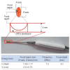
Specifications of the high-intensity focused ultrasound (HIFU) prototype design and energy. X, width of the healing region; Z, length of the healing region.
Fig. 3.
Fig. 3.
Nasal endoscopic findings in the control, radiofrequency, and high-intensity focused ultrasound (HIFU) groups. (A, B) Preoperative and postoperative findings in the control group (week 1). (C, D) Preoperative and postoperative findings in the control group (week 4). (E, F) Preoperative and postoperative findings in the radiofrequency group (week 1). (G, H) Preoperative and postoperative findings in the radiofrequency group (week 4). The red circle in (F) indicates eschar formation, bleeding, and crust formation, while the red circle in (H) indicates scar formation. (I, J) Preoperative and postoperative findings in the HIFU group (week 1). (K, L) Preoperative and postoperative findings in the HIFU group (week 4).
Fig. 4.
Fig. 4.
Computed tomography evaluation of the control, radiofrequency, and high-intensity focused ultrasound (HIFU) groups at 1 week (A) and 4 weeks (B) after turbinoplasty. The numerical values refer to the three-dimensional volume of the nasal cavity (mm3) measured preoperatively and postoperatively. Rt, right; Lt, left; NC, nasal cavity.
Fig. 5.
Fig. 5.
Pathologic evaluation of the control, radiofrequency, high-intensity focused ultrasound (HIFU)-high, and HIFU-low groups at postoperative week 1 (slide magnification: A-C, ×100; D-F, ×400; periodic acid–Schiff staining). (A) Control, (B) radiofrequency group (asterisk, epithelial disruption), (C) HIFU group (asterisk, intact epithelium), (D) control, (E) HIFU-high group, (F) HIFU-low group (asterisks, similar findings of infiltrated polymorphonuclear cells). The infiltrated polymorphonuclear cells appear more prominently in the turbinates treated with a high level of HIFU than in those treated with low level of HIFU.
Fig. 6.
Fig. 6.
Pathologic evaluation of the control, radiofrequency, and high-intensity focused ultrasound (HIFU) groups at postoperative week 4 (slide magnification: A-C, ×100; D-F, ×400; periodic acid–Schiff staining). (A) Control, (B) radiofrequency group, (C) HIFU group (asterisks, similar findings of increased fibrosis/decreased glandular structure). (D) Control, (E) HIFU-high group, (F) HIFU-low group. Similar findings of increased fibrosis and decreased glandular structure are confirmed in both the HIFU-high and HIFU-low groups.

References

    1. Stewart M, Ferguson B, Fromer L. Epidemiology and burden of nasal congestion. Int J Gen Med. 2010 Apr;3:37–45. - PMC - PubMed
    1. Jang TY, Kim YH, Shin SH. Long-term effectiveness and safety of endoscopic vidian neurectomy for the treatment of intractable rhinitis. Clin Exp Otorhinolaryngol. 2010 Dec;3(4):212–6. - PMC - PubMed
    1. Cingi C, Ure B, Cakli H, Ozudogru E. Microdebrider-assisted versus radiofrequency-assisted inferior turbinoplasty: a prospective study with objective and subjective outcome measures. Acta Otorhinolaryngol Ital. 2010 Jun;30(3):138–43. - PMC - PubMed
    1. Jenne JW, Preusser T, Gunther M. High-intensity focused ultrasound: principles, therapy guidance, simulations and applications. Z Med Phys. 2012 Dec;22(4):311–22. - PubMed
    1. Kennedy JE, Ter Haar GR, Cranston D. High intensity focused ultrasound: surgery of the future. Br J Radiol. 2003 Sep;76(909):590–9. - PubMed