Review
. 2021 Mar 29;18(1):66.
doi: 10.1186/s12985-021-01526-y.

Precision therapeutic targets for COVID-19

Affiliations
Review

Precision therapeutic targets for COVID-19

Zachary A Krumm et al. Virol J. .

Abstract

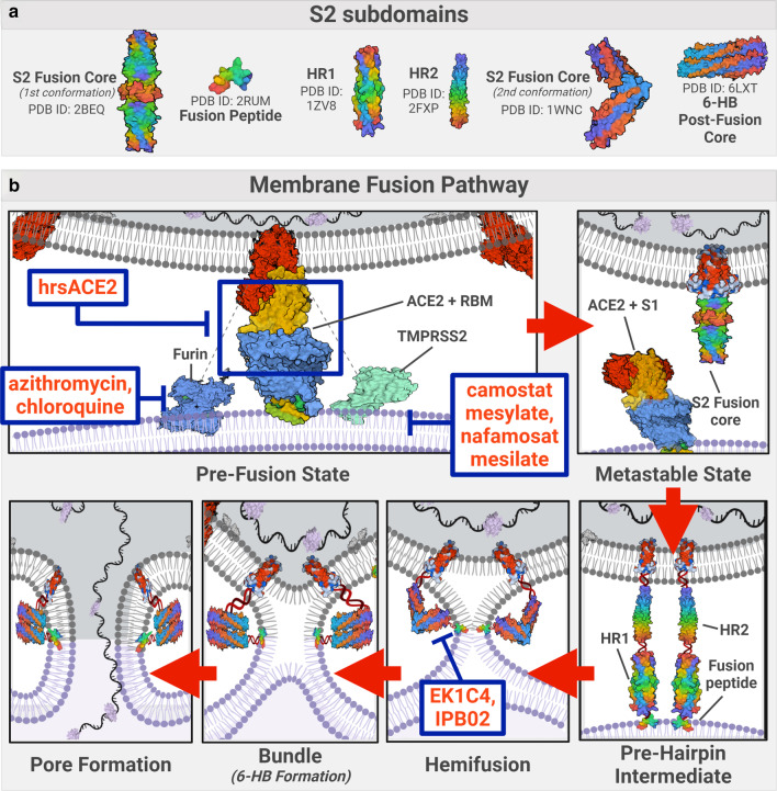
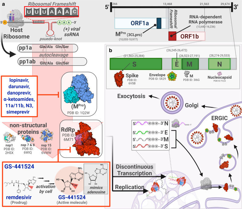
Beginning in late 2019, severe acute respiratory syndrome coronavirus 2 (SARS-CoV-2) emerged as a novel pathogen that causes coronavirus disease 2019 (COVID-19). SARS-CoV-2 has infected more than 111 million people worldwide and caused over 2.47 million deaths. Individuals infected with SARS-CoV-2 show symptoms of fever, cough, dyspnea, and fatigue with severe cases that can develop into pneumonia, myocarditis, acute respiratory distress syndrome, hypercoagulability, and even multi-organ failure. Current clinical management consists largely of supportive care as commonly administered treatments, including convalescent plasma, remdesivir, and high-dose glucocorticoids. These have demonstrated modest benefits in a small subset of hospitalized patients, with only dexamethasone showing demonstrable efficacy in reducing mortality and length of hospitalization. At this time, no SARS-CoV-2-specific antiviral drugs are available, although several vaccines have been approved for use in recent months. In this review, we will evaluate the efficacy of preclinical and clinical drugs that precisely target three different, essential steps of the SARS-CoV-2 replication cycle: the spike protein during entry, main protease (MPro) during proteolytic activation, and RNA-dependent RNA polymerase (RdRp) during transcription. We will assess the advantages and limitations of drugs that precisely target evolutionarily well-conserved domains, which are less likely to mutate, and therefore less likely to escape the effects of these drugs. We propose that a multi-drug cocktail targeting precise proteins, critical to the viral replication cycle, such as spike protein, MPro, and RdRp, will be the most effective strategy of inhibiting SARS-CoV-2 replication and limiting its spread in the general population.

Keywords: COVID-19; MPro; Main protease; RNA-dependent RNA polymerase; SARS-CoV-2; Spike protein; Therapy.


Conflict of interest statement

Dr. Todd Golde is a co-founder of Lacerta Therapeutics, Inc. and Andante Biologics Inc. and serves on their scientific advisory boards (SABs). He is on the SAB for Promis Neuroscience, Inc. In the past he has served, ad hoc, on SABs related to neurodegenerative disease programs for Eli Lilly, Novartis, Bristol Myers Squib, Abbvie, Lundbeck, Biogen and Pfizer. He has served on the medical and scientific advisory board for the Alzheimer’s Association. He serves as a scientific advisor and participates in grant reviews for BrightFocus Foundation and the American Federation for Aging Research. He is a co-inventor on multiple patents and patent applications relating to AD therapeutics. Dr. Duane A. Mitchell holds related patents that haven been licensed to iOncologi, Inc.

Figures

Fig. 1
Fig. 1
SARS-CoV-2 Viral Entry Mechanisms and Machinery. (a) SARS-CoV-2 is a lipid membrane, enveloped, plus-sense ( +) single strand (ss) RNA betacoronavirus that must undergo host lipid membrane fusion in order to gain entry into the host cell. Potential inhibitors for subsequent steps of this process are depicted. Enveloped viruses are capable of entering the host cell via (1) direct, neutral pH, plasma membrane fusion or via (2) endocytosis, where membrane fusion would rely on pH-dependent proteases and optimal intra-endosomal conditions [144, 241]. (b) Structural diagrams of key enzymes involved in viral-cellular entry. (c) Structural diagram of a spike protein (S) depicting the location of S1 and S2 subunits, following S protein cleavage, and the altered conformational states (closed and open). To initiate the entry process, S protein must undergo a conformational change from a closed to open state, which exposes the receptor binding domain (RBD) on S, allowing it to bind to angiotensin converting enzyme 2 (ACE2) on the host cell [40]. Altered S structure bound to ACE2 and S cleaved products are also shown. PDB codes for structures are referenced in Additional file 1: Table 5. Figure was created with BioRender.com
Fig. 2
Fig. 2
SARS-CoV-2 Membrane Fusion Pathway. (a) Structural diagrams of some key elements of S2 involved in membrane fusion. (b) Schematic summary of the essential steps in viral-host membrane fusion. Following the binding to ACE2, S protein must be cleaved by a protease, such as Transmembrane Serine Protease 2 (TMPRSS2), furin or cathepsin L to generate the S1 and S2 subunits, in order to release the S1 subunit; thus exposing the fusogenic core of S2 [109, 121, 242]. With its hydrophobic core exposed, S2 protein is now in a high-energy, pre-fusion, metastable state, fostered by the energetic imbalance induced by its uncovered core [150]. The S2 subunit can undergo a conformational change, extending heptad repeat 1 (HR1) and heptad repeat 2 (HR2) domains, and injecting its fusion peptide (FP) into the membrane of the host cell, forming the pre-hairpin intermediate. This pre-hairpin structure then folds back into a six helix bundle (6-HB), pulling apart the host membrane. Finally, the viral and host membranes fuse with one another, as HR1 and HR2 fold into a trimer of hairpins resulting in pore formation [152, 243]. The viral genome is then able to access the intracellular space of the host cell for transcription and replication. PDB codes for structures are referenced in Additional file 1: Table 5. Figure was created with  BioRender.com
Fig. 3
Fig. 3
Key elements of SARS-CoV-2 viral replication and some therapeutic targets. (a) ( +) RNA viruses are ‘ribosome-ready’, meaning that upon cytoplasmic entry, their genome is recognized by the host ribosome as mRNA and can immediately be translated. During translation, the viral genome employs a technique called 'ribosomal frameshifting' to produce two types of polyproteins, pp1a and pp1ab, which encode the non-structural proteins (nsps) including the viral protease MPro/nsp5 [232]. MPro first autocleaves  the polyproteins at a Gln/Ala and Gln/Ser junction, then cleaves most of the remaining proteins from the first two reading frames of the viral genome, including RdRp [232]. RdRp integrates with nsp7 and nsp8 to assemble into the polymerase holoenzyme. (b) The 3′ region of SARS-CoV-2 RNA genome encodes its structural proteins, S, Envelope (E), Membrane (M) and Nucleocapsid (N) proteins. Discontinuous transcription of the 3′ region generates a nested set of subgenomic ( −) RNAs that are copied into ( +) mRNA, resulting in the host ribosomal translation of the structural proteins [196]. RdRp is also responsible for replicating the viral genome for packaging. Replication and transcription processes are localized into interconnected, double-membraned, ER-derived vesicles called replicase-transcriptase complex (RTC) [189, 244], which centralize the machinery required for these processes and serve as a buffer to any host immune response [245]. Viral structural proteins are translated by host ribosomes from the subgenomic RNA synthesized by RdRp. After processing in the ER-Golgi Intermediate Compartment (ERGIC), the structural proteins and viral RNA are transported to budding vesicles. Finally, virus particles are assembled and released by exocytosis. PDB codes for structures are referenced in Additional file 1: Table 5. Figure was created with BioRender.com

References

    1. Wu F, Zhao S, Yu B, Chen YM, Wang W, Song ZG, et al. A new coronavirus associated with human respiratory disease in China. Nature. 2020;579:265–269. doi: 10.1038/s41586-020-2008-3. - DOI - PMC - PubMed
    1. Zhu N, Zhang D, Wang W, Li X, Yang B, Song J, et al. A novel coronavirus from patients with pneumonia in China, 2019. N Engl J Med. 2020;382:727–733. doi: 10.1056/NEJMoa2001017. - DOI - PMC - PubMed
    1. Zhou P, Yang XL, Wang XG, Hu B, Zhang L, Zhang W, et al. A pneumonia outbreak associated with a new coronavirus of probable bat origin. Nature. 2020;579:270–273. doi: 10.1038/s41586-020-2012-7. - DOI - PMC - PubMed
    1. Johns Hopkins University. COVID-19 Map—Johns Hopkins Coronavirus Resource Center. Johns Hopkins Coronavirus Resour Cent 2020:1. https://coronavirus.jhu.edu/map.html.
    1. Lu R, Zhao X, Li J, Niu P, Yang B, Wu H, et al. Genomic characterisation and epidemiology of 2019 novel coronavirus: implications for virus origins and receptor binding. Lancet. 2020;395:565–574. doi: 10.1016/S0140-6736(20)30251-8. - DOI - PMC - PubMed

MeSH terms