Review
. 2021 Jul:83:109993.
doi: 10.1016/j.cellsig.2021.109993. Epub 2021 Mar 27.

Reversion inducing cysteine rich protein with Kazal motifs and cardiovascular diseases: The RECKlessness of adverse remodeling

Affiliations
Review

Reversion inducing cysteine rich protein with Kazal motifs and cardiovascular diseases: The RECKlessness of adverse remodeling

Jacob J Russell et al. Cell Signal. 2021 Jul.

Abstract

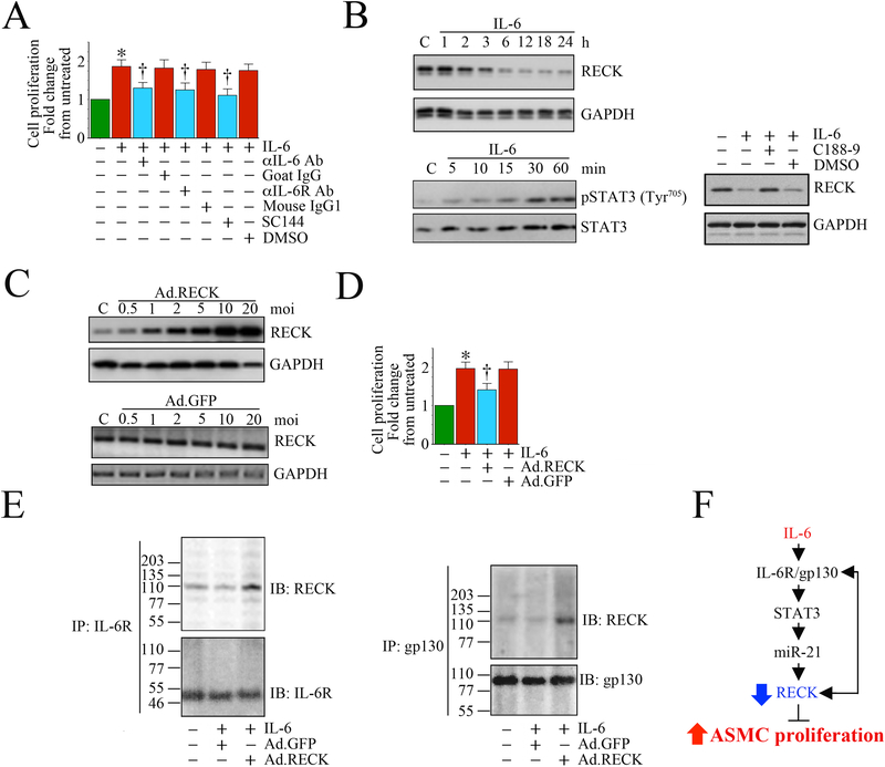
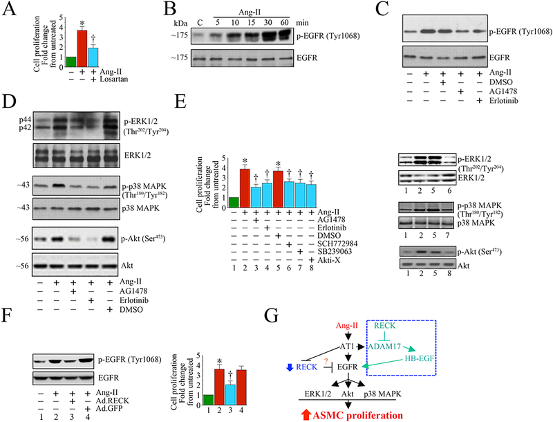
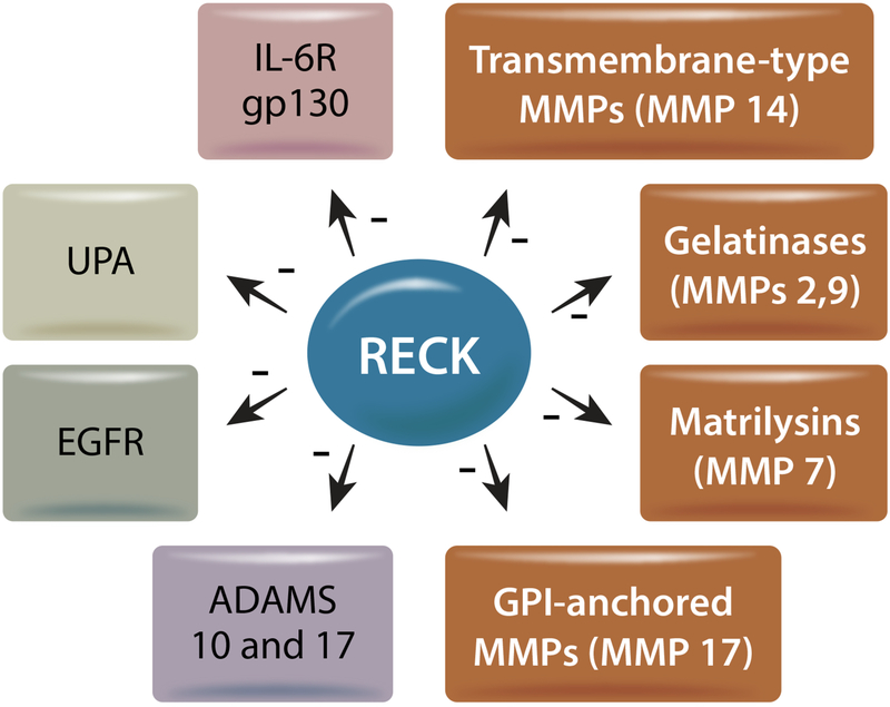
The Reversion Inducing Cysteine Rich Protein With Kazal Motifs (RECK) is a glycosylphosphatidylinositol (GPI) anchored membrane-bound regulator of matrix metalloproteinases (MMPs). It is expressed throughout the body and plays a role in extracellular matrix (ECM) homeostasis and inflammation. In initial studies, RECK expression was found to be downregulated in various invasive cancers and associated with poor prognostic outcome. Restoring RECK, however, has been shown to reverse the metastatic phenotype. Downregulation of RECK expression is also reported in non-malignant diseases, such as periodontal disease, renal fibrosis, and myocardial fibrosis. As such, RECK induction has therapeutic potential in several chronic diseases. Mechanistically, RECK negatively regulates various matrixins involved in cell migration, proliferation, and adverse remodeling by targeting the expression and/or activation of multiple MMPs, A Disintegrin And Metalloproteinase Domain-Containing Proteins (ADAMs), and A Disintegrin And Metalloproteinase With Thrombospondin Motifs (ADAMTS). Outside of its role in remodeling, RECK has also been reported to exert anti-inflammatory effects. In cardiac diseases, for example, it has been shown to counteract several downstream effectors of Angiotensin II (Ang-II) that play a role in adverse cardiac and vascular remodeling, such as Interleukin-6 (IL-6)/IL-6 receptor (IL-6R)/glycoprotein 130 (IL-6 signal transducer) signaling and Epidermal Growth Factor Receptor (EGFR) transactivation. This review article focuses on the current understanding of the multifunctional effects of RECK and how its downregulation may contribute to adverse cardiovascular remodeling.

Keywords: Adverse remodeling; EGFR; Fibrosis; Inflammation; Metallopeptidases; RECK.


Conflict of interest statement

Declaration of Competing Interest None.

Figures

Fig. 1:
Fig. 1:
Domains/motifs of canonical RECK (top) and RECK isoforms, including corresponding amino acid residues. RECKVar3 and RECKVar5 share the first 212 amino acids with canonical RECK but differ in the COOH-terminal. Illustration made in Adobe Illustrator.
Fig. 2.
Fig. 2.
Post-transcriptional regulation of RECK by microRNAs. Those in light grey suppress RECK expression, while the one in light blue promotes its expression. Illustration made in Adobe Illustrator.
Fig. 3:
Fig. 3:
Downstream effects of TGF-β1 signaling. TGF-β1 increases MMP-9 expression but suppresses RECK through ERK½ activation in cancer cells. It also upregulates MMP-2 through p38 MAPK activation. TGF-β1 has also been shown to suppress RECK expression in part via miR-21 in hepatic oval cells. Illustration made in Adobe Illustrator.
Fig. 4:
Fig. 4:
Schematic for protease mediated activation of latent TGF-β1, and the potential role RECK plays in regulating this process. Collagen Binding Domain (CBD), Latency Associated Peptide (LAP), Latent TGF-β1 Binding Protein (LTBP). Illustration made in Adobe Illustrator.
Fig. 5:
Fig. 5:. Ectopic expression of RECK suppresses IL-6 mediated aortic smooth muscle cell (ASMC) proliferation. RECK physically associates with IL-6R and gp130.
A, Recombinant human IL-6 induces human ASMC (SMC) proliferation. SMC were grown in SmGM-2 medium, and at 70% confluency, the culture medium was replaced with basal medium containing 0.5% BSA (conditioning medium). After 48 h incubation, the quiescent SMC were incubated with IL-6 (10 ng/ml) for 48 h and analyzed for proliferation by CyQuant assay (n=6). Specificity of IL-6 was verified by incubating quiescent SMC with neutralizing IL-6 or IL-6R antibody (10 mg/ml for 1 h) or the gp130-specific inhibitor SC144 (5 μM in DMSO for 24 h) prior to IL-6 addition (n=6). B, IL-6 suppresses RECK via STAT3 activation. Quiescent SMC were incubated with IL-6 (10 ng/ml) for the indicated time periods. In a subset of experiments, SMC were incubated with the STAT3 inhibitor C188–9 (10 mM in DMSO for 15 min) prior to IL-6 addition (10 ng/ml for 6 h). RECK expression (upper panel) and STAT3 phosphorylation (lower panel) were analyzed in cleared whole cell homogenates (20 μg) by Western blotting. GAPDH and total STAT3 served as loading controls. C, Adenoviral transduction of RECK (upper panel), but not control GFP (lower panel), increases RECK expression in a dose-dependent manner. Quiescent SMC were transduced with Ad.RECK (upper panel) or Ad.GFP (lower panel) at the indicated multiplicity of infection (moi) for 24 h. RECK protein expression was analyzed by Western blotting. D, Forced expression of RECK inhibits IL-6-induced SMC proliferation. SMC were transduced with Ad.RECK or Ad.GFP (moi10 for 24 h) were treated with IL-6 (10 ng/ml for 48h) and analyzed for proliferation as in A (n=6). E, RECK physically associates with IL-6R and gp130. SMC transduced with Ad.RECK or Ad.GFP and then treated with IL-6 were analyzed for IL-6R/RECK and gp30/RECK association by immunoprecipitation (IP) and immunnoblotting (IB) using soluble membrane fractions. F, Schematic showing the signaling pathway involved in IL-6/IL-6R/gp130-mediated STAT3 activation, RECK suppression and SMC proliferation. Importantly, forced expression of RECK suppressed IL-6-mediated SMC proliferation. Double head arrow: Physical association of RECK with IL-6R or gp130. *P<0.05 vs. untreated, †P<0.05 vs. IL-6.
Fig. 6:
Fig. 6:
Ectopic expression of RECK suppresses Angiotensin (Ang)-II-induced human aortic smooth muscle cell (SMC) proliferation by inhibiting EGFR activation. A, Angiotensin (Ang)-II stimulates SMC proliferation via AT1. Quiescent SMC were incubated with the AT1 antagonist Losartan potassium (10 μM) for 1 h prior to Ang-II addition (100 nM for 48 h). Cell proliferation was analyzed by CyQuant assay (n=6). B, C, Ang-II induces EGFR activation in a time-dependent manner and is inhibited by AG1478 and erlotinib. Quiescent SMC treated with Ang-II were analyzed for EGFR activation by Western blotting using cleared whole cell lysates and activation-specific antibodies. Total EGFR served as a loading control. In a subset of experiments (C), quiescent SMC were treated with the EGFR-specific inhibitors AG1478 (100 μM in DMSO for 30 min) or erlotinib (1 μM in DMSO for 1h) prior to Ang-II (100 nM for 30 min). D, E, Ang-II induces SMC proliferation via EGFR-dependent ERK½, p38 MAPK and Akt activation. Quiescent SMC were treated with AG1478 or erlotinib prior to Ang-II addition (100 nM for 1 h). Activation of ERK½, p38 MAPK, and Akt were analyzed by Western blotting using cleared whole cell lysates and activation-specific antibodies (D). In a subset of experiments, quiescent SMC incubated with EGFR inhibitors AG1478 or erlotinib, the ERK½ inhibitor SCH772984 (10 μM in DMSO for 1h), p38 MAPK inhibitor SB239063 (10 μM in DMSO for 1h) or the Akt inhibitor Akti-X (1 μM in DMSO for 1h) prior to Ang-II addition (100 nM for 48 h) were analyzed for proliferation as In A (E). The efficacy of inhibitors on respective target proteins was analyzed by Western blotting as shown on the right. F, Ectopic expression of RECK inhibits Ang-II-induced EGFR activation. SMC transduced with Ad.RECK or control GFP were incubated with Ang-II (100 nM for 30 min) were analyzed for EGFR activation by Western blotting (left hand panel). In a subset of experiments, SMC transduced with Ad.RECK or Ad.GFP were made quiescent, treated with Ang-II (100 nM for 48 h) and then analyzed for proliferation (right hand panel). G, Schema showing possible signaling pathways involved in Ang-II/AT1-mediated EGFR activation, RECK suppression and SMC proliferation. While Ang-II induced EGFR activation, it suppressed RECK expression. Further, targeting EGFR inhibits Ang-II-induced ERK½, Akt and p38 MAPK activation, and ASMC proliferation. Importantly, ectopic expression of RECK suppresses EGFR activation and inhibits Ang-II-induced SMC proliferation. RECK suppresses EGFR activation without physical association (data not shown), suggesting that RECK-mediated suppression of EGFR activation is indirect, and may involve (hypothesis) RECK inhibition of Ang-II/AT1/ADAM17-mediated cleavage and release of EGFR ligands such as HB-EGF from the cell surface, and binding to EGFR (blue box at the top right). *P<0.05 vs. untreated, †P<0.05
Fig. 7:
Fig. 7:
Regulation of RECK substrates involved in extracellular remodeling, inflammation, migration and proliferation. Illustration made in Adobe Illustrator.

References

    1. Hinz B, The extracellular matrix and transforming growth factor-β1: Tale of a strained relationship, Matrix Biology 47 (2015) 54–65. - PubMed
    1. Briet M, Schiffrin EL, Treatment of Arterial Remodeling in Essential Hypertension, Current Hypertension Reports 15(1) (2013) 3–9. - PubMed
    1. Burchfield JS, Xie M, Hill JA, Pathological Ventricular Remodeling, Circulation 128(4) (2013) 388–400. - PMC - PubMed
    1. Forrester SJ, Kawai T, O’Brien S, Thomas W, Harris RC, Eguchi S, Epidermal Growth Factor Receptor Transactivation: Mechanisms, Pathophysiology, and Potential Therapies in the Cardiovascular System, Annual Review of Pharmacology and Toxicology 56(1) (2016) 627–653. - PMC - PubMed
    1. Li L, Zhao Q, Kong W, Extracellular matrix remodeling and cardiac fibrosis, Matrix Biology 68–69 (2018) 490–506. - PubMed

Publication types