. 2021 Mar 29;148(6):dev194514.
doi: 10.1242/dev.194514.

Dynamic extrinsic pacing of the HOX clock in human axial progenitors controls motor neuron subtype specification

Affiliations

Dynamic extrinsic pacing of the HOX clock in human axial progenitors controls motor neuron subtype specification

Vincent Mouilleau et al. Development. .

Abstract

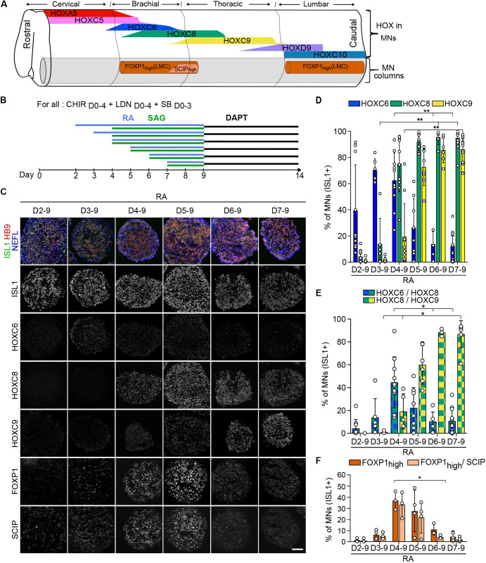
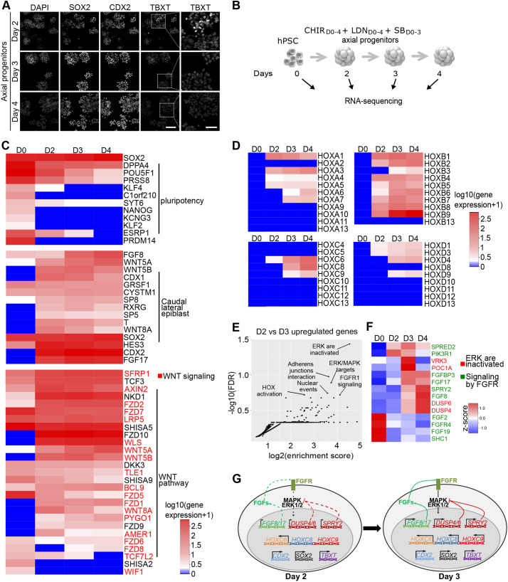
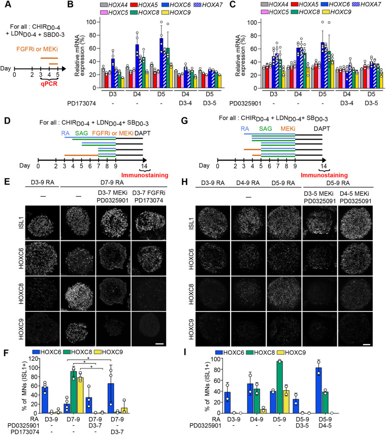
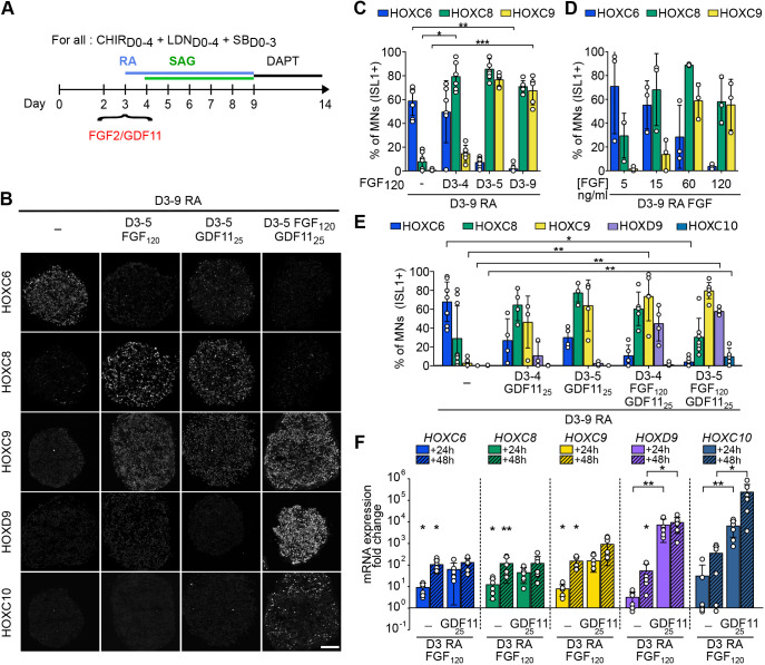
Rostro-caudal patterning of vertebrates depends on the temporally progressive activation of HOX genes within axial stem cells that fuel axial embryo elongation. Whether the pace of sequential activation of HOX genes, the 'HOX clock', is controlled by intrinsic chromatin-based timing mechanisms or by temporal changes in extrinsic cues remains unclear. Here, we studied HOX clock pacing in human pluripotent stem cell-derived axial progenitors differentiating into diverse spinal cord motor neuron subtypes. We show that the progressive activation of caudal HOX genes is controlled by a dynamic increase in FGF signaling. Blocking the FGF pathway stalled induction of HOX genes, while a precocious increase of FGF, alone or with GDF11 ligand, accelerated the HOX clock. Cells differentiated under accelerated HOX induction generated appropriate posterior motor neuron subtypes found along the human embryonic spinal cord. The pacing of the HOX clock is thus dynamically regulated by exposure to secreted cues. Its manipulation by extrinsic factors provides synchronized access to multiple human neuronal subtypes of distinct rostro-caudal identities for basic and translational applications.This article has an associated 'The people behind the papers' interview.

Keywords: Axial; HOX genes; Human; Motor neurons; Pluripotent stem cells; Spinal cord.


Conflict of interest statement

Competing interestsThe authors declare no competing or financial interests.

Figures

Fig. 1.
Fig. 1.
Human axial progenitors generate progressively more caudal motor neuron subtypes. (A) Schematic summary of data in Figs S1, S2. MNs are defined by the expression of ISL1 or HB9 (gray) are organized in motor columns in spinal ventral horns. HOX expression profiles within MNs and localization of MNs expressing high levels of FOXP1 and SCIP are represented as observed in the human embryonic spinal cord at 6.3 and 7.5 weeks of gestation. Changes in shapes indicate an increase or decrease in the number of MNs expressing a given marker. FOXP1high MNs are observed selectively in lateral motor columns (LMCs) of the brachio-thoracic and lumbar spinal cord. SCIPhigh MNs are a subset of LMC MNs in the caudal brachial spinal cord. (B) Differentiation conditions used in C to F in which the time of exposure to retinoic acid (RA)/SAG (an agonist of the sonic hedgehog pathway) is modulated. (C) Immunostaining for ISL1/HB9 (MNs), NEFL (neurons), HOX transcription factors, FOXP1 and SCIP on cryostat sections of hESC-derived EBs on day 14 of differentiation. The later RA is applied, the more caudal the MNs are. FOXP1 and SCIP MNs are mostly generated when RA is applied at day 4 and day 5, further defining the rostro-caudal identity of the MNs within HOXC8+ conditions. Scale bar: 100 µm. (D-F) Proportion of MNs (ISL1+ cells) expressing the indicated markers. Data are mean±s.d. Each circle is an independent biological replicate. (D) n=6-13, (E) n=3-13, (F) n=3 or 4. *P≤0.05, **P≤0.01 (ANOVA with Kruskal-Wallis post-hoc test).
Fig. 2.
Fig. 2.
Temporal transcriptomic analysis of hPSC-derived axial progenitors. (A) Immunostaining for axial progenitor markers on hESC-derived progenitors at day 2, day 3 and day 4 of differentiation. Scale bars: 40 µm. (B) Experimental design of RNA-seq experiment to profile the transcriptome of hESC (SA001)-derived axial progenitors. n=2 per sample. (C) Heatmap of gene expression [log10(gene expression +1)] for pluripotency genes, most common mouse NMPs markers and Wnt pathway-related genes. Genes in red are part of the ‘Formation β-catenin:TCF transactivating complex' annotation found enriched in reactome pathway analysis in Fig. S3E,F. (D) Heatmap showing temporal transcriptional changes [log10(gene expression +1)] of all HOX genes. (E) Functional enrichment analysis (Reactome pathway) of the 232 genes upregulated twofold (P<0.05) between D3 and D2: y axis, FDR (false discovery rate); x-axis, enrichment score calculated for a given Reactome pathway. (F) Heatmap based on z-score of the genes associated with the annotations ‘ERK/MAPK target' (red labels) or ‘signaling by FGFR' (green labels) in reactome analysis in 2E. (G) Schematic representation of the transcriptional and immunostaining analysis of day 2 and day 3 progenitors.
Fig. 3.
Fig. 3.
FGF and MEK pathway inhibitors stall temporal induction of HOX genes in axial progenitors and prevent specification of caudal motor neuron subtypes. (A) Differentiation conditions. PD173074 (FGFR1-3 inhibitor) or PD032590 (MEK1/2 inhibitor) were added on day 3 up to day 5 and hESC-derived progenitors were collected from day 3 to 5 for qPCR analysis. (B,C) Real-time PCR analysis of HOX mRNAs in day 3, 4 and 5 progenitors. Data represent the expression of the different genes relative to the highest expressed gene for all time points and conditions (HOXC6). MEK1/2 and FGFR inhibitors, applied from day 3, prevent the temporal increase in caudal HOX expression. (D,G) Differentiation conditions. PD173074 (FGFR1-3 inhibitor; FGFRi) or PD032590 (MEK1/2 inhibitor; MEKi) was added on day 3 up to day 7 (D) or from day 3 or day 4 until day 5 (G) and hESC-derived MNs were collected at day 14 for immunostaining analysis. Retinoic acid (RA) and SAG (an agonist of the sonic hedgehog pathway) were added at the indicated time points, between day 3 and day 7. (E,H) Immunostaining for ISL1 (MNs) and HOX transcription factors on cryostat sections of embryoid bodies on day 14 of differentiation, according to conditions presented in D for E and in G for H. MEK and FGFR inhibitors prevent the specification of HOXC8+ and HOXC9+ MNs. Instead, HOXC6+ MNs are generated. Scale bars: 100 µm. (F,I) Quantification of HOXC6, HOXC8 and HOXC9 MNs on day 14 of differentiation, according to conditions presented in D for F and in G for I. Data are mean±s.d. Each circle is an independent biological replicate: (B) n=4, (C) n=5, (F) n=3 or 4 and (I) n=2. *P≤0.05 (ANOVA with Kruskal-Wallis post-hoc test).
Fig. 4.
Fig. 4.
Dynamic pacing of HOX induction in axial progenitors by changes in extrinsic FGF2 and GDF11 levels. (A) Differentiation conditions. Extrinsic cues, FGF2, GDF11 or FGF2+GDF11 were added on day 3 of differentiation at various concentrations or for different durations. (B) Immunostaining for HOX proteins on cryostat sections of hESC-derived EBs on day 14 of differentiation. FGF2, GDF11 and FGF2/GDF11 induce more caudal MN subtypes. Scale bar: 100 µm. (C-E) Proportion of MNs (ISL1+ cells) expressing the indicated markers. The effect of the duration of FGF2 treatment (C), FGF2 concentration (D) and duration of GDF11 or FGF2+GDF11 (E) were monitored. (F) Real-time quantitative PCR analysis of the expression of HOX genes regionally expressed in human MNs in vivo. HOX mRNAs were monitored at day 4 (24 h post-treatment) and day 5 (48 h post-treatment) upon addition of FGF2 or GDF11, or a combination of FGF2 (120 ng/ml) and GDF11 (25 ng/ml). Data are expressed as fold changes to their respective control [day 4 retinoic acid (RA) 24 h or day 5 RA 48 h]. Asterisks above the horizontal lines indicate significance of statistical comparison between the indicated conditions; asterisks above the histogram bars are for statistical comparison with the control. Data are mean±s.d. Each circle is an independent biological replicate: (C) n=6 or 7, (D) n=3, (E) n=3-9, (F) n=6. *P≤0.05, **P≤0.01, ***P≤0.001 (ANOVA with Kruskal-Wallis post-hoc test).

References

    1. Abati, E., Bresolin, N., Comi, G. and Corti, S. (2019). Advances, challenges, and perspectives in translational stem cell therapy for amyotrophic lateral sclerosis. Mol. Neurobiol. 56, 6703-6715. 10.1007/s12035-019-1554-x - DOI - PubMed
    1. Aires, R., de Lemos, L., Nóvoa, A., Jurberg, A. D., Mascrez, B., Duboule, D. and Mallo, M. (2019). Tail bud progenitor activity relies on a network comprising Gdf11, Lin28, and Hox13 genes. Dev. Cell 48, 383-395.e8. 10.1016/j.devcel.2018.12.004 - DOI - PubMed
    1. Albors, A., Halley, P. A. and Storey, K. G. (2018). Lineage tracing of axial progenitors using Nkx1-2CreER T2 mice defines their trunk and tail contributions. Development 145, dev164319. 10.1242/dev.164319 - DOI - PMC - PubMed
    1. Amin, S., Neijts, R., Simmini, S., van Rooijen, C., Tan, S. C., Kester, L., van Oudenaarden, A., Creyghton, M. P. and Deschamps, J. (2016). Cdx and T Brachyury co-activate growth signaling in the embryonic axial progenitor niche. Cell Rep. 17, 3165-3177. 10.1016/j.celrep.2016.11.069 - DOI - PubMed
    1. Amoroso, M. W., Croft, G. F., Williams, D. J., O'Keeffe, S., Carrasco, M. A., Davis, A. R., Roybon, L., Oakley, D. H., Maniatis, T., Henderson, C. E.et al. (2013). Accelerated high-yield generation of limb-innervating motor neurons from human stem cells. J. Neurosci. 33, 574-586. 10.1523/JNEUROSCI.0906-12.2013 - DOI - PMC - PubMed

Publication types

MeSH terms