Case Reports
. 2021 Mar 29;14(3):e242127.
doi: 10.1136/bcr-2021-242127.

Incomplete lung torsion following spontaneous pneumothorax

Affiliations
Case Reports

Incomplete lung torsion following spontaneous pneumothorax

Konstantinos Stefanidis et al. BMJ Case Rep. .
No abstract available

Keywords: pneumothorax; radiology (diagnostics); respiratory medicine.


Conflict of interest statement

Competing interests: None declared.

Figures

Figure 1
Figure 1
(A) Axial CT image shows the normal lobar anatomy with mild emphysematous changes in the right upper lobe. (B). CT findings 3 months later following the spontaneous pneumothorax and reinflation. Axial CT image at the same level demonstrates extensive emphysema in the upper lung zone which corresponds to the displaced right lower lobe. Pulmonary blebs (asterisk) in the apical segment of the left lower lobe demonstrating the same anatomical level. RUL, right upper lobe; RLL, right lower lobe; LUL, left upper lobe; LLL, left lower lobe.
Figure 2
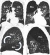
Figure 2
(A) Coronal CT image shows the normal lobar anatomy demonstrating the origin of the right upper lobe bronchus (arrow). (B). Following the spontaneous pneumothorax, coronal CT image shows the abnormal lobar orientation of the right upper and lower lobe. It also demonstrates the downward orientation of the right upper lobe bronchus (arrow). (C). Sagittal CT image show the normal lobar anatomy of the right upper and lower lobe. Curved arrows show the anticlockwise rotation of the lobes. (D). Following the spontaneous pneumothorax, sagittal CT image shows the new positioning of the right upper and lower lobe. LLL, left lower lobe; LUL, left upper lobe; RLL, right lower lobe; RUL, right upper lobe.

References

    1. Cable DG, Deschamps C, Allen MS, et al. . Lobar torsion after pulmonary resection: presentation and outcome. J Thorac Cardiovasc Surg 2001;122:1091–3. 10.1067/mtc.2001.117839 - DOI - PubMed
    1. Hennink S, Wouters MWJM, Klomp HM, et al. . Necrotizing pneumonitis caused by postoperative pulmonary torsion. Interact Cardiovasc Thorac Surg 2008;7:144–5. 10.1510/icvts.2007.158378 - DOI - PubMed
    1. Felson B. Lung torsion: radiographic findings in nine cases. Radiology 1987;162:631–8. 10.1148/radiology.162.3.3809475 - DOI - PubMed
    1. Hammer MM, Madan R. Clinical and imaging features in lung torsion and description of a novel imaging sign. Emerg Radiol 2018;25:121–7. 10.1007/s10140-017-1563-x - DOI - PubMed
    1. Irie M, Okumura N, Nakano J, et al. . Spontaneous whole-lung torsion after massive pleural effusion and atelectasis. Ann Thorac Surg 2014;97:329–32. 10.1016/j.athoracsur.2013.04.133 - DOI - PubMed

Publication types