. 2022 Apr;59(4):346-350.
doi: 10.1136/jmedgenet-2020-107434. Epub 2021 Mar 29.

Testicular Sertoli cell tumour and potentially testicular Leydig cell tumour are features of DICER1 syndrome

Affiliations

Testicular Sertoli cell tumour and potentially testicular Leydig cell tumour are features of DICER1 syndrome

Lisa Golmard et al. J Med Genet. 2022 Apr.

Abstract

DICER1 syndrome is a rare paediatric autosomal dominant inherited disorder predisposing to various benign and malignant tumours. It is caused by a germline pathogenic variant in DICER1, and the second hit for tumour development is usually a missense hotspot pathogenic variant in the DICER1 ribonuclease IIIb domain. While DICER1 predisposing variants account for about 60% of ovarian Sertoli-Leydig cell tumours, no DICER1-related testicular stromal tumours have been described. Here we report the first two cases of testicular stromal tumours in children carrying a DICER1 germline pathogenic variant: a case of Sertoli cell tumour and a case of Leydig cell tumour diagnosed at 2 and 12 years of age, respectively. A somatic DICER1 hotspot pathogenic variant was detected in the Sertoli cell tumour. This report extends the spectrum of DICER1-related tumours to include testicular Sertoli cell tumour and potentially testicular Leydig cell tumour. Diagnosis of a testicular Sertoli cell tumour should prompt DICER1 genetic testing so that patients with a DICER1 germline pathogenic variant can benefit from established surveillance guidelines. DICER1 genetic evaluation may be considered for testicular Leydig cell tumour. Our findings suggest that miRNA dysregulation underlies the aetiology of some testicular stromal tumours.

Keywords: genetic predisposition to disease; medical oncology; paediatrics.


Conflict of interest statement

Competing interests: None declared.

Figures
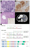
Figure 1.
Figure 1.
Case 1, testicular Sertoli cell tumor and pleuropulmonary blastoma images, and DICER1 genetic analysis. Testicular Sertoli cell tumor pathology: (A) Hematoxylin and Eosin staining (200x magnification), showing well-differentiated component with tubules surrounded by basement membrane and a central lumen. Pathology examination revealed a biphasic proliferation consisting of small undifferentiated stromal hyperchromatic cells and tubular epithelial structures with hyperchromatic cubic lining. On the periphery were images of cystic distention and a thin band of testicular tissue composed of immature seminiferous tubules; (B) Inhibin reactivity, 200x magnification. Immunohistochemistry (IHC) of the tumor cells was positive for inhibin, vimentin, CD99, WT1 and cytokeratin AE1/AE3, and negative for CEA and cytokeratin 5/6. (C) Pleuropulmonary blastoma (PPB) pathology: Hematoxylin and Eosin staining (200x magnification), showing round and spindle cell undifferentiated proliferation with marked atypia and edematous background. Bottom right insert: myogenin reactivity, 200x magnification. IHC of the tumor cells was positive for myogenin, desmin, vimentin and CD99, and negative for CK-AE1/AE3, EMA, synaptophysin, chromogranin, TTFF1, S-100 and SMA. The proliferative index evaluated by an anti-Ki67 antibody was 80%. (D) PPB image: chest computed tomography scan, showing a heterogeneous enhancing mass in the left hemithorax associated with pleural nodules and moderate pleural effusion and a cystic lesion in the left upper lung with air-fluid levels. (E) DICER1 genetic analysis. Top: electropherogram of DNA analysis showing the DICER1 heterozygous pathogenic variant c.4206+2dup (in a square) and the DICER1 homozygous common single nucleotide polymorphism c.4206+9G>T, rs1778057 (underlined). Middle: electropherograms of mRNA analysis from patient-derived lymphoblastoid cell lines that were grown with (puromycin+) or without puromycin (puromycin-), an inhibitor of nonsense-mediated mRNA decay (NMD). Δ symbol represents exon skipping. Sequences of the three transcripts are described, and Δ22-23 transcript is written in brackets for electropherogram without puromycin as this transcript leading to a premature termination codon undergoes NMD, hence the fluorescence signal of its sequence is close to background noise after culture without puromycin. Bottom: schematic representation of predicted effect on DICER1 protein, a loss of 52 amino acids in RNase IIIa domain for Δ22 transcript, and a truncated protein for Δ22-23 transcript with a black line that represents the frameshift with six different amino acids at the end of the truncated protein. * symbol for Δ22-23 protein is used as this truncated protein is expected to be present at a very low level because of NMD that leads to Δ22-23 transcript degradation.
Figure 2.
Figure 2.
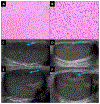
Case 2, testicular Leydig cell tumor pathology and ultrasound images. Up: Hematoxylin and Eosin staining of testicular Leydig cell tumor demonstrating polygonal cells with eosinophilic cytoplasm and oval nuclei without atypia. (A) 20x magnification. (B) 40x magnification. Tumor consisted of sheets of uniform Leydig cells with moderately eosinophilic cytoplasm and no detectable mitotic activity. IHC was positive for MART-1, synaptophysin, inhibin, and calretinin, and negative for WT1, chromogranin and OCT4. Bottom: longitudinal section of the testicular mass (arrows) from initial ultrasound (C) and follow up ultrasound (D). Transverse section of the testicular mass (arrows) from the initial ultrasound (E) and follow up ultrasound (F).

References

    1. Stewart DR, Best AF, Williams GM, Harney LA, Carr AG, Harris AK, Kratz CP, Dehner LP, Messinger YH, Rosenberg PS, Hill DA, Schultz KAP. Neoplasm Risk Among Individuals With a Pathogenic Germline Variant in DICER1. J. Clin. Oncol 2019;37:668–676. - PMC - PubMed
    1. Kock L, Wu MK, Foulkes WD. Ten years of DICER1 mutations: Provenance, distribution, and associated phenotypes. Hum. Mutat 2019;40:1939–1953. - PubMed
    1. Schultz KAP, Harris AK, Finch M, Dehner LP, Brown JB, Gershenson DM, Young RH, Field A, Yu W, Turner J, Cost NG, Schneider DT, Stewart DR, Frazier AL, Messinger Y, Hill DA. DICER1-related Sertoli-Leydig cell tumor and gynandroblastoma: Clinical and genetic findings from the International Ovarian and Testicular Stromal Tumor Registry. Gynecol. Oncol 2017;147:521–527. - PMC - PubMed
    1. de Boer CM, Eini R, Gillis AM, Stoop H, Looijenga LH, White SJ. DICER1 RNase IIIb domain mutations are infrequent in testicular germ cell tumours. BMC Res. Notes 2012;5:569. - PMC - PubMed
    1. Sabbaghian N, Bahubeshi A, Shuen AY, Kanetsky PA, Tischkowitz MD, Nathanson KL, Foulkes WD. Germ-line DICER1 mutations do not make a major contribution to the etiology of familial testicular germ cell tumours. BMC Res. Notes 2013;6:127. - PMC - PubMed

Publication types