. 2021 Mar 29;12(1):1946.
doi: 10.1038/s41467-021-21963-1.

Profiling PRMT methylome reveals roles of hnRNPA1 arginine methylation in RNA splicing and cell growth

Affiliations

Profiling PRMT methylome reveals roles of hnRNPA1 arginine methylation in RNA splicing and cell growth

Wen-Juan Li et al. Nat Commun. .

Abstract

Numerous substrates have been identified for Type I and II arginine methyltransferases (PRMTs). However, the full substrate spectrum of the only type III PRMT, PRMT7, and its connection to type I and II PRMT substrates remains unknown. Here, we use mass spectrometry to reveal features of PRMT7-regulated methylation. We find that PRMT7 predominantly methylates a glycine and arginine motif; multiple PRMT7-regulated arginine methylation sites are close to phosphorylations sites; methylation sites and proximal sequences are vulnerable to cancer mutations; and methylation is enriched in proteins associated with spliceosome and RNA-related pathways. We show that PRMT4/5/7-mediated arginine methylation regulates hnRNPA1 binding to RNA and several alternative splicing events. In breast, colorectal and prostate cancer cells, PRMT4/5/7 are upregulated and associated with high levels of hnRNPA1 arginine methylation and aberrant alternative splicing. Pharmacological inhibition of PRMT4/5/7 suppresses cancer cell growth and their co-inhibition shows synergistic effects, suggesting them as targets for cancer therapy.


Conflict of interest statement

The authors declare no competing interests.

Figures
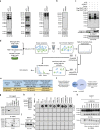
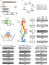
Fig. 1
Fig. 1. Proteome-wide profiling of arginine methylation regulated by PRMT7.
a, b HEK293 cells transfected with control siRNA (siCTL) or siRNA against PRMT7 (siPRMT7) were analyzed by immunoblotting. MMA mono-methyl-arginine, sDMA symmetric di-methyl-arginine, aDMA asymmetric di-methyl-arginine. ACTIN was served as a loading control. c HEK293 cells transfected with siCTL or siPRMT7 together with or without Flag-tagged, wild-type (WT) or enzymatic dead mutant (MT) PRMT7 were analyzed by immunoblotting. d Experimental flowchart for identification of arginine methylation sites regulated by PRMT7 or responsive to PRMT7 inhibitor SGC3027 in HEK293 cells (see detail in “Methods”). e The number of mono-methyl arginine (Rme1) sites detected (column 1), Rme1 sites could be quantified (column 2), Rme1 sites with methylation signals decreased at least two-fold (column 3) or abolished (column 4) when knocking down of PRMT7 was shown. The number of proteins encompass all these methylation sites was also shown (bottom lane). f The overlap between PRMT7 methylome and proteins of which abundance was decreased at least two-fold when PRMT7 was knocked down is shown. g In vitro methylation assay was performed by mixing purified PRMT7 with CCT7, TFG, YBX1, CKMT1B, hnRNPK, hnRNPA2B1 or △Np63α, followed by immunoblotting with anti-MMA antibody (top panel). Methylation was indicated by white asterisk. The expression of proteins was examined by coomassie blue staining (C.B.S) and indicated by black asterisk (bottom panel). h The expression of purified PRMT7 was examined by C.B.S and indicated by black asterisk. i In vitro methylation assay was performed by mixing purified PRMT7 with synthetic short peptides from CCT7, TFG, YBX1, CKMT1B, hnRNPK, hnRNPA2B1, and hnRNPA1 proteins. Amino (N)-terminal of histone H3 and H4 were also included. The reactions were subjected to dot blotting. aa, amino acid. j The expression of purified PRMT7 was examined by immunoblotting. k In vitro methylation assay was performed by mixing purified PRMT7 with hnRNPA1 wild-type (WT) or mutants including R/K (7) (all five arginine (R) residues methylated by PRMT7 were replaced by lysine (K)), R194K, R206K, R218K, R225K, and R232K. The reactions were subjected to immunoblotting. Source data are provided as a Source data file.
Fig. 2
Fig. 2. Characterization of PRMT7-regulated arginine methylation sites.
a Motif analysis was performed for PRMT7-regulated arginine methylation sites using iceLogo. b The distribution of proteins in PRMT7 methylome with different number of arginine methylation sites. The proteins with more than two methylation sites were shown in oval. c The percentage of PRMT7-regulated arginine methylation sites with different number of phosphorylation sites (n = 0, 1, 2, 3 or ≥4) in vicinity (±3 amino acid (AA) window) was shown by pie chart. d Rates of somatic mutations at all arginine sites in the proteome or PRMT7-regulated arginine sites in human cancers (p = 9.14e−4 by the Fisher’s exact test). e Rates of somatic mutations in the vicinity of all arginine sites in the proteome or PRMT7-regulated arginine sites in human cancers (±5 nucleotides) (p = 1.6e−20 by the Fisher’s exact test). fh Gene ontology (GO) (f), KEGG pathway (g), and CORUM (h) analysis using Metascape for PRMT7 methylome. Representative terms from the top 20 enriched GO term clusters were shown.
Fig. 3
Fig. 3. Comparison among PRMT7, 4, and 5-regulated arginine methylation revealed that these three PRMTs all methylate proteins with implications in RNA biology.
a The number of arginine methylation sites detected (the sum of mono- and di-methylation sites after removing duplicates) (column 2), arginine methylation sites could be quantified (column 3), arginine methylation sites with methylation signals decreased at least two-fold (column 4) or abolished (column 5) in PRMT4-, PRMT5- or PRMT7-knockdown experiments (lane 2, 3, and 4) following mass spectrometry analysis as described in Fig. 1d. The number of proteins encompass all these methylation sites was also shown (bottom three lanes). For comparison, data shown for PRMT7 in Fig. 1e was also included here. b, c Motif analysis was performed for PRMT4- (b) and PRMT5- (c) regulated arginine methylation sites using iceLogo. d, f Rates of somatic mutations at all arginine sites in the proteome and PRMT4- (d) or PRMT5- (f) regulated methylation sites in human cancers (p = 6.54e−08 (d) and p = 0.1684 (f) by the Fisher’s exact test). e, g Rates of somatic mutations in the vicinity of all arginine sites in the proteome or PRMT4- (e) or PRMT5- (g) regulated methylation sites in human cancers (±5 nucleotides) (p = 3.89e−108 (e) and p = 2.57e−45 (g) by the Fisher’s exact test). h, i KEGG pathway analysis using Metascape for PRMT4 (h) and PRMT5 (i) methylome. Representative terms from the top 20 enriched GO term clusters were shown.
Fig. 4
Fig. 4. RNA-seq profiling revealed that PRMT4, 5, and 7 exhibited a global impact on RNA alternative splicing.
a HEK293 cells transfected with siCTL or siPRMT4, siPRMT5 or siPRMT7 were subjected to RNA-seq analysis, and the number of alternative splicing events regulated is shown (|ΔPSI| ≥ 0.5). Cassette exons (SE); intron retain (IR); mutually exclusive exons (MXE); alternative 5′ splice sites (5′ ASS) and alternative 3′ splice sites (3′ ASS). b The number of exon inclusion (EI) and skipping (ES) induced is shown. c All alternative splicing events regulated by PRMT4, 5, and 7 were classified into three categories, events regulated by one (light green), any two out of three (light black) or all three (light purple) PRMTs. d Overlap between EI and ES induced by PRMT4, 5, or 7 is shown. e Overlap among PRMT4-, 5-, and 7-regulated cassette exons is shown. f HEK293 cells transfected with siCTL or two individual siPRMT4 (siPRMT4-1 and siPRMT4-2), siPRMT5 (siPRMT5-1 and siPRMT5-2), or siPRMT7 (siPRMT7-1 and siPRMT7-2) were subjected to analysis of the expression of both short and long isoforms of representative genes as indicated. The length of the alternatively spliced exon, as well as the expected length of the PCR products, was shown as indicated. DNA marker (M) was included on the left (bp: base pair). The position of the cassette exon in each gene was as follows: PPARA (NM_005036, exon3); SREBF2 (NR_103834, exon18); RAB27B (NM_001375327, exon3); WDPCP (NM_015910, exon6). F: forward primer; R: reverse primer. The translation start sites (ATG) of the two isoforms of RAB27B were indicated by asterisks. n = 3 biological replicates and representative data are shown. gi HEK293 cells transfected with siCTL or siPRMT4 (g), siPRMT5 (h), or siPRMT7 (i) in the presence or absence of wild-type (WT) or enzymatic dead mutant (MT) PRMT4 (g), PRMT5 (h), or PRMT7 (i) were subjected to alternative splicing analysis as described in (f). Source data are provided as a Source data file.
Fig. 5
Fig. 5. PRMT4-, 5-, and 7-mediated hnRNPA1 methylation was involved in the regulation of alternative splicing events co-regulated by the three PRMTs.
a Overlap among PRMT4, 5, and 7 methylome is shown. Splicing factors among substrates commonly regulated by PRMT4, 5, and 7 are listed. b Metascape CORUM, analysis was performed for substrates commonly regulated by PRMT4, 5, and 7. c Motif analysis was performed for cassette exons commonly regulated by PRMT4, 5, and 7 using CentriMo (v5.0.5). d HEK293 cells transfected with siCTL or sihnRNPA1 were subjected to alternative splicing analysis as described in Fig. 4f. e Schematic representation of the domain architecture of hnRNPA1. Arginine (R) sites methylated by PRMT4, 5, and/or 7 in the RGG domain were highlighted in red. The PRMTs that methylated each arginine and the methylation status were indicated at the bottom. (RRM: RNA recognition motif; RGG: arginine-glycine-glycine). f HEK293 cells transfected with siCTL or sihnRNPA1 in the presence or absence of wild type (WT) or methylation mutants including R/K (4), R/K (5), R/K (7), and R/K (457) were subjected to alternative splicing analysis as described in Fig. 4f. g HEK293 cells transfected with siCTL or siPRMT4, 5, or 7 were subjected to RNA-IP assay using control IgG or anti-hnRNPA1 antibody. n = 3 biological replicates (mean ± SEM, *P < 0.05, **P < 0.01, ***P < 0.001 by unpaired Student t-test, two-tailed). h HEK293 cells transfected with empty vector (CTL) or Flag-tagged, wild-type (WT) or methylation mutants as described in (f) were subjected to RNA-IP assay using anti-Flag antibody. n = 3 biological replicates (mean ± SEM, *P < 0.05, **P < 0.01, ***P < 0.001 by unpaired Student t-test, two-tailed). Source data are provided as a Source Data file.
Fig. 6
Fig. 6. PRMT4, 5, 7, and hnRNPA1 arginine methylation were over-presented in multiple types of cancers.
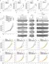
ac Clinical samples of breast cancer (BRCA) (n = 10), colorectal cancer (CRC) (n = 10) and prostate cancer (PC) (n = 10) were subjected to RNA extraction and RT-qPCR analysis to examine the expression of PRMT4 (a), PRMT5 (b), and PRMT7 (c). Pearson correlation coefficient of the expression of PRMT4, PRMT5, and PRMT7 between normal and tumor samples is 0.60, −0.50, and −0.17 in BRCA, 0.12, 0.21, and −0.05 in CRC, and 0.48, 0.74, and 0.69 in PC, respectively. (*P < 0.05, **P < 0.01, ***P < 0.001 by paired Student t-test, two-tailed). d Arginine methylation sites in RGG domain of hnRNPA1 protein that could be detected and quantified in clinical samples of BRCA, CRC, and PC were shown. eg Clinical samples of BRCA (e), CRC (f), and PC (g) were subjected to alternative splicing analysis as described in Fig. 4f. Source data are provided as a Source data file.
Fig. 7
Fig. 7. PRMT4, 5, 7, and hnRNPA1 arginine methylation were required for the growth of multiple types of cancer cells.
ac, f MCF7 cells were transfected with siCTL or siPRMT4, 5, or 7 followed by cell proliferation assay (ac) and alternative splicing analysis (f). n = 3 biological replicates for cell proliferation assay (mean ± SD, ***P < 0.001, day 4 by unpaired Student t-test, two-tailed). n = 3 biological replicates for alternative splicing analysis and representative data are shown. d, g MCF7 cells were transfected with siCTL or sihnRNPA1 followed by cell proliferation assay (d) or alternative splicing analysis (g). n = 3 biological replicates for cell proliferation assay (mean ± SD, ***P < 0.001, day 4 by unpaired Student t-test, two-tailed). e, h MCF7 cells transfected with siCTL or sihnRNPA1 in the presence or absence of wild type (WT) or methylation deficient mutants including R/K (4), R/K (5), R/K (7), and R/K (457) were subjected to cell proliferation assay (e) or alternative splicing analysis (h). n = 3 biological replicates for cell proliferation assay (mean ± SD, ***P < 0.001, day 4 by unpaired Student t-test, two-tailed). i, j MCF7 cells were transfected with siCTL or siPRMT4, siPRMT5, siPRMT7, or sihnRNPA1 in the presence or absence of Flag-tagged WDPCP (i) or RAB27B (j), both short (S) and long (L) isoforms, followed by cell proliferation assay. n = 3 biological replicates (mean ± SD, *P < 0.05, **P < 0.01, ***P < 0.001, N.S: non-significant, day 4 by unpaired Student t-test, two-tailed). Source data are provided as a Source data file.
Fig. 8
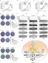
Fig. 8. Pharmacological inhibition of PRMT4, 5, and 7 alters RNA alternative splicing and suppresses cancer cell growth.
ac, d, f, h MCF7 (a, d), HCT 116 (b, f), or LNCaP (c, h) cells were treated with EZM2302 (10 μM), GSK591 (10 μM), or SGC3027 (10 μM) alone or in combination (+++) for duration as indicated followed by proliferation assay (ac) or colony formation assay (d, f, h). n = 3 biological replicates for cell proliferation assay (mean ± SD, **P < 0.01, ***P < 0.001, day 4 by unpaired Student t-test, two-tailed). e, g, i Quantification of the crystal violet dye as shown in (d), (f), and (h). Absorbance was measured three time. jl MCF7 (j), HCT 116 (k), or LNCaP (l) cells were treated with EZM2302 (10 μM), GSK591 (10 μM), or SGC3027 (10 μM) alone or in combination for 24 h as indicated followed by alternative splicing analysis. n = 2 biological replicates and representative data are shown. m A working model of PRMT4-, 5-, and 7-mediated arginine methylation in alternative splicing regulation and cancer cell growth. Global profiling of PRMT4-, 5-, and 7-mediated arginine methylation revealed that PRMT4-, 5-, and 7-methylome shared a group of proteins with implications in mRNA splicing, and RNA-seq analysis confirmed the global impact of these three PRMTs on gene alternative splicing. Exemplified by hnRNPA1, a critical regulator of gene alternative splicing, PRMT4-, 5-, and 7-mediated arginine methylation was shown to be involved in hnRNPA1 binding with pre-mRNA and, therefore, the regulation of those alternative splicing events commonly regulated by PRMT4, 5, and 7. In cancers, such as breast, colorectal, and prostate cancer, PRMT4, 5, and 7 were found to be overexpressed, leading to elevated levels of arginine methylation on their substrates including hnRNPA1, and therefore aberrant alternative splicing changes, which potentially drive cancer cell growth. Pharmacological inhibition of PRMT4, 5, and 7 exhibited great potential in suppressing the growth of cancer cells. Source data are provided as a Source data file.

References

    1. Feinberg AP, Koldobskiy MA, Gondor A. Epigenetic modulators, modifiers and mediators in cancer aetiology and progression. Nat. Rev. Genet. 2016;17:284–299. doi: 10.1038/nrg.2016.13. - DOI - PMC - PubMed
    1. Turner BM. Reading signals on the nucleosome with a new nomenclature for modified histones. Nat. Struct. Mol. Biol. 2005;12:110–112. doi: 10.1038/nsmb0205-110. - DOI - PubMed
    1. Paik WK, Kim S. Omega-N-methylarginine in protein. J. Biol. Chem. 1970;245:88–92. doi: 10.1016/S0021-9258(18)63425-8. - DOI - PubMed
    1. Baldwin GS, Carnegie PR. Specific enzymic methylation of an arginine in the experimental allergic encephalomyelitis protein from human myelin. Science. 1971;171:579–581. doi: 10.1126/science.171.3971.579. - DOI - PubMed
    1. Jarrold, J. & Davies, C. C. PRMTs and Arginine methylation: cancer’s best-kept secret? Trends Mol. Med. 10.1016/j.molmed.2019.05.007 (2019). - PubMed

Publication types

MeSH terms

Substances