Right Ventricular Strain Is Common in Intubated COVID-19 Patients and Does Not Reflect Severity of Respiratory Illness
- PMID: 33783269
- PMCID: PMC8267080
- DOI: 10.1177/08850666211006335
Right Ventricular Strain Is Common in Intubated COVID-19 Patients and Does Not Reflect Severity of Respiratory Illness
Abstract
Background: Right ventricular (RV) dysfunction is common and associated with worse outcomes in patients with coronavirus disease 2019 (COVID-19). In non-COVID-19 acute respiratory distress syndrome, RV dysfunction develops due to pulmonary hypoxic vasoconstriction, inflammation, and alveolar overdistension or atelectasis. Although similar pathogenic mechanisms may induce RV dysfunction in COVID-19, other COVID-19-specific pathology, such as pulmonary endothelialitis, thrombosis, or myocarditis, may also affect RV function. We quantified RV dysfunction by echocardiographic strain analysis and investigated its correlation with disease severity, ventilatory parameters, biomarkers, and imaging findings in critically ill COVID-19 patients.
Methods: We determined RV free wall longitudinal strain (FWLS) in 32 patients receiving mechanical ventilation for COVID-19-associated respiratory failure. Demographics, comorbid conditions, ventilatory parameters, medications, and laboratory findings were extracted from the medical record. Chest imaging was assessed to determine the severity of lung disease and the presence of pulmonary embolism.
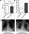
Results: Abnormal FWLS was present in 66% of mechanically ventilated COVID-19 patients and was associated with higher lung compliance (39.6 vs 29.4 mL/cmH2O, P = 0.016), lower airway plateau pressures (21 vs 24 cmH2O, P = 0.043), lower tidal volume ventilation (5.74 vs 6.17 cc/kg, P = 0.031), and reduced left ventricular function. FWLS correlated negatively with age (r = -0.414, P = 0.018) and with serum troponin (r = 0.402, P = 0.034). Patients with abnormal RV strain did not exhibit decreased oxygenation or increased disease severity based on inflammatory markers, vasopressor requirements, or chest imaging findings.
Conclusions: RV dysfunction is common among critically ill COVID-19 patients and is not related to abnormal lung mechanics or ventilatory pressures. Instead, patients with abnormal FWLS had more favorable lung compliance. RV dysfunction may be secondary to diffuse intravascular micro- and macro-thrombosis or direct myocardial damage.
Trial registration: National Institutes of Health #NCT04306393. Registered 10 March 2020, https://clinicaltrials.gov/ct2/show/NCT04306393.
Keywords: COVID-19; acute respiratory distress syndrome; cardiac dysfunction; right ventricle; strain.
Conflict of interest statement
Figures


References
-
- Zochios V, Parhar K, Tunnicliffe W, Roscoe A, Gao F. The right ventricle in ARDS. Chest. 2017;152(1):181–193. doi:10.1016/j.chest.2017.02.019 - PubMed
MeSH terms
Associated data
Grants and funding
LinkOut - more resources
Full Text Sources
Other Literature Sources
Medical

