

The presence of contrast agent increases organ radiation dose in contrast-enhanced CT
- PMID: 33783569
- PMCID: PMC8452580
- DOI: 10.1007/s00330-021-07763-7
The presence of contrast agent increases organ radiation dose in contrast-enhanced CT
Abstract
Objectives: Routine dosimetry calculations do not account for the presence of iodine in organs and tissues during CT acquisition. This study aims to investigate the impact of contrast agent (CA) on radiation dose.
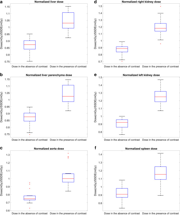
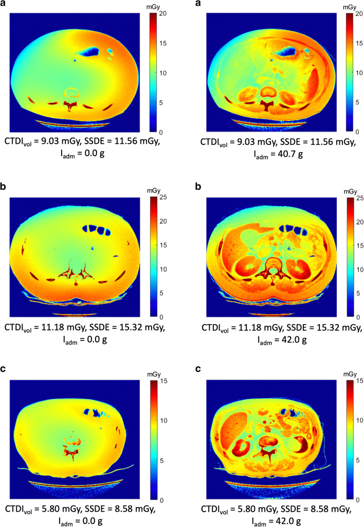
Methods: First, relation between absorbed radiation dose and iodine concentrations was investigated using a cylindrical water phantom with iodine-saline dilution insertions. Subsequently, a retrospective study on abdominal dual-energy CT (DECT) patient data was performed to assess the increase of the local absorbed radiation dose compared to a non-contrast scan. Absorbed doses were estimated with Monte Carlo simulations using the individual CT voxel data of phantom and patients. Further, organ segmentations were performed to obtain the dose in liver, liver parenchyma, left kidney, right kidney, aorta, and spleen.
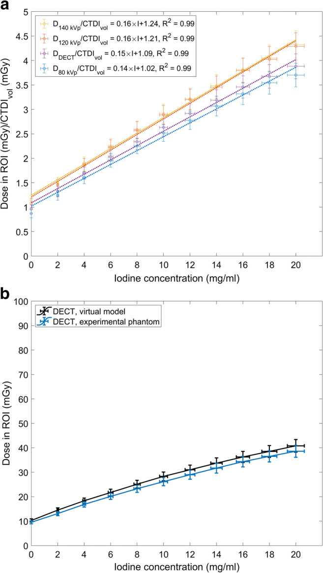
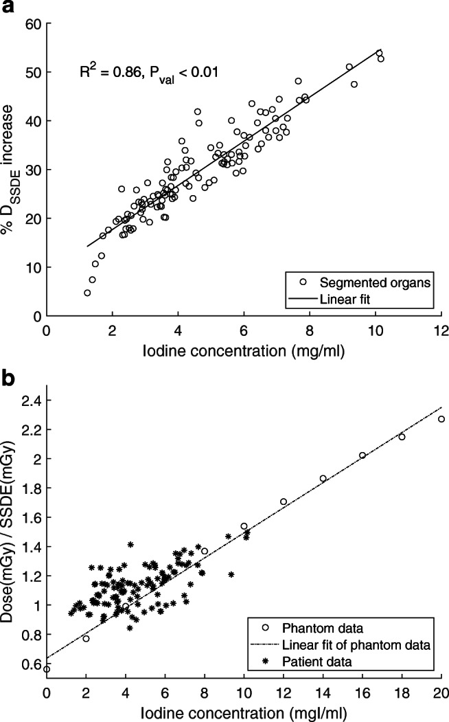
Results: In the phantom study, a linear relation was observed between the radiation dose normalized by computed tomography dose index (CTDI) and CA concentrations Iconc (mg/ml) for three tube voltages; [Formula: see text] = 0.14 × Iconc + 1.02, [Formula: see text] = 0.16 × Iconc + 1.21, [Formula: see text] = 0.16 × Iconc + 1.24, and for DECT acquisition; [Formula: see text] = 0.15 × Iconc + 1.09. Similarly, a linear relation was observed between the dose increase and the organ iodine contents (R2 = 0.86 and pvalue < 0.01) in the patient study. The relative doses increased in the liver (21 ± 5%), liver parenchyma (20 ± 5%), right kidney (37 ± 7%), left kidney (39 ± 7%), aorta (34 ± 6%) and spleen (26 ± 4%). In addition, the local dose distributions changed based on patient's anatomy and physiology.
Conclusions: Compared to a non-contrast scan, the organ doses increase by 30% in contrast-enhanced abdominal CT. This study suggests considering CA in dosimetry calculations, epidemiological studies, and organ dose estimations while developing new CT protocols.
Key points: • The presence of contrast media increases radiation absorption in CT, and this increase is related to the iodine content in the organs. • The increased radiation absorption due to contrast media can lead to an average 30% increase in absorbed organ dose. • Iodine should be considered in CT radiation safety studies.
Keywords: Humans; Iodine; Phantoms, Imaging; Radiation dosage; Tomography, X-ray Computed.
© 2021. The Author(s).
Conflict of interest statement
The authors of this manuscript declare no relationships with any companies whose products or services may be related to the subject matter of the article.
Figures

References
-
- American College of Radiology . ACR manual on contrast media. Reston: American College of Radiology; 2020.
-
- Van der Molen AJ, Reimer P, Dekkers IA, et al. Post-contrast acute kidney injury. Part 2: risk stratification, role of hydration and other prophylactic measures, patients taking metformin and chronic dialysis patients: Recommendations for updated ESUR Contrast Medium Safety Committee guidelines. Eur Radiol. 2018;28:2856–2869. doi: 10.1007/s00330-017-5247-4. - DOI - PMC - PubMed
MeSH terms
Substances
Grants and funding
LinkOut - more resources
Full Text Sources
Other Literature Sources
Medical
