Review
. 2021 May;24(2):199-211.
doi: 10.1007/s10456-021-09783-9. Epub 2021 Mar 30.

First blood: the endothelial origins of hematopoietic progenitors

Affiliations
Review

First blood: the endothelial origins of hematopoietic progenitors

Giovanni Canu et al. Angiogenesis. 2021 May.

Abstract

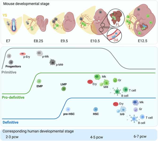
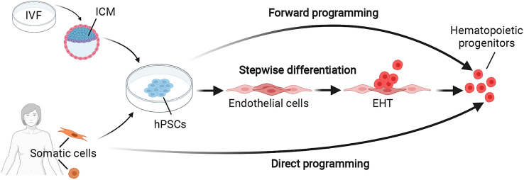
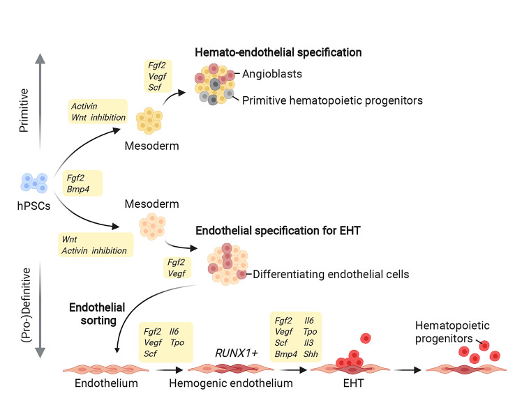
Hematopoiesis in vertebrate embryos occurs in temporally and spatially overlapping waves in close proximity to blood vascular endothelial cells. Initially, yolk sac hematopoiesis produces primitive erythrocytes, megakaryocytes, and macrophages. Thereafter, sequential waves of definitive hematopoiesis arise from yolk sac and intraembryonic hemogenic endothelia through an endothelial-to-hematopoietic transition (EHT). During EHT, the endothelial and hematopoietic transcriptional programs are tightly co-regulated to orchestrate a shift in cell identity. In the yolk sac, EHT generates erythro-myeloid progenitors, which upon migration to the liver differentiate into fetal blood cells, including erythrocytes and tissue-resident macrophages. In the dorsal aorta, EHT produces hematopoietic stem cells, which engraft the fetal liver and then the bone marrow to sustain adult hematopoiesis. Recent studies have defined the relationship between the developing vascular and hematopoietic systems in animal models, including molecular mechanisms that drive the hemato-endothelial transcription program for EHT. Moreover, human pluripotent stem cells have enabled modeling of fetal human hematopoiesis and have begun to generate cell types of clinical interest for regenerative medicine.

Keywords: EndoHT; Endothelium; Erythro-myeloid progenitor; Fetal liver; Hematopoiesis; Hematopoietic stem cell; Tissue‐resident macrophage; Yolk sac.


Conflict of interest statement

The authors have no conflicts or competing interests to report.

Figures

Fig. 1
Fig. 1
Ontogeny of the hematopoietic system. Top panel: Hematopoietic development proceeds in three spatiotemporally overlapping waves termed primitive, pro-definitive, and definitive hematopoiesis, indicated with gray, green, and blue colors, respectively. Each wave produces distinct hematopoietic progenitors, which are shown in the top panel at their site of origin and their destination in the embryo at the relevant developmental stages. Pro-definitive progenitors arising in the yolk sac (YS) and hematopoietic stem cells (HSCs) arising in the dorsal aorta (DA) co-exist in the fetal liver (FL), as shown at higher magnification for E10.5. Middle panel: Each hematopoietic wave generates a unique, essential, and complementary set of circulating and tissue-resident hematopoietic cells. The primitive wave produces erythrocytes (p-Ery), megakaryocytes (p-Mk), and macrophages (p-MΦ) that remain in the yolk sac or invade the embryo to generate microglia. The pro-definitive wave generates erythro-myeloid progenitors (EMPs) and lympho-myeloid progenitors (LMPs). The definitive wave generates pre-HSCs, which mature into HSCs capable of self-renewal. Both pro-definitive and definitive wave progenitors travel to the liver, where they produce erythrocytes (Ery), megakaryocytes (Mk), granulocytes (Gr), T cells and B cells as well as monocyte-derived macrophages (MΦ). EMP-derived MΦs colonize the embryo and constitute the majority of tissue-resident MΦs at birth. Bottom panel: Hematopoietic development is thought to follow similar principles in human, with the corresponding developmental stages shown
Fig. 2
Fig. 2
Strategies for hematopoietic cell production in vitro. Human pluripotent stem cells (hPSCs), including embryonic stem cells and induced pluripotent stem cells, have been used to model human hematopoiesis in vitro and to generate differentiated blood cells. Embryonic stem cells are derived from the inner cell mass (ICM) of donated, surplus human pre-implantation embryos generated for in vitro fertilization (IVF). Induced pluripotent stem cells are derived from reprogrammed adult somatic cells, usually fibroblasts or circulating blood cells. Both types of hPSCs are able to self-renew in appropriate culture conditions and can be induced to differentiate into hematopoietic cells. Forward programming (top): The enforced expression of several key transcription factors activates the hematopoietic program that converts hPSCs into hematopoietic cells. Stepwise differentiation (middle): The sequential administration of cytokines and small molecules induces the developmental steps that drive hematopoietic differentiation in vivo. Direct programming (bottom): The enforced expression of several key transcription factors in adult somatic cells converts them directly into hematopoietic cells
Fig. 3
Fig. 3
Induction of distinct hematopoietic waves using hPSCs. Modulating key signalling pathways early during stepwise hPSC differentiation enables the production of primitive versus (pro-)definitive hematopoietic cells (only key steps shared between various different protocols are shown). FGF and BMP induce hPSC differentiation towards mesoderm. When combined with activin activation and WNT inhibition, mesodermal cells differentiate further into primitive hematopoietic cells. Instead, WNT activation with activin inhibition induces mesodermal cells to differentiate further into endothelial cells, including hemogenic endothelial cells that express RUNX1 and undergo EHT to produce (pro-)definitive hematopoietic cells

References

    1. Goldie LC, Nix MK, Hirschi KK. Embryonic vasculogenesis and hematopoietic specification. Organogenesis. 2008;4:257–263. doi: 10.4161/org.4.4.7416. - DOI - PMC - PubMed
    1. Gritz E, Hirschi KK. Specification and function of hemogenic endothelium during embryogenesis. Cell Mol Life Sci. 2016;73:1547–1567. doi: 10.1007/s00018-016-2134-0. - DOI - PMC - PubMed
    1. Ferkowicz MJ, Yoder MC. Blood island formation: longstanding observations and modern interpretations. Exp Hematol. 2005;33:1041–1047. doi: 10.1016/j.exphem.2005.06.006. - DOI - PubMed
    1. Palis J, Robertson S, Kennedy M, et al. Development of erythroid and myeloid progenitors in the yolk sac and embryo proper of the mouse. Development. 1999;126:5073–5084. doi: 10.1242/dev.126.22.5073. - DOI - PubMed
    1. Hoeffel G, Ginhoux F. Fetal monocytes and the origins of tissue-resident macrophages. Cell Immunol. 2018;330:5–15. doi: 10.1016/j.cellimm.2018.01.001. - DOI - PubMed

Publication types

MeSH terms