. 2021 May 10;6(9):e143940.
doi: 10.1172/jci.insight.143940.

HIV-1 integration sites in CD4+ T cells during primary, chronic, and late presentation of HIV-1 infection

Affiliations

HIV-1 integration sites in CD4+ T cells during primary, chronic, and late presentation of HIV-1 infection

Yik Lim Kok et al. JCI Insight. .

Abstract

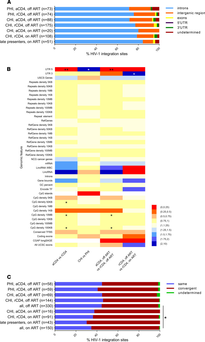
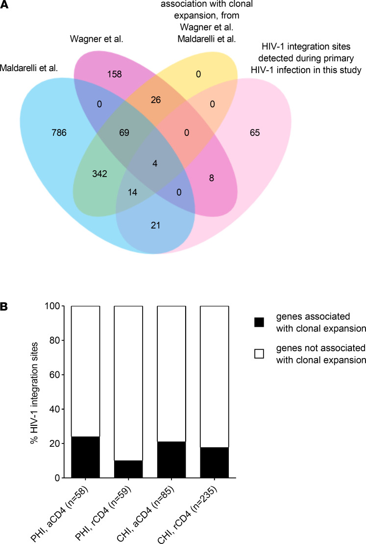
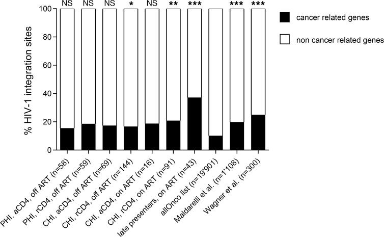
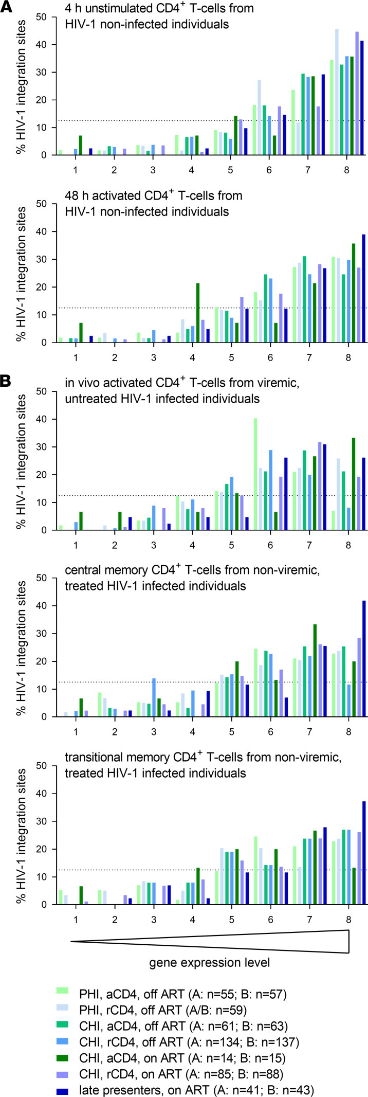
HIV-1 is capable of integrating its genome into that of its host cell. We examined the influence of the activation state of CD4+ T cells, the effect of antiretroviral therapy (ART), and the clinical stage of HIV-1 infection on HIV-1 integration site features and selection. HIV-1 integration sites were sequenced from longitudinally sampled resting and activated CD4+ T cells from 12 HIV-1-infected individuals. In total, 589 unique HIV-1 integration sites were analyzed: 147, 391, and 51 during primary, chronic, and late presentation of HIV-1 infection, respectively. As early as during primary HIV-1 infection and independent of the activation state of CD4+ T cells collected on and off ART, HIV-1 integration sites were preferentially detected in recurrent integration genes, genes associated with clonal expansion of latently HIV-1-infected CD4+ T cells, cancer-related genes, and highly expressed genes. The preference for cancer-related genes was more pronounced at late stages of HIV-1 infection. Host genomic features of HIV-1 integration site selection remained stable during HIV-1 infection in both resting and activated CD4+ T cells. In summary, characteristic HIV-1 integration site features are preestablished as early as during primary HIV-1 infection and are found in both resting and activated CD4+ T cells.

Keywords: AIDS/HIV; Molecular biology; T cells; Virology.


Conflict of interest statement

Conflict of interest: DLB received honoraria from Gilead Sciences, Merck, and ViiV. HFG received unrestricted research grants from Gilead Sciences and Roche; fees for data and safety monitoring board membership from Merck; and consulting/advisory board membership fees from Merck, ViiV, and Gilead. KJM received travel grants and honoraria from Gilead Sciences, Merck Sharp & Dohme, and ViiV. The University of Zurich has received research grants from Gilead Sciences, Roche, and Merck Sharp & Dohme for studies of which KJM serves as principal investigator.

Figures

Figure 1
Figure 1. Genomic features of HIV-1 integration site libraries from longitudinally sampled resting and activated CD4+ T cells obtained from HIV-1–infected individuals during primary and chronic HIV-1 infection and during periods on and off ART and PBMCs from late presenters.
(A) Genomic distribution of HIV-1 integration sites. Undetermined HIV-1 integration sites were caused by splice variants. (B) Pairwise comparison of genomic features associated with HIV-1 integration sites in the different cell populations. The Benjamini-Hochberg method was used to correct for multiple hypothesis testing. (C) The orientation of HIV-1 proviruses relative to the host gene transcription orientation. Only intragenic HIV-1 integration sites were considered. P values were calculated using the 2-tailed Fisher’s exact test: *, P < 0.05, **, P < 0.01.
Figure 2
Figure 2. Comparison of HIV-1 integration sites identified during primary HIV-1 infection with HIV-1 integration sites identified by Wagner et al. and Maldarelli et al.
(A) HIV-1 integration sites identified during primary HIV-1 infection were compared with HIV-1 integration sites described by Wagner et al. and Maldarelli et al. (27, 28), with particular focus on those described to be associated with clonal expansion of latently HIV-1–infected, resting CD4+ T cells. (B) HIV-1 integration sites in genes associated with clonal expansion of latently HIV-1–infected, resting CD4+ T cells as described by Wagner et al. and Maldarelli et al. (27, 28).
Figure 3
Figure 3. Cancer-related genes are overrepresented in HIV-1 integration sites during all clinical stages of HIV-1 infection, particularly in resting CD4+ T cells.
The distribution of HIV-1 integration sites in resting and activated CD4+ T cells during primary and chronic HIV-1 infection on and off ART was analyzed in comparison with cancer-related genes (allOnco list) in the Gene Expression Omnibus (GEO) database (40). For further comparison, the list of HIV-1 integration sites described by Wagner et al. and Maldarelli et al. (27, 28) were included. P values were calculated using the 2-tailed χ2 test: ns, not significant; *, P < 0.05; **, P < 0.01; ***, P < 0.001.
Figure 4
Figure 4. Highly expressed genes are preferentially targeted by HIV-1 during all clinical stages of HIV-1 infection in both activated and resting CD4+ T cells.
The distribution of HIV-1 integration sites in resting and activated CD4+ T cells during primary and chronic HIV-1 infection on and off ART was analyzed in comparison with gene expression levels in different cell populations. The following gene expression data sets were obtained from the GEO database (40): (A) GSE60235, 4-hour unstimulated and 48-hour activated CD4+ T cells (41); (B) GSE9927, in vivo activated CD4+ T cells from viremic, untreated HIV-1–infected individuals (42); and GSE66214, HIV-1 central memory and transitional memory CD4+ T cells from nonviremic, treated HIV-1–infected individuals. Accordingly to their expression levels, the genes were distributed into 8 bins by expression levels, each bin containing 12.5% of all genes (12.5% is marked with a dotted line).

Similar articles

Cited by

References

    1. Deeks SG, et al. International AIDS Society global scientific strategy: towards an HIV cure 2016. Nat Med. 2016;22(8):839–850. doi: 10.1038/nm.4108. - DOI - PMC - PubMed
    1. Siliciano JD, Siliciano RF. Recent developments in the effort to cure HIV infection: going beyond N = 1. J Clin Invest. 2016;126(2):409–414. doi: 10.1172/JCI86047. - DOI - PMC - PubMed
    1. Siliciano JD, et al. Long-term follow-up studies confirm the stability of the latent reservoir for HIV-1 in resting CD4+ T cells. Nat Med. 2003;9(6):727–728. doi: 10.1038/nm880. - DOI - PubMed
    1. Wong JK, et al. Recovery of replication-competent HIV despite prolonged suppression of plasma viremia. Science. 1997;278(5341):1291–1295. doi: 10.1126/science.278.5341.1291. - DOI - PubMed
    1. Finzi D, et al. Identification of a reservoir for HIV-1 in patients on highly active antiretroviral therapy. Science. 1997;278(5341):1295–1300. doi: 10.1126/science.278.5341.1295. - DOI - PubMed

Publication types