. 2021 Aug 1;204(3):312-325.
doi: 10.1164/rccm.202008-3100OC.

CD148 Deficiency in Fibroblasts Promotes the Development of Pulmonary Fibrosis

Affiliations

CD148 Deficiency in Fibroblasts Promotes the Development of Pulmonary Fibrosis

Konstantin Tsoyi et al. Am J Respir Crit Care Med. .

Abstract

Rationale: CD148/PTRJ (receptor-like protein tyrosine phosphatase η) exerts antifibrotic effects in experimental pulmonary fibrosis via interactions with its ligand syndecan-2; however, the role of CD148 in human pulmonary fibrosis remains incompletely characterized.Objectives: We investigated the role of CD148 in the profibrotic phenotype of fibroblasts in idiopathic pulmonary fibrosis (IPF).Methods: Conditional CD148 fibroblast-specific knockout mice were generated and exposed to bleomycin and then assessed for pulmonary fibrosis. Lung fibroblasts (mouse lung and human IPF lung), and precision-cut lung slices from human patients with IPF were isolated and subjected to experimental treatments. A CD148-activating 18-aa mimetic peptide (SDC2-pep) derived from syndecan-2 was evaluated for its therapeutic potential.Measurements and Main Results: CD148 expression was downregulated in IPF lungs and fibroblasts. In human IPF lung fibroblasts, silencing of CD148 increased extracellular matrix production and resistance to apoptosis, whereas overexpression of CD148 reversed the profibrotic phenotype. CD148 fibroblast-specific knockout mice displayed increased pulmonary fibrosis after bleomycin challenge compared with control mice. CD148-deficient fibroblasts exhibited hyperactivated PI3K/Akt/mTOR signaling, reduced autophagy, and increased p62 accumulation, which induced NF-κB activation and profibrotic gene expression. SDC2-pep reduced pulmonary fibrosis in vivo and inhibited IPF-derived fibroblast activation. In precision-cut lung slices from patients with IPF and control patients, SDC2-pep attenuated profibrotic gene expression in IPF and normal lungs stimulated with profibrotic stimuli.Conclusions: Lung fibroblast CD148 activation reduces p62 accumulation, which exerts antifibrotic effects by inhibiting NF-κB-mediated profibrotic gene expression. Targeting the CD148 phosphatase with activating ligands such as SDC2-pep may represent a potential therapeutic strategy in IPF.

Keywords: CD148; fibroblast; idiopathic pulmonary fibrosis; nuclear factor-kappa-B; syndecan-2.


Figures

Figure 1.
Figure 1.
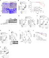
CD148 is downregulated in idiopathic pulmonary fibrosis (IPF) lung and contributes to profibrotic phenotype of IPF-derived lung fibroblasts. (A) CD148 protein expression levels were determined in lung homogenates from control patients (n = 5) and patients with IPF (n = 5). Densitometry (by ImageJ software). (B) Lung specimens from control patients (n = 9) and patients with IPF (n = 10) were homogenized and subjected to total RNA isolation. Ptprj/CD148 (receptor-like protein tyrosine phosphatase η) mRNA concentrations were assessed using quantitative PCR (qPCR). (C) Single-cell suspensions of control (n = 6) and IPF (n = 7) cells were enriched for fibroblasts (see Figure E1). mRNA expression of CD148 in fibroblast-enriched cell populations was assessed using qPCR. (D) Representative fluorescence microscopy images of CD148 (Cy3, red), Vimentin (GFP, green), and DAPI (blue) in control (n = 4) and IPF lung tissue (n = 5). Scale bars, 100 μm. Enlarged areas (clockwise from top right panel) represent Vimentin, CD148, DAPI, and merged image. CD148 fluorescence intensity was quantified by ImageJ. (E and F) Lung fibroblasts from nondisease (control) and IPF samples were transfected with scramble (Scr) or shCD148. (E) CD148 protein concentrations were measured by Western blot (n = 3). (F) mRNA concentrations of Fn (fibronectin) and Col1a1 (collagen 1a1) were measured using qPCR (n = 5). (G) Scr and shCD148 transfected cells were seeded in 24-well plates and then treated with Fas ligand (FasL; 200 ng/ml) for 24 hours. After treatment, cell viability was determined using the 3-[4,5-dimethylthiazol-2-yl]-2,5-diphenyl tetrazolium bromide (MTT) assay (n = 5). (H) IPF-derived lung fibroblasts were stably transfected with empty vector (EV; pLenti-GIII-HA) or pLenti-GIII-CD148-HA (CD148-HA). The cells were lysed and subjected to Western blot to measure hemagglutinin tag (HA) (n = 3). (I) In EV- and CD148-HA–transfected cells, the expression of Fn and Col1a1 were measured using qPCR (n = 5). (J) EV- and CD148-HA–transfected cells were seeded at equal amounts in 24-well plates and treated with FasL (200 ng/ml) for 24 hours. Cell viability was determined using the MTT assay (n = 5). Data are mean ± SEM. *P < 0.05 by Mann-Whitney’s unpaired nonparametric test (E and H), Student’s unpaired t test (B and C), or one-way ANOVA (FJ).
Figure 2.
Figure 2.
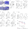
CD148 deficiency in fibroblasts worsens pulmonary fibrosis in bleomycin (BLM)-treated mice and increases fibroblast activation in response to TGF-β1. CD148 fibroblast-specific knockout mice (Ptprjfl/fl Col1a2Cre−ER(T)+/0) were developed as described in Methods. (A) Ptprjfl/fl Col1a2Cre−ER(T)+/0 mice or Col1a2Cre−ER(T)+/0 (wild-type [WT]) mice were exposed to BLM to induce lung fibrosis. At Day 21, mouse lungs were harvested and stained with Masson’s trichrome (n = 3 for saline and n = 5 for BLM groups). (B) Hydroxyproline content was measured in the left lung of Ptprjfl/fl Col1a2Cre−ER(T)+/0 mice (n = 12) and Col1a2Cre−ER(T)+/0 (WT) mice (n = 12) exposed to BLM and Ptprjfl/fl Col1a2Cre−ER(T)+/0 (n = 6) and Col1a2Cre−ER(T)+/0 (n = 6) exposed to saline at Day 21. (C) Relative survival at Days 0–21 after BLM. (D and E) α-SMA and CD148 expression in harvested lungs were measured by Western blot (n = 3–5 for each condition). (F and G) Gene expression of Fn (fibronectin) and Col1a1 (collagen 1a1) and Ctgf (connective tissue growth factor) in harvested lungs was measured using quantitative PCR (qPCR) (n = 5 for each condition). (H and I) Mouse lung fibroblasts from Ptprjfl/fl Col1a2Cre−ER(T)+/0 mice (Ptprj−/−) and Col1a2Cre−ER(T)+/0 (WT) mice were isolated and treated with 4-hydroxytamoxifen (4-OHT; 1 μM) for 24 hours. After 4-OHT treatment, cells were exposed to TGF-β1 (10 ng/ml) for an additional 24 hours. After stimulation, cells were harvested. (H) Western blot (n = 4 for each condition). (I) mRNA concentrations of Col1a1 and Fn were measured by qPCR (n = 5 for each condition). (J) Cells were mixed with collagen 1. Gel contractility was measured at 0, 12, and 24 hours after TGF-β1 (10 ng/ml) stimulation (n = 6 for each condition). (K) Cell death was induced by FasL (200 ng/ml). At 24 hours, cell viability was measured with 3-[4,5-dimethylthiazol-2-yl]-2,5-diphenyl tetrazolium bromide assay (n = 5 for each condition). Data are mean ± SEM. *P < 0.05 and **P < 0.01 by one-way ANOVA. FasL = Fas ligand.
Figure 3.
Figure 3.
CD148 deficiency enhances PI3K/Akt/mTOR signaling, which results in low autophagy and high p62 expression in lung fibroblasts. (A) Mouse lung fibroblasts were isolated from Ptprjfl/fl Col1a2Cre−ER(T)+/0 mice (Ptprj−/−) and Col1a2Cre−ER(T)+/0 mice (wild-type [WT]) and then treated with 4-hydroxytamoxifen (4-OHT; 1 μM) for 24 hours. After 4-OHT treatment, cells were exposed to TGF-β1 (10 ng/ml) for 24 hours. After stimulation, cells were harvested, and the expression of phospho (p)-p85 (Tyr458), p-Akt (Ser473), p-mTOR (Ser2448), p-p70 S6 kinase (Thr389), p-S6 ribosomal protein (Ser235/236), and CD148 (n = 4 for each condition) in lysates was determined by Western immunoblotting and corresponding densitometry (ImageJ software). Data were normalized to corresponding dephosphorylated forms or GAPDH. (B) WT and Ptprj−/− cells were stimulated with TGF-β1 (10 ng/ml) for 24 hours. Then cells were lysed, and LC3 (light chain-3B)-I, LC3-II, and p62 were measured by Western blot (n = 4). (C) WT and Ptprj−/− cells were incubated in starvation media (Hank’s buffered salt solution without calcium/magnesium, containing 1% regular medium) for 24 hours. Then, autophagy flux was measured by LC3-II accumulation in the absence or presence of lysosomal acidification inhibitor chloroquine (25 μM) at 2, 4, and 6 hours (n = 4). (D and E) Lung fibroblasts from LC3-GFP transgenic mice were transfected with Scr or shCD148 (lentivirus). Cells were starved for 24 hours in the presence or absence of TGF-β1 (10 ng/ml), and then cells were treated with chloroquine (25 μM) for 4 hours. After treatment, cells were fixed and digital images (three images per sample) were taken using fluorescent microscope. Representative images are shown in E. LC3 puncta positive cells were quantified using ImageJ (D). Data are mean ± SEM. *P < 0.05 by one-way ANOVA. CQ = chloroquine; Scr = scramble.
Figure 4.
Figure 4.
CD148 deficiency enhances PI3K/Akt/mTOR signaling, which results in enhanced NF-κB activation in lung fibroblasts. (A) Wild-type (WT) and Ptprj−/− cells were stimulated with TGF-β1 (10 ng/ml) for 24 hours. Cells were lysed and phospho (p)-IKKα/β, p-IκB, and IκB were measured by Western blot (n = 4). GAPDH was the standard. Bar graphs at right are the quantitation of corresponding proteins. (B) WT and Ptprj−/− cells were stimulated with TGF-β1 (10 ng/ml) for 24 hours. Cells were subjected to cytosol and nuclear protein fractionation. The p65 (NF-κB subunit) nuclear translocation was measured by Western blot (n = 3). β-actin and PCNA were used as cytosolic and nuclear markers, respectively. Bar graphs at right are the quantitation of corresponding proteins. The p65 cytosolic/nuclear ratio is shown (far right). Data are mean ± SEM. *P < 0.05 by Kruskal-Wallis nonparametric test (C and D) WT or Ptprj−/− cells were transfected with scramble (Scr) or shp62 (lentivirus). Cells were stimulated with TGF-β1 (10 ng/ml) in the presence or absence or wortmannin (wort; 50 nM) or rapamycin (rapa; 1 μM). At 24 hours, cells were harvested and subjected to quantitative PCR (qPCR) for Col1a1 (collagen 1a1) or Acta2 (n = 5). (E) WT or Ptprj−/− cells were transfected with NF-κB luciferase reporter plasmid in the presence or absence of Scr or shp62 or of wort (50 nM) or rapa (1 μM). Then, cells were treated with TGF-β1 (10 ng/ml) for 4 hours. Luciferase activity was measured as described in Methods (n = 4 or 7). Data are mean ± SEM. *P < 0.05 by one-way ANOVA. (F) WT or Ptprj−/− cells were stimulated with TGF-β1 (10 ng/ml) in the presence or absence of the NF-κB inhibitor Bay 11–7082 (10 μM). mRNA concentrations of Col1a1 were measured by qPCR (n = 5 for each condition). Data are mean ± SEM. *P < 0.05 by one-way ANOVA.
Figure 5.
Figure 5.
SDC2-ED 18-aa peptide (SDC2-pep) inhibits pulmonary fibrosis in vivo, upregulates autophagy by the downregulation of PI3K/Akt/mTOR signaling, and inhibits extracellular matrix gene expression in mouse fibroblasts via CD148. (AC) Ptprjfl/fl Col1a2Cre−ER(T)+/0 mice (fibroblast-specific CD148-deficient) and Col1a2Cre−ER(T)+/0 (wild-type [WT]) mice were exposed to bleomycin (BLM). SDC2-pep (0.5 mg/kg) was intranasally delivered into WT and Ptprjfl/fl Col1a2Cre−ER(T)+/0 mice 10 days after BLM injury. Treatments were repeated at Days 12, 14, 16, and 18. (A) At 24 days after BLM exposure, lungs were harvested and stained with Masson’s trichrome (n = 3 for saline and n = 5 for BLM groups). (B) Hydroxyproline content was measured in the left lung of mice exposed to BLM (n = 11) or saline (n = 5). Gene expression of (C) Fn (fibronectin) and Col1a1 (collagen 1a1) and (D) Ctgf (connective tissue growth factor) in harvested lungs were measured using quantitative PCR (qPCR) (n = 5 for each condition). (E and F) Mouse lung fibroblasts from Ptprjfl/fl Col1a2Cre−ER(T)+/0 mice (Ptprj−/−) and Col1a2Cre−ER(T)+/0 mice (WT) were isolated and treated with 4-hydroxytamoxifen (4-OHT; 1 μM) for 24 hours. After 4-OHT treatment, cells were exposed to ±TGF-β1 (10 ng/ml) for an additional 24 hours in the absence or presence of SDC2-pep (5 μM). (E) Expression of phospho (p)-AKT/Akt, p-mTOR/mTOR, p62/GAPDH p-IKKα/β/IKKα, α-SMA/GAPDH, and CD148/GAPDH was determined by Western immunoblotting (n = 4) and corresponding densitometry (ImageJ software). Data are mean ± SEM. *P < 0.05 by one-way ANOVA. (F) mRNA concentrations of Col1a1 and Fn were measured by qPCR (n = 5 for each condition). (G) After treatment, cells were mixed with collagen 1. Gel contractility was measured at 0, 12, and 24 hours after TGF-β1 stimulation (n = 6 for each condition). Data are mean ± SEM. *P < 0.05 by one-way ANOVA. NS = not significant.
Figure 6.
Figure 6.
SDC2-ED 18-aa peptide (SDC2-pep) attenuates profibrotic gene expression in human idiopathic pulmonary fibrosis (IPF) fibroblasts. (A) IPF-derived lung fibroblasts were transfected with scramble (Scr) or shCD148. Then cells were treated with SDC2-pep (5 μM) for 24 hours. After incubation, phospho (p)-Akt (Ser473), p-mTOR (Ser2448), LC3 (light chain-3B), p62, and CD148 were measured by Western blot (n = 4). Band intensities were quantified using ImageJ software and were expressed as a ratio of band intensity relative to GAPDH. Data are mean ± SEM. *P < 0.05 by one-way ANOVA. (B) mRNA concentrations of Fn (fibronectin) and Col1a1 (collagen 1a1) were measured by quantitative PCR (n = 5). (C) Scr- or shCD148-transfected control or IPF-derived lung fibroblasts were mixed with collagen 1. Gel contractility were measured at 0, 12, and 24 hours in the presence or absence of SDC2-pep (5 μM) (n = 5/group). (D) Scr- or shCD148-transfected control or IPF-derived lung fibroblasts were treated with FasL (200 ng/ml) for 24 hours in the presence or absence of SDC2-pep (5 μM) (n = 7). Cell viability was measured using 3-[4,5-dimethylthiazol-2-yl]-2,5-diphenyl tetrazolium bromide assay. Data are mean ± SEM. *P < 0.05 by one-way ANOVA. FasL = Fas ligand.
Figure 7.
Figure 7.
SDC2-ED 18-aa peptide (SDC2-pep) and CD148 overexpression attenuate profibrotic gene expression in precision-cut lung slices (PCLS) derived from idiopathic pulmonary fibrosis (IPF) lungs and from wild-type lungs subjected to profibrotic stimuli. (AD) PCLS from IPF lungs were transfected with scramble (Scr), shCD148, or empty vector (EV) and CD148-HA. Then, PCLS were treated with SDC2-pep (5 μM) for 72 hours. After incubation, PCLS were digested for total RNA isolation. The expression of (A) Fn (fibronectin) and Col1a1 (collagen 1a1), (B) Acta2, and (C) Ptprj (CD148) were measured by quantitative PCR (qPCR) (n = 7 for each condition). (D) mRNA concentrations of Col1a1, Fn, Acta2, and Ptprj (CD148) were measured by qPCR in EV- and CD148-overexpressing PCLS (CD148-HA). (EH) PCLS from control lungs were transfected with Scr and shCD148 (lentivirus). Then, PCLSs were treated with profibrotic mix (FGF basic, 25 ng/ml; PDGF-BB, 10 ng/ml; and TGF-β1, 10 ng/ml) 72 hours with or without SDC2-pep (5 μM). After incubation, slices were digested for total RNA isolation. The expression of (E) Fn, (F) Col1a1, (G) Acta2, and (H) Ptprj were measured by qPCR. For AH, n = 6 for each condition. Data are mean ± SEM. *P < 0.05 by one-way ANOVA.
Figure 8.
Figure 8.
Schema depicting proposed antifibrotic effects of CD148 in fibroblasts. SDC2 (syndecan-2) via binding its receptor protein tyrosine phosphatase CD148/PTPRJ activates an antifibrotic pathway dependent on downregulation of TGF-β1–dependent signaling. TGF-β1 stimulates profibrotic effects via its receptor TGFβ-I/-II complex, which activates a PI3K/AKT/mTOR-dependent signaling pathway, culminating in the suppression of autophagy. The autophagy pathway, driven by LC3 (light chain-3B)-dependent formation of autophagosomes, directs the lysosomal degradation of autophagosome-sequestered cargo. Impaired autophagy is a characteristic feature of pulmonary fibrosis, which leads to aberrant accumulation of the autophagy substrate and cargo adaptor protein p62. Accumulated p62 promotes phosphorylation of the IKK (inhibitor of κ-B kinase) complex, leading to phosphorylation and dissociation of I-κB from the p65 subunit of NF-κB. The latter promotes p65/p50 assembly and migration of the NF-κB complex to the nucleus, where it stimulates profibrotic gene expression. NF-κB has also been implicated in apoptosis resistance and myofibroblast differentiation, characteristic of the profibrotic phenotype. Sequences (upper right) depict an 18-aa peptide region of the SDC2 ectodomain (SDC2-pep) with a high degree of homology between human and mouse sequences. SDC2-pep was tested in the current study as a therapeutic ligand of CD148/PTPRJ and found to have antifibrotic effects in idiopathic pulmonary fibrosis and models of pulmonary fibrosis. ECM = extracellular matrix.

Comment in

References

    1. Martinez FJ, Collard HR, Pardo A, Raghu G, Richeldi L, Selman M, et al. Idiopathic pulmonary fibrosis. Nat Rev Dis Primers. 2017;3:17074. - PubMed
    1. Chambers RC, Mercer PF. Mechanisms of alveolar epithelial injury, repair, and fibrosis. Ann Am Thorac Soc. 2015;12:S16–S20. - PMC - PubMed
    1. Richeldi L, du Bois RM, Raghu G, Azuma A, Brown KK, Costabel U, et al. INPULSIS Trial Investigators. Efficacy and safety of nintedanib in idiopathic pulmonary fibrosis. N Engl J Med. 2014;370:2071–2082. - PubMed
    1. Richeldi L, Costabel U, Selman M, Kim DS, Hansell DM, Nicholson AG, et al. Efficacy of a tyrosine kinase inhibitor in idiopathic pulmonary fibrosis. N Engl J Med. 2011;365:1079–1087. - PubMed
    1. Harrod TR, Justement LB. Evaluating function of transmembrane protein tyrosine phosphatase CD148 in lymphocyte biology. Immunol Res. 2002;26:153–166. - PubMed

Publication types

MeSH terms

LinkOut - more resources