. 2021 Apr 6;118(14):e2026656118.
doi: 10.1073/pnas.2026656118.

Structural analyses of an RNA stability element interacting with poly(A)

Affiliations

Structural analyses of an RNA stability element interacting with poly(A)

Seyed-Fakhreddin Torabi et al. Proc Natl Acad Sci U S A. .

Abstract

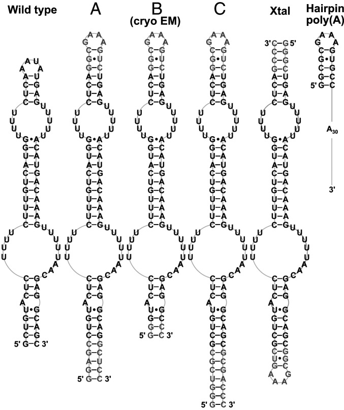
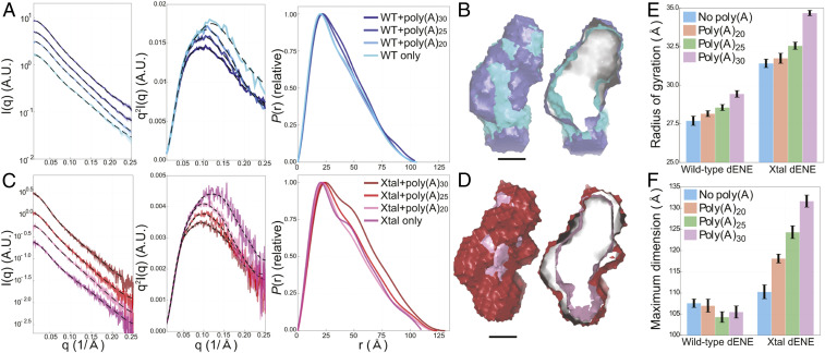
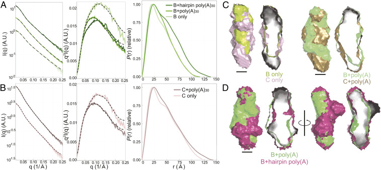
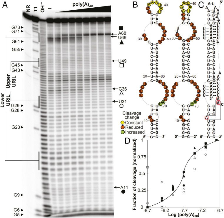
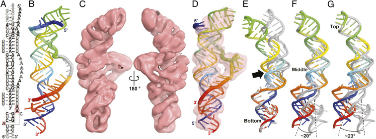
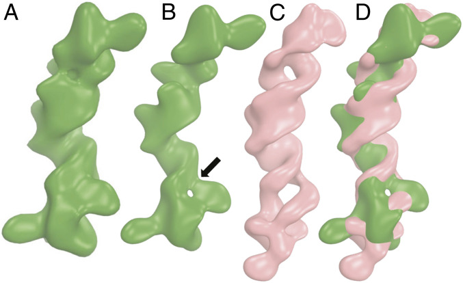
Cis-acting RNA elements are crucial for the regulation of polyadenylated RNA stability. The element for nuclear expression (ENE) contains a U-rich internal loop flanked by short helices. An ENE stabilizes RNA by sequestering the poly(A) tail via formation of a triplex structure that inhibits a rapid deadenylation-dependent decay pathway. Structure-based bioinformatic studies identified numerous ENE-like elements in evolutionarily diverse genomes, including a subclass containing two ENE motifs separated by a short double-helical region (double ENEs [dENEs]). Here, the structure of a dENE derived from a rice transposable element (TWIFB1) before and after poly(A) binding (∼24 kDa and ∼33 kDa, respectively) is investigated. We combine biochemical structure probing, small angle X-ray scattering (SAXS), and cryo-electron microscopy (cryo-EM) to investigate the dENE structure and its local and global structural changes upon poly(A) binding. Our data reveal 1) the directionality of poly(A) binding to the dENE, and 2) that the dENE-poly(A) interaction involves a motif that protects the 3'-most seven adenylates of the poly(A). Furthermore, we demonstrate that the dENE does not undergo a dramatic global conformational change upon poly(A) binding. These findings are consistent with the recently solved crystal structure of a dENE+poly(A) complex [S.-F. Torabi et al., Science 371, eabe6523 (2021)]. Identification of additional modes of poly(A)-RNA interaction opens new venues for better understanding of poly(A) tail biology.

Keywords: RNA stability; RNA triple helix; SAXS; cryo-EM; poly(A).


Conflict of interest statement

The authors declare no competing interest.

Figures

Fig. 1.
Fig. 1.
Schematic diagrams of the secondary structures of the TWIFB1 dENE constructs (including wild type, A, B, C, and Xtal) and the hairpin poly(A) used in SAXS experiments. The B dENE is also used in our cryo-EM studies. Shown in black are wild-type sequences while nonnative residues in other dENE constructs are shown in gray.
Fig. 2.
Fig. 2.
Comparative accumulation of β-globin reporter transcripts containing different poly(A) lengths stabilized by the dENE. (A) Schematic of the β-globin reporter constructs containing a cytomegalovirus (CMV) promoter, a human intronless β-globin gene (βΔ1,2), and the dENE, followed by either a bovine growth hormone poly(A) site (BGH pA) (Middle) or a poly(A) sequence of different lengths as indicated on the x-axis of the graph in B and mascRNA (Bottom). The arrowhead indicates the RNase P cleavage site upstream of mascRNA. (B) Northern blot analyses of transcripts expressed in HEK293T cells upon transfection of different β-globin reporter constructs. Blots (Top) were probed for β-globin and neomycin resistance (NeoR) sequences. NeoR serves as a transfection and loading control. Quantification of the Northern blots (Bottom) with the β-globin signals normalized to those of NeoR. Accumulation of βΔ1,2 was set at 1. Accumulations relative to no ENE control are the average of at least three independent experiments ± SD.
Fig. 3.
Fig. 3.
In-line probing analyses of poly(A) RNAs complexed with dENE variants. (A) Sequence and secondary structure model for the wild-type and mutant (M1 to M3) dENEs used in the experiment. (B) In-line probing analyses of 32P 5ʹ end-labeled poly(A)30 or poly(A)36 in free form (—) or complexed with the wild-type (WT) or M1 dENE. The dotted line indicates the directionality of poly(A) hydrolysis products. (C) In-line probing analyses of poly(A)30 complexed with the WT, M2, and M3 dENEs. NR and OH designate no reaction and partial digestion with alkali, respectively. The 7-nt 3ʹ-end protected region of the dENE-bound poly(A) is marked by brackets. The 5ʹ-end region of poly(A), whose cleavage pattern responds to the upper stem mutations, is boxed in blue.
Fig. 4.
Fig. 4.
SAXS experimental data, Kratky plots, and P(r)s for two dENE constructs and their complexes: wild-type dENE (A) and Xtal dENE (C) constructs. A.U., arbitrary units. The reconstructions of the wild-type, Xtal dENE constructs and their poly(A)30 complexes are shown in B and D (cross-sections on the Right). (Scale bars: 20 Å.) The color coding presented in A and C apply to B and D as well. The histograms compare the obtained values for Rg (E) and Dmax (F) for the wild-type and Xtal dENE constructs. The error bars in E were obtained directly from the Guinier analysis. The error bars in F were computed by sampling 25 values close to Dmax, with individual weights based on the metric α (18).
Fig. 5.
Fig. 5.
SAXS experimental data, Kratky plots, and P(r)s of two designed dENE constructs and their complexes: construct B (A) and construct C (B) (see Fig. 1). (C) The aligned comparisons of the SAXS reconstructions of B (lime green) and C (light purple) in the absence of poly(A) and the aligned reconstructions of their corresponding complexes: B+poly(A) (green) and C+poly(A) (gold). Using these reconstructions, we identified the longer stem in construct C and its poly(A)-bound complex and, therefore, established the orientation of both constructs. (Scale bars: 20 Å.) The hairpin of the hairpin poly(A) can be located using the known orientation of B+poly(A) and aligning it with B+hairpin poly(A) (magenta). The first two alignments in D demonstrate that these two complexes have similar lengths while the other alignments, rotated 90 degrees, reveal that the added hairpin extrudes from the complex near the top. These models indicate that the 5ʹ-to-3ʹ directionality of poly(A) is from top to bottom of construct B and that the interaction of the 3ʹ poly(A) terminus is with the lower ENE domain. Cross-sections of each surface model are on the Right.
Fig. 6.
Fig. 6.
In-line probing analyses of the wild-type dENE. (A) In-line probing analyses of 32P 5ʹ end-labeled dENE in the absence (—) and presence of varying concentrations of poly(A) (2 to 320 nM) at room temperature (22 °C). Lane NR shows no reaction, lane T1 indicates RNase T1-digested RNA (G-specific cleavage), and lane OH shows alkaline-mediated partial digestion. (B) Sequence and secondary structure models for the wild-type dENE (derived from in-line probing data in the absence of poly(A) on the Left and the predicted from comparative sequence analysis (11) on the Right). Nucleotide linkages that undergo constant, reduced, or increased cleavage upon poly(A) binding as determined by their relative band intensities in A are circled in yellow, red, or green, respectively. (C) Schematic of the poly(A)-bound dENE structure derived from the crystal structure (14). Long-range interactions between the A-triad nucleotides (boxed in red) are indicated by solid gray lines. (D) Dependence of the fraction of cleavage at selected sites (marked in A) in the lower dENE domain (closed symbols) and the upper dENE domain (open symbols) on poly(A) concentration. Calculation of fraction modulated is described in Materials and Methods. Data from selected cleavage sites were plotted against the logarithm of poly(A) concentration showing an apparent KD of ∼15 to 30 nM. The solid line indicates a theoretical binding curve for a 1:1 dENE-poly(A) interaction with a KD of 20 nM.
Fig. 7.
Fig. 7.
Cryo-EM structure of the B dENE construct complexed with a 28-mer poly(A) at 5.6-Å resolution. (A) Schematic of the B dENE+poly(A)28 structure derived from the crystal structure (14). Five disordered adenylates in the crystal structure are indicated by outlined letters. Non-native sequences are in gray. Dashed lines indicate regions of poly(A) interaction with the minor grooves of the stems. Long-range interactions between the A-triad nucleotides (boxed in red) are indicated by solid gray lines. (B) Computational atomic model derived from the crystal structure of the Xtal dENE+poly(A)28 complex (14) in rainbow colors (5ʹ, blue; 3ʹ, red). The disordered adenylated are added arbitrarily to the structure without any modeling (gray). (C) Two views of the cryo-EM density contoured at 3σ. (D) Real-space refined fitting of the crystallographic model to the cryo-EM density map. The cryo-EM (rainbow) and crystallographic (gray) structures were aligned using the bottom one-third (E), the middle one-third (F), or the top one-third of the structure (G) of the structure, which shows displacement of the opposite ends by ∼23° rotation, and a bend between the lower dENE domain and the middle stem (black arrow). Alignment of the poly(A)-bound structures is shown in SI Appendix, Fig. S14.
Fig. 8.
Fig. 8.
Cryo-EM structure of the B dENE construct without poly(A) at 8.7-Å resolution. (A and B) Density maps displayed at two contour levels (2.5σ, A; 3.5σ, B). The arrow indicates a hole in the lower dENE domain. (C) The dENE+poly(A) complex cryo-EM map with 8.7-Å resolution low-pass filtering, with poly(A) density subtracted (contoured at 3σ, salmon) and (D) its alignment with the free dENE map (contoured at 3.5σ, green). The two holes in C are in the upper and the lower URILs that form major-groove triple helices with poly(A). Upon poly(A) binding, local conformational changes occur in the lower dENE domain.

References

    1. Chen C. Y., Shyu A. B., Mechanisms of deadenylation-dependent decay. Wiley Interdiscip. Rev. RNA 2, 167–183 (2011). - PMC - PubMed
    1. Meyer S., Temme C., Wahle E., Messenger RNA turnover in eukaryotes: Pathways and enzymes. Crit. Rev. Biochem. Mol. Biol. 39, 197–216 (2004). - PubMed
    1. Geisberg J. V., Moqtaderi Z., Fan X., Ozsolak F., Struhl K., Global analysis of mRNA isoform half-lives reveals stabilizing and destabilizing elements in yeast. Cell 156, 812–824 (2014). - PMC - PubMed
    1. Wu X., Bartel D. P., Widespread influence of 3′-end structures on mammalian mRNA processing and stability. Cell 169, 905–917.e11 (2017). - PMC - PubMed
    1. Conrad N. K., Steitz J. A., A Kaposi’s sarcoma virus RNA element that increases the nuclear abundance of intronless transcripts. EMBO J. 24, 1831–1841 (2005). - PMC - PubMed

Publication types