. 2021 Mar 30;12(2):e00115-21.
doi: 10.1128/mBio.00115-21.

Species-Specific Deamidation of RIG-I Reveals Collaborative Action between Viral and Cellular Deamidases in HSV-1 Lytic Replication

Affiliations

Species-Specific Deamidation of RIG-I Reveals Collaborative Action between Viral and Cellular Deamidases in HSV-1 Lytic Replication

Huichao Huang et al. mBio. .

Abstract

Retinoic acid-inducible gene I (RIG-I) is a sensor that recognizes cytosolic double-stranded RNA derived from microbes to induce host immune response. Viruses, such as herpesviruses, deploy diverse mechanisms to derail RIG-I-dependent innate immune defense. In this study, we discovered that mouse RIG-I is intrinsically resistant to deamidation and evasion by herpes simplex virus 1 (HSV-1). Comparative studies involving human and mouse RIG-I indicate that N495 of human RIG-I dictates species-specific deamidation by HSV-1 UL37. Remarkably, deamidation of the other site, N549, hinges on that of N495, and it is catalyzed by cellular phosphoribosylpyrophosphate amidotransferase (PPAT). Specifically, deamidation of N495 enables RIG-I to interact with PPAT, leading to subsequent deamidation of N549. Collaboration between UL37 and PPAT is required for HSV-1 to evade RIG-I-mediated antiviral immune response. This work identifies an immune regulatory role of PPAT in innate host defense and establishes a sequential deamidation event catalyzed by distinct deamidases in immune evasion.IMPORTANCE Herpesviruses are ubiquitous pathogens in human and establish lifelong persistence despite host immunity. The ability to evade host immune response is pivotal for viral persistence and pathogenesis. In this study, we investigated the evasion, mediated by deamidation, of species-specific RIG-I by herpes simplex virus 1 (HSV-1). Our findings uncovered a collaborative and sequential action between viral deamidase UL37 and a cellular glutamine amidotransferase, phosphoribosylpyrophosphate amidotransferase (PPAT), to inactivate RIG-I and mute antiviral gene expression. PPAT catalyzes the rate-limiting step of the de novo purine synthesis pathway. This work describes a new function of cellular metabolic enzymes in host defense and viral immune evasion.

Keywords: HSV-1 UL37; RIG-I; deamidation; glutamine amidotransferase; herpesvirus; immune evasion; innate immune defense; phosphoribosyl pyrophosphate amidotransferase.


Figures

FIG 1
FIG 1
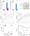
Antiviral activity of mouse and human RIG-I in HSV-1 infection. (A and B) RIG-I wild-type or knockout mouse embryonic fibroblasts (MEFs) were infected with HSV-1 (MOI = 2) and total RNA was extracted to analyze Ifnb and Isg56 gene expression (A). Viral titer was determined by plaque assay (B). (C and D) The expression of human and mouse RIG-I (hRIG and mRIG) was reconstituted via lentivirus infection in Rig-i−/− MEFs analyzed by immunoblotting (IB) (C), and HSV-1 replication (MOI = 0.01) was determined as in panel D. (E and F) Age- and gender-matched RIG-I wild-type and knockout mice were infected with HSV-1 (1,000 PFU) via the ocular route. Viral load in eye swabs was determined by plaque assay (E). Alternatively, mice were infected with lethal dose of HSV-1 (5 × 107 PFU) intravenously, and mouse survival was recorded over time (F). Data are presented as mean ± standard deviation (SD). Significance was calculated using an unpaired (paired for Fig. 1B and D) two-tailed Student’s t test. **, P < 0.01; ***, P < 0.001; NS, nonsignificant. Two-way analysis of variance (ANOVA) test and log-rank (Mantel-Cox) test were used to calculate the P values for Fig. 1E and F, respectively, using GraphPad Prism.
FIG 2
FIG 2
Comparative analysis indicates that N495 of human RIG-I dictates the deamidation by HSV-1 UL37. (A) Structure and surface of the helicase domain of hRIG-I with N495 and N549 highlighted (PDB identifier 3TMI). (B) Sequences flanking N495 and N549 of RIG-I across multiple species were aligned, with N495 and N549 (of human RIG-I) highlighted in red boxes. (C) 293T cells stably expressing hRIG-I (top) or mRIG-I were infected with HSV-1 (MOI = 2) and whole-cell lysates were analyzed by two-dimensional gel electrophoresis or regular SDS-PAGE, followed by immunoblotting with indicated antibodies. (D) Alignment of hRIG-I and mRIG-I sequences flanking N495 of hRIG-I and K496 of mRIG-I (top). Highlighted residues were mutated to create the hRIG-I containing N495K mutation (hRIG-I-K495) and the mRIG-I containing K496N mutation (mRIG-I-N496) (bottom). (E) 293T cells stably expressing hRIG-I-K495 or mRIG-I-N496 were infected with HSV-1 (multiplicity of infection [MOI] = 2). At 16 h postinfection, whole-cell lysates were prepared and analyzed by two-dimensional gel electrophoresis, followed by immunoblotting with indicated antibodies. (F and G) 293T cells stably expressing mRIG-I-N496 were infected with HSV-1 carrying UL37 or UL37C819S mutant (MOI = 2) (F) or were transfected with empty vector or vector containing UL37 or UL37C819S mutant (G). At 16 h postinfection or 30 h posttransfection, whole-cell lysates were analyzed as above to assess the charge status of mRIG-I-N496 with indicated antibodies.
FIG 3
FIG 3
hRIG-I-K495 is more potent than the hRIG-I wild-type in host defense in response to HSV-1 infection. (A) 293T cells stably expressing FLAG-RIG-I (293T/FLAG-RIG-I) were transfected with plasmids containing the V5-tagged hRIG-I wild type or RIG-I-K495. At 24 h posttransfection, cells were infected with HSV-1 (MOI = 2) for 5 h. Whole-cell lysates were precipitated with anti-FLAG (RIG-I). Precipitated proteins and WCLs were analyzed by immunoblotting with indicated antibodies. (B) 293T/FLAG-RIG-I cells were infected with HSV-1 (MOI = 2) for 5 h. WCLs were precipitated with anti-FLAG (RIG-I), precipitated proteins and whole-cell lysates (WCLs) were analyzed by immunoblotting with indicated antibodies. (C and D) 293T/FLAG-RIG-I cells were infected with HSV-1 (MOI = 2) for 10 (C) and 15 h (D). WCLs were prepared and analyzed by immunoblotting with indicated antibodies for phosphorylation of TBK-1 and IRF3 (C) and for IRF3 dimerization by native gel electrophoresis (D). (E and F) Control 293T cells (vector) or those stably expressing the hRIG-I wild type (WT) or hRIG-I-K495 were infected with Sendai virus (100 hemagglutinating units [HAU]/ml) (E) or HSV-1 (MOI = 2) (F) for the indicated time. Total RNA was extracted and analyzed by reverse transcription and real-time PCR with primers specific for Ifnb and Ccl5. (G and H) 293T cells as described in panel E were infected with HSV-1 (MOI = 0.1). Total RNA was extracted and reverse transcribed, and quantified by real-time PCR with primers specific for HSV-1 UL29 and UL54 genes (G). Viral titer was determined by plaque assay on Vero monolayer (H). Data are presented as mean ± SD. Significance was calculated using an unpaired, two-tailed Student’s t test. **, P < 0.01; ***, P < 0.001; NS, nonsignificant.
FIG 4
FIG 4
RIG-I-D495 is deamidated at N549 in cells. (A). RIG-I-deficient mouse embryonic fibroblasts (MEFs) were “reconstituted” with lentivirus containing the hRIG-I wild type (WT), hRIG-I-D495, hRIG-I-D549, and hRIG-I-DD. Whole-cell lysates (WCLs) were analyzed with immunoblotting with anti-FLAG (RIG-I) and β-actin. (B and C) Ifnb (C) and Cxcl10 (D) abundance in “reconstituted” Rig-i−/− MEFs as described in panel A and infected with Sendai virus (100 hemagglutinating units [HAU]/ml) for the indicated time. (D) 293T cells stably expressing the human RIG-I wild type, hRIG-I-D495, hRIG-I-D549, or hRIG-I-DD were infected with HSV-1 (MOI = 2) for 16 h, and WCLs were analyzed by two-dimensional gel electrophoresis and immunoblotting. (E) Percentage of the N549 deamidated peptides of the human RIG-I wild type or hRIG-I-D495 that were purified from stable 239T cells and analyzed by tandem mass spectrometry. Data are presented as mean ± SD. Significance was calculated using an unpaired two-tailed Student’s t test. *, P < 0.05; **, P < 0.01; ***, P < 0.001; NS, nonsignificant.
FIG 5
FIG 5
PPAT targets N549 of human RIG-I for deamidation. (A) 293T cells were transiently transfected with plasmids containing glutathione S-transferase (GST)-tagged RIG-I-D495 and FLAG-tagged GATs (CTPS1 and PPAT). Whole-cell lysates (WCLs) were precipitated with anti-FLAG (GAT). Precipitated proteins and WCLs were analyzed by immunoblotting with indicated antibodies. (B) 293T cells stably expressing hRIG-I-D495 were infected with lentivirus containing short hairpin RNA (shRNA) control (shCon) or PPAT shRNA (shPPAT) for 48 h, then infected with HSV-1 (MOI = 2) for 12 h. WCLs were analyzed by two-dimensional gel electrophoresis and immunoblotting. (C) 293T cells were mock infected or infected with HSV-1 (MOI = 1) for 12 h. WCLs were precipitated with anti-RIG-I or control antibody, precipitated proteins, and WCLs were analyzed by immunoblotting with indicated antibodies. (D) WCLs of 293T cells stably expressing the hRIG-I wild type, hRIG-I-D495, or hRIG-I-D549 were precipitated with anti-FLAG (RIG-I). WCLs and precipitated proteins were analyzed by immunoblotting with the indicated antibodies. (E) In vitro deamidation reaction purified substrate (GST-hRIG-I or GST-hRIG-I-D495), with deamidase (PPAT or PPAT-ED) were analyzed by two-dimensional gel electrophoresis and immunoblotting with anti-GST (RIG-I) antibody.
FIG 6
FIG 6
PPAT is crucial for HSV-1 to evade RIG-I-dependent innate immune response. (A) 293T cells stably expressing FLAG-hRIG-I were infected with lentivirus containing control shRNA (shCon) or PPAT shRNA (shPPAT) for 48 h, and with HSV-1 (MOI = 2) for 12 h. Whole-cell lysates (WCLs) were analyzed by two-dimensional gel electrophoresis (RIG-I, left) or regular SDS-PAGE (PPAT, right) and immunoblotting. (B and C) 293T cells were infected with lentivirus containing shCon or shPPAT, and with HSV-1 as described in panel A. WCLs were prepared at 12 h postinfection and analyzed by immunoblotting with the indicated antibodies (B). (C) Total RNA was extracted at the indicated time points, and cDNA was generated and analyzed by real-time PCR with primers specific for IFNB and CCL5 (C). (D and E) Viral titer (D), HSV-1 UL29 and UL54 abundance (E) in cells as described in panel A and infected with HSV-1 (MOI = 0.01 and 2, respectively) for the indicated time. (F) A schematic model illustrating the species-specific deamidation of RIG-I and the collaborative action between viral UL37 deamidase and the cellular PPAT deamidase in HSV-1 lytic replication. Data are presented as mean ± SD. Significance was calculated using unpaired (paired for Fig. 6D), two-tailed Student's t test. *, P < 0.05; **, P < 0.01; ***, P < 0.001; NS, nonsignificant.

Similar articles

Cited by

References

    1. Akira S, Uematsu S, Takeuchi O. 2006. Pathogen recognition and innate immunity. Cell 124:783–801. doi:10.1016/j.cell.2006.02.015. - DOI - PubMed
    1. Yoneyama M, Kikuchi M, Natsukawa T, Shinobu N, Imaizumi T, Miyagishi M, Taira K, Akira S, Fujita T. 2004. The RNA helicase RIG-I has an essential function in double-stranded RNA-induced innate antiviral responses. Nat Immunol 5:730–737. doi:10.1038/ni1087. - DOI - PubMed
    1. Schlee M, Roth A, Hornung V, Hagmann CA, Wimmenauer V, Barchet W, Coch C, Janke M, Mihailovic A, Wardle G, Juranek S, Kato H, Kawai T, Poeck H, Fitzgerald KA, Takeuchi O, Akira S, Tuschl T, Latz E, Ludwig J, Hartmann G. 2009. Recognition of 5′ triphosphate by RIG-I helicase requires short blunt double-stranded RNA as contained in panhandle of negative-strand virus. Immunity 31:25–34. doi:10.1016/j.immuni.2009.05.008. - DOI - PMC - PubMed
    1. Goubau D, Schlee M, Deddouche S, Pruijssers AJ, Zillinger T, Goldeck M, Schuberth C, Van der Veen AG, Fujimura T, Rehwinkel J, Iskarpatyoti JA, Barchet W, Ludwig J, Dermody TS, Hartmann G, Reis e Sousa C. 2014. Antiviral immunity via RIG-I-mediated recognition of RNA bearing 5′-diphosphates. Nature 514:372–375. doi:10.1038/nature13590. - DOI - PMC - PubMed
    1. Luo D, Ding SC, Vela A, Kohlway A, Lindenbach BD, Pyle AM. 2011. Structural insights into RNA recognition by RIG-I. Cell 147:409–422. doi:10.1016/j.cell.2011.09.023. - DOI - PMC - PubMed

Publication types

MeSH terms

Substances

LinkOut - more resources