. 2021 Mar 30;12(2):e00279-21.
doi: 10.1128/mBio.00279-21.

Cryptococcus neoformans - Infected Macrophages Release Proinflammatory Extracellular Vesicles: Insight into Their Components by Multi-omics

Affiliations

Cryptococcus neoformans - Infected Macrophages Release Proinflammatory Extracellular Vesicles: Insight into Their Components by Multi-omics

Lei Zhang et al. mBio. .

Abstract

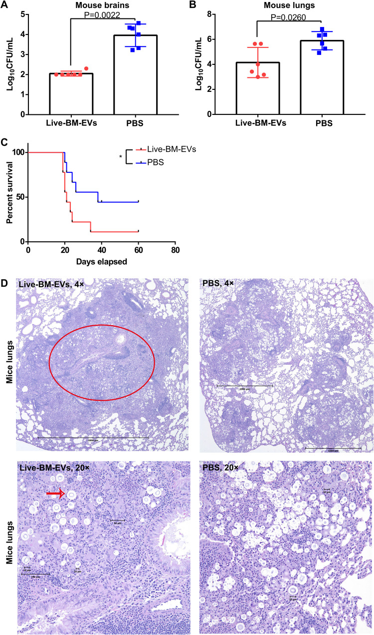
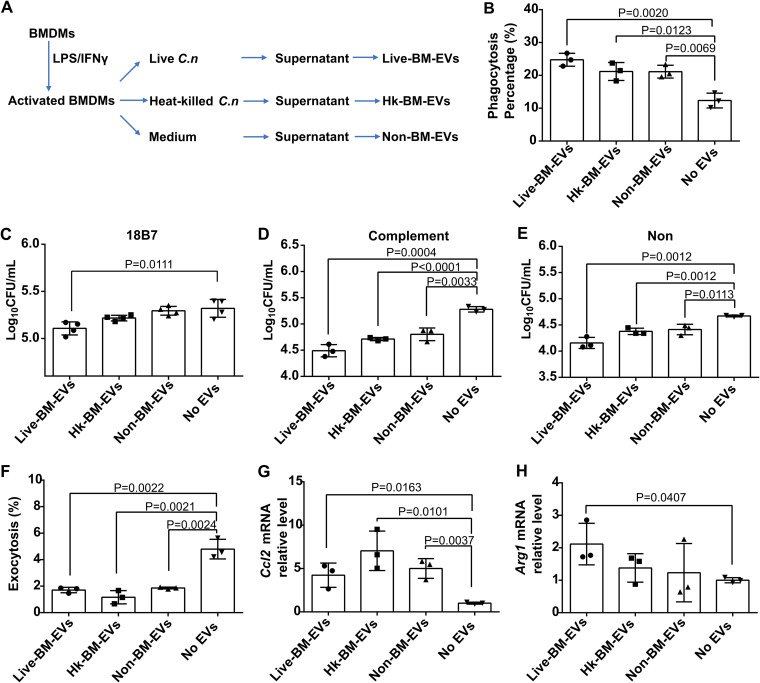
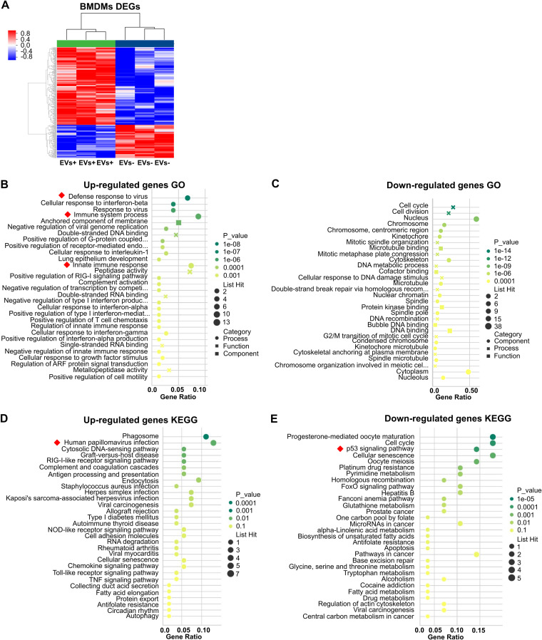
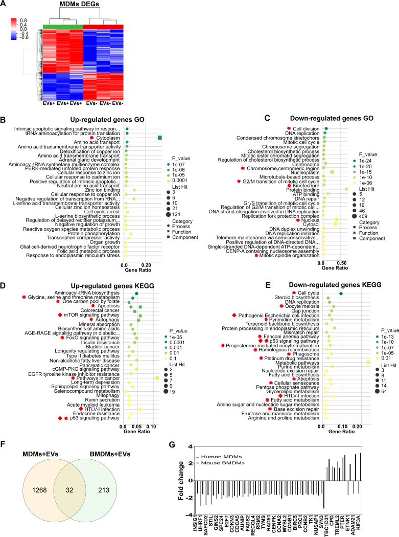
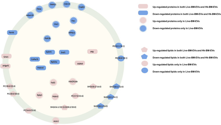
Cryptococcus neoformans causes deadly mycosis in immunocompromised individuals. Macrophages are key cells fighting against microbes. Extracellular vesicles (EVs) are cell-to-cell communication mediators. The roles of EVs from infected host cells in the interaction with Cryptococcus remain uninvestigated. Here, EVs from viable C. neoformans-infected macrophages reduced fungal burdens but led to shorter survival of infected mice. In vitro, EVs induced naive macrophages to an inflammatory phenotype. Transcriptome analysis showed that EVs from viable C. neoformans-infected macrophages activated immune-related pathways, including p53 in naive human and murine macrophages. Conserved analysis demonstrated that basic cell biological processes, including cell cycle and division, were activated by infection-derived EVs from both murine and human infected macrophages. Combined proteomics, lipidomics, and metabolomics of EVs from infected macrophages showed regulation of pathways such as extracellular matrix (ECM) receptors and phosphatidylcholine. This form of intermacrophage communication could serve to prepare cells at more distant sites of infection to resist C. neoformans infection.IMPORTANCECryptococcus neoformans causes cryptococcal meningitis, which is frequent in patients with HIV/AIDS, especially in less-developed countries. The incidence of cryptococcal meningitis is close to 1 million each year globally. Macrophages are key cells that protect the body against microbes, including C. neoformans Extracellular vesicles are a group of membrane structures that are released from cells such as macrophages that modulate cell activities via the transfer of materials such as proteins, lipids, and RNAs. In this study, we found that Cryptococcus neoformans-infected macrophages produce extracellular vesicles that enhance the inflammatory response in Cryptococcus-infected mice. These Cryptococcus neoformans-infected macrophage vesicles also showed higher fungicidal biological effects on inactivated macrophages. Using omics technology, unique protein and lipid signatures were identified in these extracellular vesicles. Transcriptome analysis showed that these vesicles activated immune-related pathways like p53 in naive macrophages. The understanding of this intermacrophage communication could provide potential targets for the design of therapeutic agents to fight this deadly mycosis.

Keywords: Cryptococcus neoformans; extracellular vesicles; lipidomics; metabolomics; proteomics.


Figures

FIG 1
FIG 1
Administration of live-BM-EVs (EVs from live C. neoformans-infected activated BMDMs) decreased fungal burdens in both lungs and brains but led to shorter survival in murine experimental cryptococcosis. (A) CFU of mouse brains in EV-treated and nontreated groups. (B) CFU of mouse lungs in EV-treated and nontreated groups. Each point represents CFU from one mouse. (C) Survival analysis of EV-treated and nontreated groups (n = 9). *, P < 0.05. (D) Representative histopathology of lungs in EV-treated and nontreated groups. The red circle indicates a granuloma in an infected lung, and the red arrow points at cryptococcal giant cells. Data are shown as the means ± SD from at least six mice per group. An unpaired t test was used to calculate P values. A log rank (Mantel-Cox) test was used to compare the survival differences.
FIG 2
FIG 2
All three types of activated BMDM-EVs triggered increased antifungal activity of naive BMDMs during C. neoformans (C.n) infection, while live-BM-EVs (EVs from live C. neoformans-infected activated BMDMs) had the highest potential. (A) Flowchart of harvesting of the three types of EV samples. (B) Phagocytosis percentages of naive BMDMs after incubation with three types of EV samples. At least 100 macrophages were counted for each group. (C to E) Fungicidal activity of naive BMDMs after incubation with three types of EV samples with opsonin 18B7 (C), complement (D), or none (E). (F) Nonlytic exocytosis of naive BMDMs after incubation with three types of EV samples. (G and H) Macrophage polarization, as measured by mRNA levels of Ccl2 (G) and Arg1 (H), in naive BMDMs after incubation with three types of EV samples. Non-BM-EVs, EVs from activated BMDMs without C. neoformans infection; No EVs, EV-nontreated macrophages; Hk, heat killed. Data are shown as the means ± SD from at least three independent experiments, each performed in triplicate. An unpaired t test was used to calculate P values.
FIG 3
FIG 3
Transcriptional changes in naive BMDMs incubated with live-BM-EVs. EV-nontreated BMDMs were used as a control. (A) Volcano plot of differentially expressed genes (DEGs) in EV-treated and untreated groups. Red, upregulated; blue, downregulated (n = 3 for each group). (B and C) GO analysis of upregulated (B) and downregulated (C) DEGs. (D and E) KEGG analysis of upregulated (D) and downregulated (E) DEGs. Red squares indicate immune-related pathways.
FIG 4
FIG 4
Transcriptional changes of naive human MDMs (monocyte-derived macrophages) incubated with live-M-EVs (EVs from live C. neoformans-infected activated human peripheral MDMs). (A) Differentially expressed genes (DEGs) in EV-treated and untreated groups. Red, upregulated; blue, downregulated (n = 3 for each group). (B and C) GO analysis of upregulated (B) and downregulated (C) DEGs. (D and E) KEGG analysis of upregulated (D) and downregulated (E) DEGs. EGFR, epidermal growth factor receptor. (F) Shared DEGs between MDMs and BMDMs identified by Venn analysis. (G) Fold changes of shared DEGs between MDMs and BMDMs. Red squares indicate immune-related pathways. Red asterisks indicate shared GO and KEGG pathways.
FIG 5
FIG 5
Size distributions of BMDM-EVs under different cryptococcal stimulations. The major size distribution of BMDM-EVs was around 40 to 60 nm in diameter. (A) Representative TEM image of BMDM-EVs. (B and C) Frequency distributions of live-BM-EVs (EVs from live C. neoformans-infected activated BMDMs) analyzed by DLS (B) (n = 192) and TEM (C) (n = 59). (D and E) Frequency distributions of Hk-BM-EVs (EVs from heat-killed C. neoformans-infected activated BMDMs) analyzed by DLS (D) (n = 329) and TEM (E) (n = 61). (F and G) Frequency distributions of non-BM-EVs (EVs from activated BMDMs without C. neoformans infection) analyzed by DLS (F) (n = 289) and TEM (G) (n = 74). HV, high voltage.
FIG 6
FIG 6
Multi-omics analysis of BMDM-EVs under different cryptococcal stimulations. (A) Protein and cholesterol levels of three types of EV samples. Lower protein and lipid levels are shown in Hk-BM-EVs (EVs from heat-killed C. neoformans-infected activated BMDMs). *, P < 0.05 by ANOVA. (B) Ratios of protein to cholesterol in three types of EV samples. (C) Differentially expressed proteins in EVs (n = 3 for each type of EV). (D) Pathway analysis of differentially expressed proteins in EVs. (E) Differentially expressed lipids in EVs (n = 3 for each type of EV). Live-BM-EVs, EVs from live C. neoformans-infected activated BMDMs; Non-BM-EVs, EVs from activated BMDMs without C. neoformans infection.
FIG 7
FIG 7
Differential protein composition in EVs from BMDMs after infection with C. neoformans. (A and B) Differential protein expression in live-BM-EVs (EVs from live Cryptococcus-infected BMDMs) (A) and Hk-BM-EVs (EVs from heat-killed Cryptococcus-infected BMDMs) (B), compared with non-BM-EVs (n = 3) (statistical analysis by ANOVA followed by Dunnett’s multiple-comparison test). Log2FC, log2 fold change. (C and D) Venn analysis of upregulated (C) and downregulated (D) proteins in live-BM-EVs and Hk-BM-EVs. *, P < 0.05; **, P < 0.01; ***, P < 0.001.
FIG 8
FIG 8
Differential lipid composition in EVs from BMDMs under different cryptococcal stimulations. (A to C) Species percentages of lipids determined by positive and negative ionization modes in live-BM-EVs (EVs from live Cryptococcus-infected BMDMs) (A) and Hk-BM-EVs (EVs from heat-killed Cryptococcus-infected BMDMs) (B and C) (n = 3). (D to F) Fold changes of dysregulated lipids determined by positive and negative ionization modes in live-BM-EVs (D) and Hk-BM-EVs (E and F) (statistical analysis by ANOVA followed by Dunnett’s multiple-comparison test). (G and H) Venn analysis of upregulated (G) and downregulated (H) lipids in live-BM-EVs and Hk-BM-EVs. *, P < 0.05; **, P < 0.01; ***, P < 0.001.
FIG 9
FIG 9
Simplified model of components in EVs from cryptococcus-infected macrophages. Red shapes indicate upregulated proteins or lipids. Blue shapes indicate downregulated proteins or lipids. Components in non-BM-EVs were used as controls. Rectangles indicate shared proteins in infection BM-EVs. Asterisks indicate proteins differentially regulated in live-BM-EVs. Pentagons indicate shared lipids. Circles indicate lipids identified only in live-BM-EVs. Both membrane-associated and intracellular components were found in EVs from cryptococcus-infected macrophages.

Similar articles

Cited by

References

    1. Coelho C, Bocca AL, Casadevall A. 2014. The tools for virulence of Cryptococcus neoformans. Adv Appl Microbiol 87:1–41. doi:10.1016/B978-0-12-800261-2.00001-3. - DOI - PubMed
    1. Rajasingham R, Smith RM, Park BJ, Jarvis JN, Govender NP, Chiller TM, Denning DW, Loyse A, Boulware DR. 2017. Global burden of disease of HIV-associated cryptococcal meningitis: an updated analysis. Lancet Infect Dis 17:873–881. doi:10.1016/S1473-3099(17)30243-8. - DOI - PMC - PubMed
    1. Bicanic T, Harrison TS. 2004. Cryptococcal meningitis. Br Med Bull 72:99–118. doi:10.1093/bmb/ldh043. - DOI - PubMed
    1. McQuiston TJ, Williamson PR. 2012. Paradoxical roles of alveolar macrophages in the host response to Cryptococcus neoformans. J Infect Chemother 18:1–9. doi:10.1007/s10156-011-0306-2. - DOI - PMC - PubMed
    1. Wager CL, Wormley F, Jr.. 2014. Classical versus alternative macrophage activation: the ying [sic] and the yang in host defense against pulmonary fungal infections. Mucosal Immunol 7:1023–1035. doi:10.1038/mi.2014.65. - DOI - PubMed

Publication types

LinkOut - more resources