. 2022 Jan;43(1):50-63.
doi: 10.1038/s41401-021-00639-y. Epub 2021 Mar 30.

Harmine is an effective therapeutic small molecule for the treatment of cardiac hypertrophy

Affiliations

Harmine is an effective therapeutic small molecule for the treatment of cardiac hypertrophy

Jie Huang et al. Acta Pharmacol Sin. 2022 Jan.

Abstract

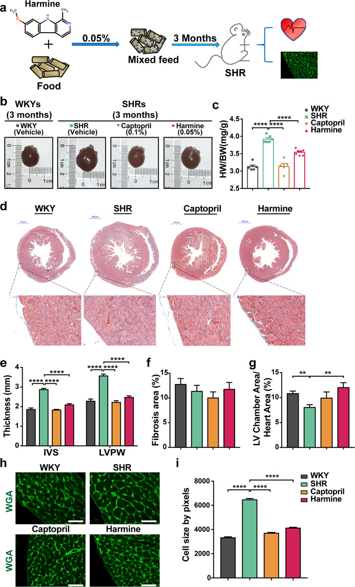
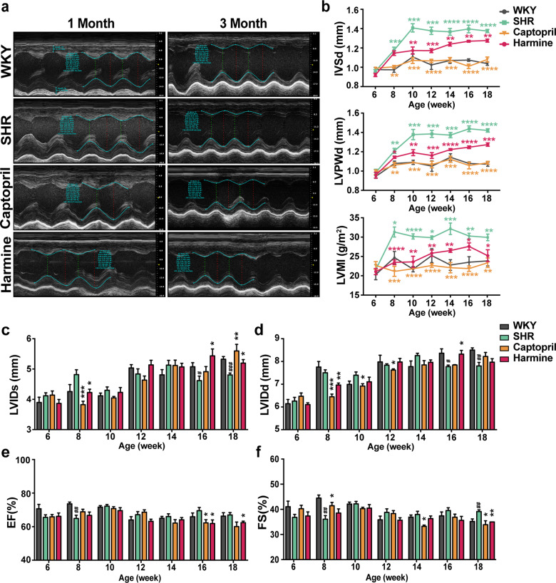
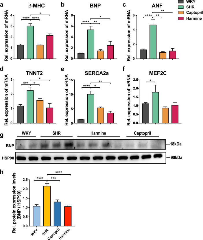
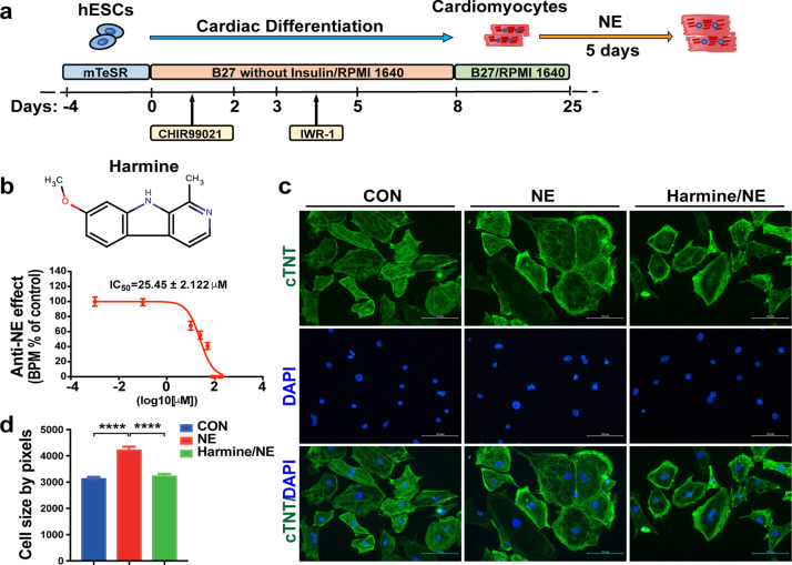
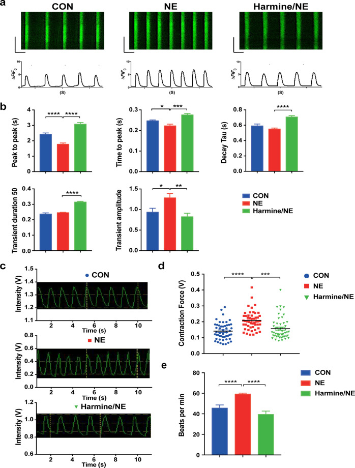
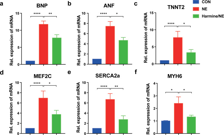
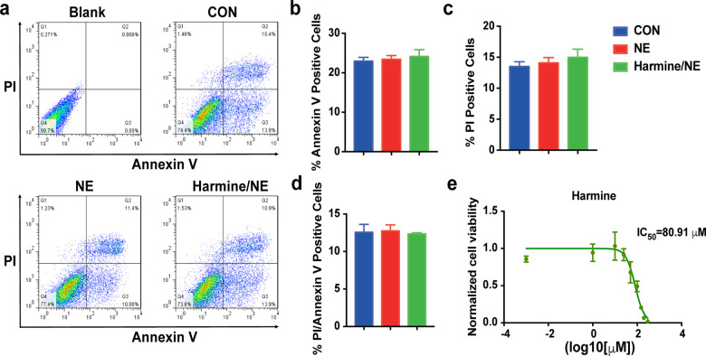
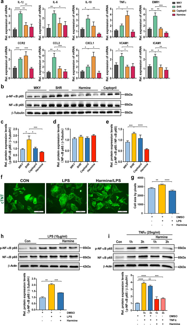
Harmine is a β-carboline alkaloid isolated from Banisteria caapi and Peganum harmala L with various pharmacological activities, including antioxidant, anti-inflammatory, antitumor, anti-depressant, and anti-leishmanial capabilities. Nevertheless, the pharmacological effect of harmine on cardiomyocytes and heart muscle has not been reported. Here we found a protective effect of harmine on cardiac hypertrophy in spontaneously hypertensive rats in vivo. Further, harmine could inhibit the phenotypes of norepinephrine-induced hypertrophy in human embryonic stem cell-derived cardiomyocytes in vitro. It reduced the enlarged cell surface area, reversed the increased calcium handling and contractility, and downregulated expression of hypertrophy-related genes in norepinephrine-induced hypertrophy of human cardiomyocytes derived from embryonic stem cells. We further showed that one of the potential underlying mechanism by which harmine alleviates cardiac hypertrophy relied on inhibition of NF-κB phosphorylation and the stimulated inflammatory cytokines in pathological ventricular remodeling. Our data suggest that harmine is a promising therapeutic agent for cardiac hypertrophy independent of blood pressure modulation and could be a promising addition of current medications for cardiac hypertrophy.

Keywords: NE; SHR; cardiac hypertrophy; hESCs-derived cardiomyocytes; harmine; inflammatory.


Conflict of interest statement

The authors declare no competing interests.

Figures

Fig. 1
Fig. 1. Harmine alleviates pressure overload-induced cardiac hypertrophy in SHRs.
a Schematic overview of harmine treatments. SHRs at 6 weeks of ages were given the mixed feed with 0.05% harmine or 0.1% captopril for additional 3 months. WKYs and SHRs group as blank control contained no pharmaceutical additives (WKYs, n = 6; SHRs, n = 6; Captopril as positive control, n = 5; Harmine, n = 7). b Macroscopic view of heart size. Scale bar: 2000 μm. c Quantification of heart weight over body weight ratio in each group. d Representative Masson’s trichrome staining images of heart tissues (upper scale bars: 2 mm), and high-magnification views of LV wall (bottom, scale bars: 50 μm). e Statistics of the average thickness of IVS and LVPW in each group. f Quantification the ratio of myocardial fibrosis area. g The ratios of LV chamber area to whole heart area. h Heart tissue slices from each group were stained with wheat germ agglutinin (WGA). Scale bars, 50 μm. i Quantification of CMs cross-sectional area with WGA staining images (n > 400). Data are mean ± SEM. **P < 0.01, ****P < 0.0001 (two-tailed Student’s t test).
Fig. 2
Fig. 2. Harmine alleviates cardiac hypertrophy and cardiac dysfunction in SHRs.
a Representative images showing the M-mode echocardiograms of rats treated for 1 month and 3 months. b Changes ininterventricular septum thickness at diastole (IVSd), left ventricular posterior wall end-diastolic thicknesses (LVPWd), and left ventricular mass index (LVMI) over time measured by echocardiography. c–f Measurement of left ventricular diastolic inner diameter (LVIDd), left ventricular systolic inner diameter (LVIDs), ejection fraction (EF), and fractional shortening (FS) (WKY group: n = 6; SHR group: n = 6; Captopril group: n = 5; harmine group: n = 7). All data are expressed as mean ± SEM. *P < 0.05, **P < 0.01, ***P < 0.001, ****P < 0.0001 by two-tailed Student’s t test compared with SHR group. #P < 0.05, ##P < 0.01, ###P < 0.001, ####P < 0.0001 by two-tailed Student’s t test compared with WKY group.
Fig. 3
Fig. 3. Downregulation of cardiac hypertrophy-related genes and proteins in SHRs after treatment of harmine.
a–f qPCR analyses of hypertrophic-related genes in rat cardiac tissues (normalized to 18S expression). g, h Immunoblotting for BNP in isolated cardiac tissues. Data are expressed as mean ± SEM. *P < 0.05, **P < 0.01, ***P < 0.001, ****P < 0.0001 (two-tailed Student’s t test).
Fig. 4
Fig. 4. Harmine reverses the effect of NE on cell size of hESC-derived cardiomyocytes.
a Schematic illustrating the differentiation protocol for H7 hESC line into cardiomyocytes in chemically defined medium. b The dose-dependent effect of harmine on beating rates of NE (20 μM) treated cardiomyocytes for 24 h. IC50 value for harmine was 25.45 ± 2.122 μM (n = 3 for each dose). c Representative immunostaining images of hESC-derived cardiomyocytes stained with cTnT (green) for sarcomere maker and DAPI (blue) for nuclei. Scale bars, 100 μm. d Stimulation with NE (20 μM) for 3 days leads to an increase in cell size, while a reduction after pre-treated with harmine (10 μM) for 3 h and then co-treated with NE for 3 days (n > 400 per group). Data are mean ± SEM. ****P < 0.0001; by two-tailed Student’s t test.
Fig. 5
Fig. 5. The contractility and calcium imaging analyses of hESC-derived cardiomyocytes after treatment of harmine.
a Representative calcium transients recorded in single cardiomyocyte after harmine treatment (25 μM; NE 20 μM). b Quantification of calcium handling parameters (peak to peak, time to peak, decay time, transient duration 50, transient amplitude (ΔF/F0)) for each group. n > 20 cells recorded for each group in every experiment. c Representative traces of contraction movements from single cardiomyocyte recorded by FelixGX detection system. d Quantification of relative contractility in single cardiomyocyte 24 h after harmine (25 μM) or/and NE (20 μM) treatment. n > 50 cells recorded for each group. e Quantification of beating frequency followed harmine and NE treatment (n > 18 for each group). Data are mean ± SEM of three independent experiments. *P < 0.05, **P < 0.01, ***P < 0.001, ****P < 0.0001 by two-tailed Student’s t test.
Fig. 6
Fig. 6. Downregulation of cardiac hypertrophy-related genes in hESC-derived cardiomyocytes after treatment of harmine.
a–f RT-PCR analyses of relative mRNA expression of hypertrophic-related genes in hESC-derived cardiomyocytes after treated with each compound for 3 days (harmine 25 μM; NE 20 μM). Control group was treated with DMSO. All data were normalized to β-actin expression and expressed as mean ± SEM. *P < 0.05, **P < 0.01, ****P < 0.0001 (n = 3 independent experiments, two-tailed Student’s t test).
Fig. 7
Fig. 7. Harmine has no effect on apoptosis and cell viability of hESC-derived cardiomyocytes.
a Representative flow cytometric analyses of apoptosis in hESC-derived cardiomyocytes following treatment of harmine (25 μM) and NE (20 μM). Cells were double labeled with Annexin V-FITC and propidum iodide after harvesting. The upper-right quadrant represents the annexin V+/PI+ cells; the lower-right quadrant represents the annexin V+/PI cells; the upper-left quadrant represents the annexin V/PI+ cells. b–d Quantification of the ratio of annexin V+/PIcells, annexin V/PI+ cells and annexin V+/PI+ cells. e Dose–response curves demonstrating the effect of harmine on cell viability of hESC-derived cardiomyocytes using CellTiter-Glo Assay (n = 4 conducted per concentration). Data are mean ± SEM of three independent experiments.
Fig. 8
Fig. 8. Harmine attenuates inflammatory response and NF-κB activation both in vivo and vitro.
a qPCR analyses of inflammatory-related genes mRNA levels in rat cardiac tissues (normalized to 18S expression). b Representative Western blot analysis of phosphorylated NF-κB p65 and NF-κB p65 protein expression in the heart tissues of WKYs and SHRs after 3-month treatment. c–e Quantification of phosphorylated NF-κB p65 and NF-κB p65 protein expression normalized to β-Tubulin, and phosphorylated NF-κB p65/NF-κB p65 ratio by densitometric analysis. f Representative images of hESC-derived cardiomyocytes stained with cTnT (green) antibody. The hESC-derived cardiomyocytes were treated with LPS (100 ng/mL) for 3 days or were pre-treated with harmine (10 μM) for 3 h and then co-treated with LPS (100 ng/mL) for 3 days (n > 200 per group). Scale bars, 100 μm. g Quantification of cell size changes in each group. h-i Representative immunoblots of phosphorylated NF-κB p65 (p-NF-κB p65) and NF-κB p65 in hESC-derived cardiomyocytes following treatment of LPS (10 μg/mL) for 3 h, or TNF-α (25;ng/mL) for 1 h and 3 h (upper), and quantification of relative protein expression by densitometry (bottom). In all harmine-treated group, hESC-derived cardiomyocytes were pre-treated with harmine (25 μM) for 0.5 h before LPS or TNF-α addition. β-Actin was used as loading control. Data are expressed as mean ± SEM. *P < 0.05, **P < 0.01, ***P < 0.001, ****P < 0.0001 (two-tailed Student’s t test).

References

    1. Hill JA, Olson EN. Cardiac plasticity. N Engl J Med. 2008;358:1370–80. - PubMed
    1. Frey N, Katus HA, Olson EN, Hill JA. Hypertrophy of the heart: a new therapeutic target? Circulation. 2004;109:1580–9. - PubMed
    1. Berenji K, Drazner MH, Rothermel BA, Hill JA. Does load-induced ventricular hypertrophy progress to systolic heart failure? Am J Physiol Heart Circ Physiol. 2005;289:H8–h16. - PubMed
    1. Lorell BH, Carabello BA. Left ventricular hypertrophy: pathogenesis, detection, and prognosis. Circulation. 2000;102:470–9. - PubMed
    1. Maron BJ, Maron MS. Hypertrophic cardiomyopathy. Lancet. 2013;381:242–55. - PubMed

MeSH terms