Metaplastic breast carcinoma: a retrospective study of 26 cases
- PMID: 33786152
- PMCID: PMC7994148
Metaplastic breast carcinoma: a retrospective study of 26 cases
Abstract
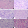
Metaplastic breast carcinoma is a rare invasive breast cancer. Metaplastic breast carcinoma is mainly characterized by an epithelial or mesenchymal cell population mixed with adenocarcinoma. We collected 26 cases of metaplastic breast carcinoma in the First Affiliated Hospital of Bengbu Medical College from 2008 to 2014. Tumor size, tumor grade, vascular invasion, ER/PR status, histologic classification, and HER2/neu status were assessed for all cases and the literature was reviewed. Clinicopathologic characteristics of patients diagnosed with metaplastic breast carcinomas and its key points of differential diagnosis were discussed. All patients were female, with the median age of 50 years. The mean tumor size was 3.2 cm. 4 subtypes of metaplastic breast carcinomas were documented. Fibromatosis-like metaplastic carcinomas are typically characterized by wavy, intertwined, gentle spindle cells. When the tumor components are almost squamous cell carcinoma components and the primary squamous cell carcinoma of other organs and tissues are excluded, we can diagnose breast squamous cell carcinoma. In spindle cell carcinoma, atypical spindle cells are arranged in many ways and are usually accompanied by inflammatory cell infiltrate. Cancer with interstitial differentiation has mixed malignant epithelial and mesenchymal differentiation, and the mesenchymal components are diverse. Most tumors are triple negative. At present, surgical resection combined with chemotherapy or radiation therapy is the most effective and acceptable method for treating metaplastic breast carcinoma.
Keywords: Metaplastic breast carcinoma; immunohistochemistry; pathology.
IJCEP Copyright © 2021.
Conflict of interest statement
None.
Figures


References
-
- McKinnon E, Xiao P. Metaplastic carcinoma of the breast. Arch Pathol Lab Med. 2015;139:819–822. - PubMed
-
- Graziano L, Graziano PF, Bitencourt AG, Soto DB, Hiro A, Nunes CC. Metaplastic squamous cell carcinoma of the breast: a case report and literature review. Rev Assoc Med Bras (1992) 2016;62:618–621. - PubMed
-
- Tzanninis IG, Kotteas EA, Ntanasis-Stathopoulos I, Kontogianni P, Fotopoulos G. Management and outcomes in metaplastic breast cancer. Clin Breast Cancer. 2016;16:437–443. - PubMed
-
- Jha A, Agrawal V, Tanveer N, Khullar R. Metaplastic breast carcinoma presenting as benign breast lump. J Cancer Res Ther. 2017;13:593–596. - PubMed
LinkOut - more resources
Full Text Sources
Research Materials
Miscellaneous
