

Case Reports
doi: 10.1016/j.hrcr.2020.11.023.
eCollection 2021 Mar.
The spatial and temporal visualization of the entire atriofascicular fiber conduction during antidromic reciprocating tachycardia
Affiliations
- PMID: 33786309
- PMCID: PMC7987897
- DOI: 10.1016/j.hrcr.2020.11.023
Item in Clipboard
Case Reports
The spatial and temporal visualization of the entire atriofascicular fiber conduction during antidromic reciprocating tachycardia
HeartRhythm Case Rep.
.
No abstract available
Keywords: Atriofascicular antidromic tachycardia; Atriofascicular fiber; High-resolution mapping catheter; Ventricular insertion.
Figures

A: Twelve-lead electrocardiogram during tachycardia with a left bundle branch block morphology with a cycle length of 292 ms. B: Twelve-lead electrocardiogram during sinus rhythm. No overt preexcitation was seen. C: Intracardiac electrograms during atriofascicular antidromic tachycardia over an accessory pathway inserting into the right bundle branch (RBB). The RBB electrogram preceded His bundle activation during tachycardia. Note that an extra atrial stimulation delivered during tachycardia at the timing of atrioventricular nodal refractoriness advanced preexcited ventricular, retrograde His, and atrial electrograms without affecting the retrograde activation sequence.
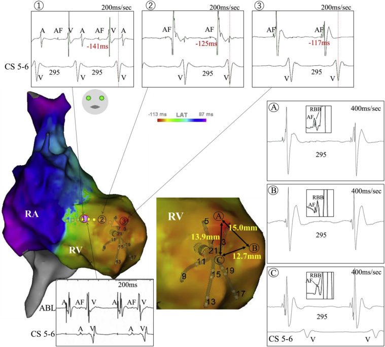
Activation maps during atriofascicular antidromic tachycardia over an atriofascicular fiber. Atriofascicular potentials are excluded from the annotation. Serial atriofascicular fiber potentials were recorded, extending from the right lateral tricuspid annulus to the apical anterolateral right ventricle adjacent to the earliest myocardial activation site (1–3). A dotted arrow shows the direction of the atriofascicular fiber conduction and the pink dot indicates the site of successful ablation. Fused atriofascicular fiber and distal right bundle branch (RBB) potentials were seen at multiple sites in a surrounding area over 1 cm2 wide adjacent to the earliest ventricular activation site, demonstrating an arborized connection between atriofascicular fibers and the distal RBB (A–C). A = atrial potential; AF = atriofascicular potential; CS = coronary sinus; V = ventricular myocardial potential.
References
-
- Haissaguerre M., Cauchemez B., Marcus F. Characteristics of the ventricular insertion sites of accessory pathways with antegrade decremental conduction properties. Circulation. 1995;91:1077–1085. - PubMed
-
- Kothari S., Gupta A.K., Lokhandwala Y.Y. Atriofascicular pathways: Where to ablate? Pacing Clin Electrophysiol. 2006;29:1226–1233. - PubMed
-
- Morita N., Kobayashi Y., Katoh T., Takano T. Anatomic and electrophysiologic evaluation of a right lateral atrioventricular Mahaim fiber. Pacing Clin Electrophysiol. 2005;28:1138–1141. - PubMed
Publication types
LinkOut - more resources
Full Text Sources
Other Literature Sources
