Review
. 2021 Mar 20;6(2):e10213.
doi: 10.1002/btm2.10213. eCollection 2021 May.

Cytosolic delivery of nucleic acids: The case of ionizable lipid nanoparticles

Affiliations
Review

Cytosolic delivery of nucleic acids: The case of ionizable lipid nanoparticles

Michele Schlich et al. Bioeng Transl Med. .

Abstract

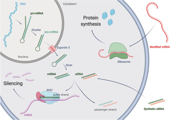
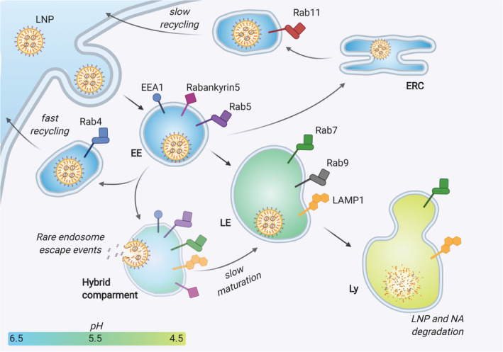
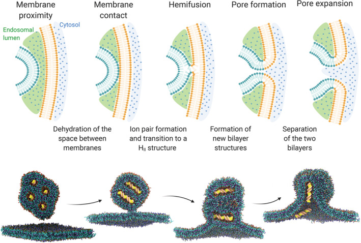
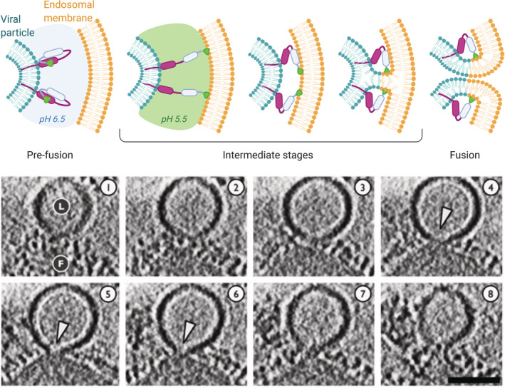
Ionizable lipid nanoparticles (LNPs) are the most clinically advanced nano-delivery system for therapeutic nucleic acids. The great effort put in the development of ionizable lipids with increased in vivo potency brought LNPs from the laboratory benches to the FDA approval of patisiran in 2018 and the ongoing clinical trials for mRNA-based vaccines against SARS-CoV-2. Despite these success stories, several challenges remain in RNA delivery, including what is known as "endosomal escape." Reaching the cytosol is mandatory for unleashing the therapeutic activity of RNA molecules, as their accumulation in other intracellular compartments would simply result in efficacy loss. In LNPs, the ability of ionizable lipids to form destabilizing non-bilayer structures at acidic pH is recognized as the key for endosomal escape and RNA cytosolic delivery. This is motivating a surge in studies aiming at designing novel ionizable lipids with improved biodegradation and safety profiles. In this work, we describe the journey of RNA-loaded LNPs across multiple intracellular barriers, from the extracellular space to the cytosol. In silico molecular dynamics modeling, in vitro high-resolution microscopy analyses, and in vivo imaging data are systematically reviewed to distill out the regulating mechanisms underlying the endosomal escape of RNA. Finally, a comparison with strategies employed by enveloped viruses to deliver their genetic material into cells is also presented. The combination of a multidisciplinary analytical toolkit for endosomal escape quantification and a nature-inspired design could foster the development of future LNPs with improved cytosolic delivery of nucleic acids.

Keywords: LNPs; RNA delivery; endosomal escape; intracellular delivery; ionizable lipids; mRNA; siRNA.


Figures

FIGURE 1
FIGURE 1
RNA interference: a miRNA gene is transcribed into primary miRNA (pri‐miRNA) that is further processed by Drosha to form pre‐miRNA. Exportin‐5 translocates the pre‐miRNA into the cytoplasm were it is processed by Dicer into mature miRNA. siRNAs can be obtained directly by chemical synthesis and ‐with the help of a carrier or chemical modifications‐ can reach the cytoplasm through endocytosis. In the cytosol, the guide (antisense) strand of mature miRNA or siRNA will be assembled into the RNA‐induced silencing complex (RISC). The passenger (sense) strand will be discarded. The mature RISC will find the target mRNA sequences through complementary base pairing with the guide strand. As few as 7 complementary bases (seed region) are sufficient for miRNA‐mediated RNAi, while full complementarity is usually required for siRNA‐induced silencing. Depending on the triggering molecule (siRNA or miRNA), the translation of the target gene could be repressed due to mRNA degradation or translocation to the P bodies. mRNA therapy: once introduced in the cytosol through an appropriate delivery method, a modified, exogenous mRNA could hijack the cell's ribosomes to be translated into a functional protein
FIGURE 2
FIGURE 2
Schematic representation of the endocytic pathway, showing the different possible fates of an internalized LNP. The endocytosed LNP engulfed in an early endosome (EE) can be sent back towards the cell membrane and excreted either directly (fast recycling) or through other intracellular organelles such as the endocytic recycling compartment (ERC) (slow recycling). Alternatively, the EE matures to late endosome (LE), gradually modifying its receptors and enzymatic pool and decreasing its pH. The endosomal escape events were suggested to occur at an intermediate, hybrid compartment stage between EE and LE (see also the section Cell‐based Studies). Eventually, the LE fuses with the lysosome (Ly), whose enzymes can dismantle and degrade the entrapped LNPs and their NA payload. On the surface of endo‐lysosomal vesicles, the figure shows the main stage‐defining markers employed in the works analyzed in this review: EEA1, early endosome antigen 1; RabX, Ras‐related protein RabX (X=4, 5, 7, 9, 11); LAMP1, Lysosome‐associated membrane glycoprotein 1
FIGURE 3
FIGURE 3
Top: the molecular structure hypothesis: the geometry of the lipid molecule dictates the structure of its aggregates. Cone lipids (e.g. 1‐Stearoyl‐sn‐glycero‐3‐phosphocholine) form micelles, cylindrical lipids (e.g. 1,2‐distearoyl‐sn‐glycero‐3‐phosphocholine, DSPC) form bilayers and inverted‐cone lipids (e.g. 1,2‐Dioleoyl‐sn‐glycero‐3‐phosphoethanolamine, DOPE) form hexagonal phases (HII). Bottom: the geometry of lipids might change upon mixing and ion pair formation, with consequences on the macrostructure. Protonated ionizable lipids interact with anionic lipids adopting an inverted cone shape, which promotes the formation HII phase. Non‐bilayer phases are associated with membrane fusion
FIGURE 4
FIGURE 4
Top: different stages of the fusion process of two oppositely charged lipid membranes (e.g. an endosome and a LNP). Bottom: computational simulation of the fusion between a cationic lipoplex loaded with DNA (yellow) and a negatively charged membrane. This panel has been adapted and reproduced with permission from 91
FIGURE 5
FIGURE 5
Top: a representation of the general mechanism of fusion between an enveloped virus and the endosomal membrane, mediated by a pH‐dependent fusion protein. In the pre‐fusion stage, the hydrophobic segment responsible for membrane binding is hidden within the protein. An external trigger (in the case of this example the pH acidification) induces a conformational change exposing the hydrophobic peptide that anchors the endosomal membrane. The protein then tends to refold to a more stable conformation, and by doing so it increases the proximity of the viral envelope and the endosomal membrane, promoting the mixing of lipids and membrane fusion. Bottom: cryo‐electron tomography sections (5.3 nm‐thick) of a fusion event between influenza virus (F) and a liposome used as a model membrane (L) at pH 5.5. White arrowheads indicate the formation of fusion pores. Scalebar 50 nm. This panel has been adapted and reproduced with permission from 111

References

    1. Tatum EL. Molecular biology, nucleic acids, and the future of medicine. Cell Ther Transplant. 2010;1:74‐79. 10.3205/ctt-2009-en-000042.01. - DOI - PubMed
    1. Wolff JA, Malone RW, Williams P, et al. Direct gene transfer into mouse muscle in vivo. Science. 1990;247(4949):1465‐1468. 10.1126/science.1690918. - DOI - PubMed
    1. Lee RC, Feinbaum RL, Ambros V. The C. elegans Heterochronic gene lin‐4 encodes small RNAs with antisense complementarity to lin‐14. Cell. 1993;75:843‐854. - PubMed
    1. Pasquinelli AE, Reinhart BJ, Slack F, et al. Conservation of the sequence and temporal expression of let‐7 heterochronic regulatory RNA. Nature. 2000;408:86‐89. 10.1038/35040556. - DOI - PubMed
    1. Fire A, Xu S, Montgomery MK, Kostas SA, Driver SE, Mello CC. Potent and specific genetic interference by double‐stranded RNA in Caenorhabditis elegans . Nature. 1998;391:806‐811. - PubMed