. 2021 Apr;18(2):1064-1080.
doi: 10.1007/s13311-021-01024-7. Epub 2021 Mar 30.

Ferulic Acid Ameliorates Alzheimer's Disease-like Pathology and Repairs Cognitive Decline by Preventing Capillary Hypofunction in APP/PS1 Mice

Affiliations

Ferulic Acid Ameliorates Alzheimer's Disease-like Pathology and Repairs Cognitive Decline by Preventing Capillary Hypofunction in APP/PS1 Mice

Ni-Ya Wang et al. Neurotherapeutics. 2021 Apr.

Abstract

Brain capillaries are crucial for cognitive functions by supplying oxygen and other nutrients to and removing metabolic wastes from the brain. Recent studies have demonstrated that constriction of brain capillaries is triggered by beta-amyloid (Aβ) oligomers via endothelin-1 (ET1)-mediated action on the ET1 receptor A (ETRA), potentially exacerbating Aβ plaque deposition, the primary pathophysiology of Alzheimer's disease (AD). However, direct evidence is still lacking whether changes in brain capillaries are causally involved in the pathophysiology of AD. Using APP/PS1 mouse model of AD (AD mice) relative to age-matched negative littermates, we identified that reductions of density and diameter of hippocampal capillaries occurred from 4 to 7 months old while Aβ plaque deposition and spatial memory deficit developed at 7 months old. Notably, the injection of ET1 into the hippocampus induced early Aβ plaque deposition at 5 months old in AD mice. Conversely, treatment of ferulic acid against the ETRA to counteract the ET1-mediated vasoconstriction for 30 days prevented reductions of density and diameter of hippocampal capillaries as well as ameliorated Aβ plaque deposition and spatial memory deficit at 7 months old in AD mice. Thus, these data suggest that reductions of density and diameter of hippocampal capillaries are crucial for initiating Aβ plaque deposition and spatial memory deficit at the early stages, implicating the development of new therapies for halting or curing memory decline in AD.

Keywords: APP/PS1 mouse; Alzheimer’s disease; Aβ plaque; Endothelin-1; Ferulic acid (FA); Hippocampus.


Conflict of interest statement

Some authors are listed as the inventors of a CN Patent (application no. 202010395578.7). The other authors declare that they have no competing interests.

Figures
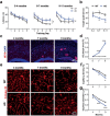
Fig. 1
Fig. 1
Reduced hippocampal capillaries precede Aβ plaque deposition and spatial memory deficit in APP/PS1 mice. (a) Spatial learning indicated by latency (s) onto a hidden platform in WT (black circle) vs. AD mice (blue circle) was not different until 6 to 7 months old. (b) Spatial memory was not different until 6 to 7 months old. (c) Representative images for hippocampal Aβ plaques using the antibody 6E10 in AD mice. (d) Aβ plaque area (%) indicated a sharp increase of Aβ plaque load since 7 months old in AD mice. (e) Representative images for hippocampal capillaries using the antibody Collagen IV in AD and WT mice. (f, g) Capillary density (%) and diameter (μm) suggested an age-dependent reduction of hippocampal capillaries in AD mice relative to WT mice. Data are presented as mean ± SEM. *P < 0.05, **P < 0.01, ***P < 0.001
Fig. 2
Fig. 2
ET1 induces a tiny hypoperfusion and early Aβ plaque deposition at 5 months old. (a) Procedures: ET1 or vehicle injection (1 µL, 0.1 µL/min, 1 µg/µL) into one side of hippocampal CA1. (b) Lectin perfusion and imaging revealed a hypoperfusion insult in the ET1 group but not the vehicle group. (c) Representative images for Aβ plaque using the antibody D54D2 (Aβ) or Thioflavine S (Ths) in AD mice. (d, e) Aβ+ or Ths+ area (μm2) indicated early Aβ plaque deposition in the ET1 group but not the vehicle group in AD mice, nearly undetectable in both the groups in WT mice. Data are presented as mean ± SEM. ***P < 0.001
Fig. 3
Fig. 3
FA prevents the reduction of the CBF. (a) Chemical structure of ferulic acid (FA). (b) Experimental procedures and representative images of laser speckle imaging for the blood flows in the jugular vein. (c) ET1 (1 µg/mL, 100 µL) induced strong vasoconstriction as indicated by the reduction of the blood flows, while following application of FA (2 mg/mL, 100 µL) but not vehicle (Saline) induced vasodilation in C57 mice at 2 months old. (d) Relative changes (%) of the blood flows before and after adding FA or saline for 10 min indicated that FA counteracted the ET1-mediated action. (e) Representative images of laser speckle imaging in AD mice at 6 months old after intraperitoneal injection (i.p.) of FA (10 mg/kg). (f) The CBF was increased by FA (10 mg/kg, i.p.) in AD or C57 mice. (g, h) The FA treatment rescued the CBF reduction in AD mice that was observed in the vehicle group, relative to WT mice, by using laser speckle imaging at 7 months old. The same region of interest (ROI) (red circle) was defined for all groups
Fig. 4
Fig. 4
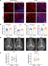
FA prevents the reduction of hippocampal capillaries in AD mice at 7 months old. (a) Representative images for immunostaining of the blood vessel (Collagen IV) in the hippocampus and cortex in AD mice after the FA or vehicle treatment. (b, c) Quantification of capillary density (%) (b) and diameter (μm) (c) in the hippocampus and the cortex suggested that the both were significantly increased by the FA treatment relative to the vehicle control. (d) Representative 2D time-of-flight images for blood vessels in AD mice before and after the FA treatment or vehicle control. (e) Comparison of the time-of-flight magnetic resonance angiography between before and after the treatment suggested that the FA treatment but not the vehicle control significantly increased the density of the whole-brain blood vessels. Data are presented as mean ± SEM. *P < 0.05, **P < 0.01
Fig. 5
Fig. 5
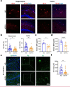
FA reduced Aβ plaque deposition in AD mice at 7 months old and ET1-induced early Aβ deposition. (a) Representative images for immunostaining of the Aβ antibody (6E10) in AD mice at 7 months old after the FA (AD-FA) or vehicle treatment (AD) for 30 days. (b) Quantification of the Aβ area (μm2) in the hippocampus and the cortex suggested a significant reduction of Aβ plaques by the FA treatment relative to the vehicle control. (c, d) The enzyme-linked immunosorbent assay test suggested a significant reduction of the Aβ 1–42 (e) and a downtrend of the Aβ 1–40 (f) concentration from AD mice after the FA treatment relative to the vehicle control. (e) Representative images for immunostaining of the early Aβ plaques (D54D2) initiated by the injection of ET1 into the hippocampal CA1 region in AD mice at 5 months old and underwent the ferulic acid (FA) treatment (AD-FA) or vehicle treatment (AD) for 30 days, started from the same day with the ET1 injection. (f) Quantification of the Aβ area (µm2) suggested that the FA treatment significantly reduced the ET1-induced early Aβ plaque deposition relative to the vehicle treatment
Fig. 6
Fig. 6
FA repairs spatial memory deficit in AD mice at 7 months old. (a) Spatial learning, as indicated by latency in escaping onto a hidden platform during 5-day training, suggested that the FA treatment but not vehicle treatment prevented learning deficit in AD mice relative to WT at 7 months old. (b) Spatial memory, as indicated by the time spent in the target quadrant (s) during the probe test 24 h after the final training, was impaired in AD mice after the vehicle treatment. This impaired spatial memory was effectively prevented by the FA treatment, to the levels without difference from that in WT mice. (c) Mean of the swimming speed during a training trial on the first day was not different among the groups. (d) Representative tracking traces during the probe test of the Morris water maze for measuring spatial memory. Data are presented as mean ± SEM. *P < 0.05, **P < 0.01
Fig. 7
Fig. 7
The schematic diagram for a feedforward cycle among capillary, Aβ plaque, and memory loss in AD. Decreased CD/CBF and increased Aβ aggregation could form a feedforward cycle (red and green arrows), leading to memory loss and Aβ plaque deposition that trigger other cascades of the pathophysiology. This feedforward cycle at the initial stages could be antagonized by FA via targeting the ETRA (yellow box)

Similar articles

Cited by

References

    1. Nativio R, Donahue G, Berson A, Lan Y, Amlie-Wolf A, Tuzer F, et al. Dysregulation of the epigenetic landscape of normal aging in Alzheimer's disease. Nat Neurosci. 2018;21(4):497–505. doi: 10.1038/s41593-018-0101-9. - DOI - PMC - PubMed
    1. Glenner GG, Wong CW. Alzheimer’s disease: initial report of the purification and characterization of a novel cerebrovascular amyloid protein. Biochem Biophys Res Commun. 1984;120(3):885–890. doi: 10.1016/S0006-291X(84)80190-4. - DOI - PubMed
    1. Beyreuther K, Masters CL. Amyloid precursor protein (APP) and beta A4 amyloid in the etiology of Alzheimer’s disease: precursor-product relationships in the derangement of neuronal function. Brain pathology (Zurich, Switzerland). 1991;1(4):241–251. doi: 10.1111/j.1750-3639.1991.tb00667.x. - DOI - PubMed
    1. Hardy JA, Higgins GA. Alzheimer's disease: the amyloid cascade hypothesis. Science. 1992;256(5054):184–185. doi: 10.1126/science.1566067. - DOI - PubMed
    1. Duyckaerts C, Delatour B, Potier MC. Classification and basic pathology of Alzheimer disease. Acta Neuropathol. 2009;118(1):5–36. doi: 10.1007/s00401-009-0532-1. - DOI - PubMed

Publication types

MeSH terms