. 2021 May 1;125(5):1720-1734.
doi: 10.1152/jn.00509.2020. Epub 2021 Mar 31.

Differential neural plasticity of individual fingers revealed by fMRI neurofeedback

Affiliations

Differential neural plasticity of individual fingers revealed by fMRI neurofeedback

Ethan Oblak et al. J Neurophysiol. .

Abstract

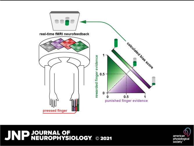
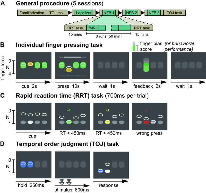
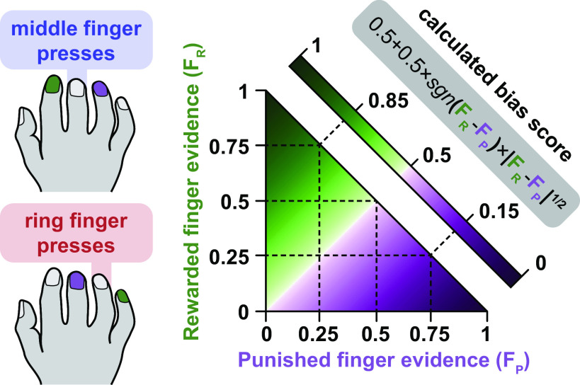
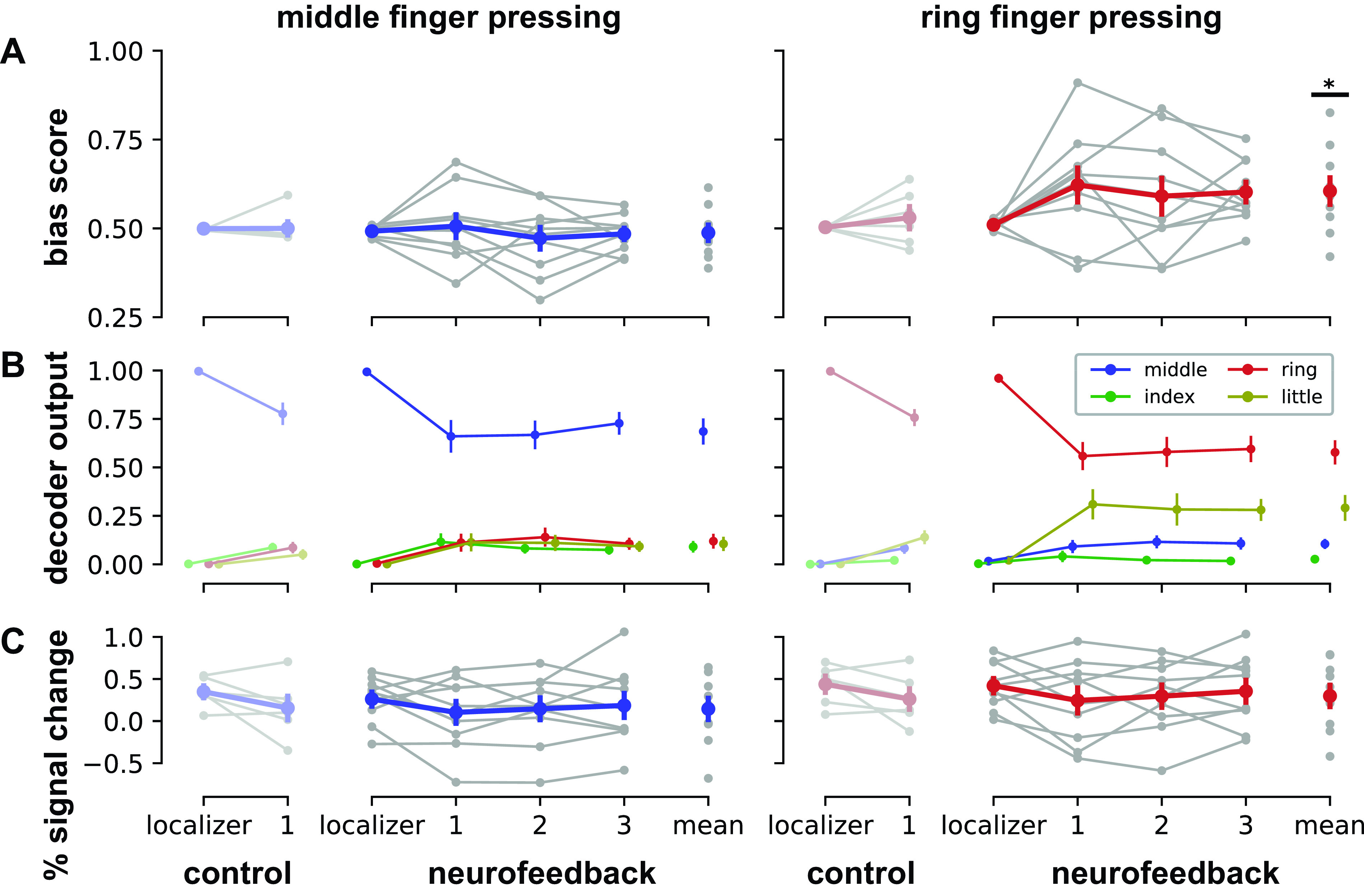
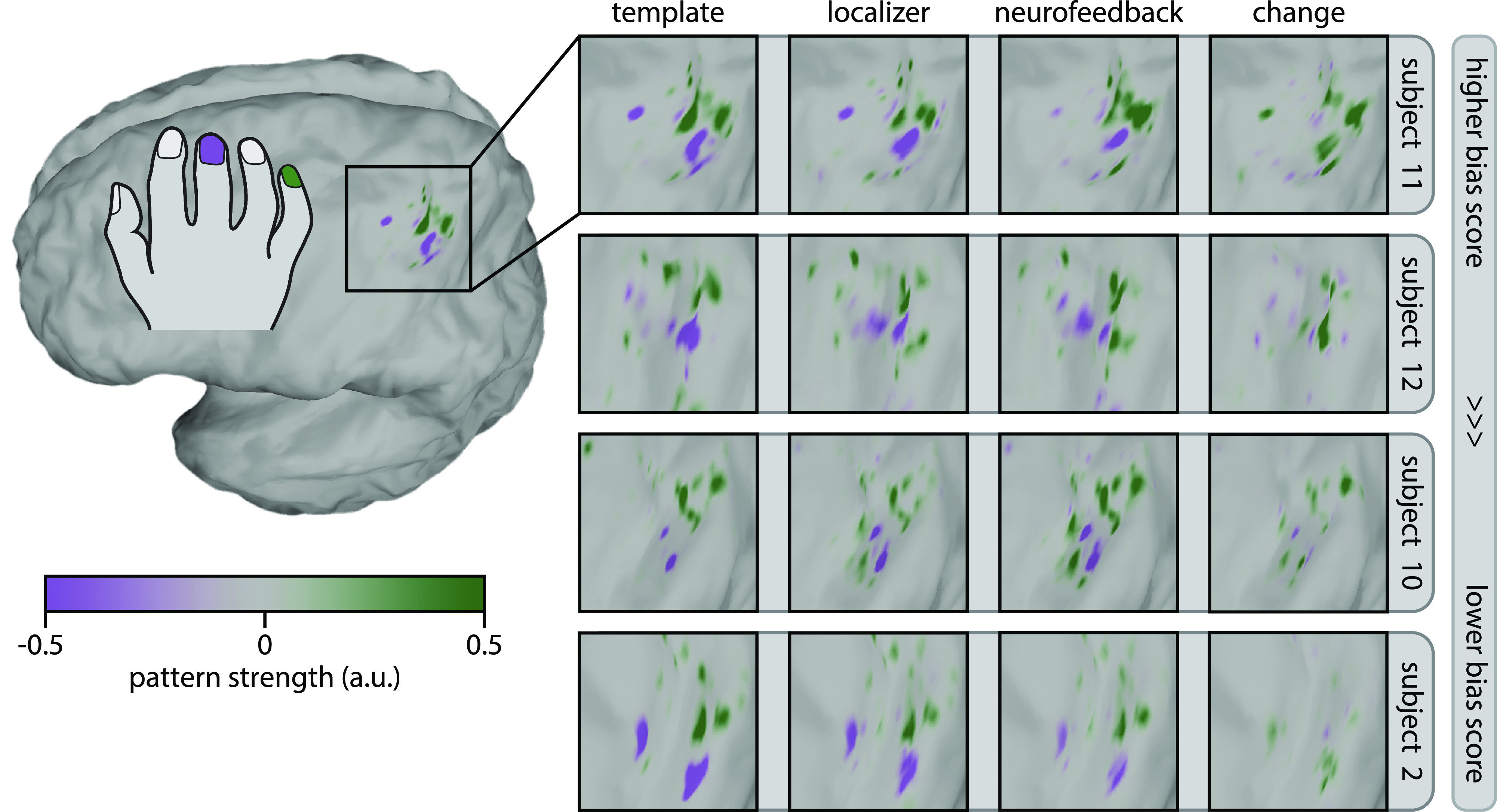
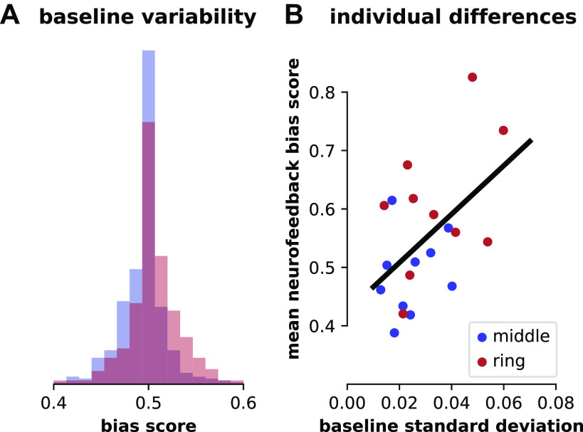
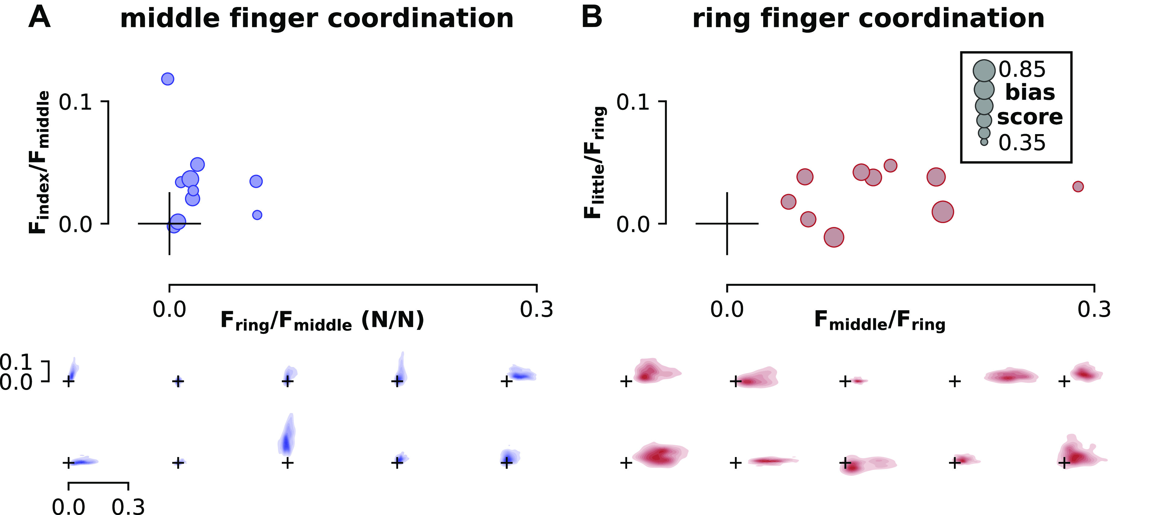
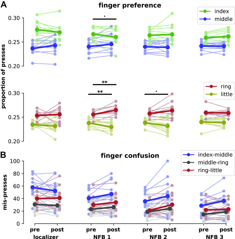
Previous work has shown that functional magnetic resonance imaging (fMRI) activity patterns associated with individual fingers can be shifted by temporary impairment of the hand. Here, we investigated whether these neural activity patterns could be modulated endogenously and whether any behavioral changes result from this modulation. We used decoded neurofeedback in healthy individuals to encourage participants to shift the neural activity pattern in sensorimotor cortex of the middle finger toward the index finger, and the ring finger toward the little finger. We first mapped the neural activity patterns for all fingers of the right hand in an fMRI pattern localizer session. Then, in three subsequent neurofeedback sessions, participants were rewarded after middle/ring finger presses according to their activity pattern overlap during each trial. A force-sensitive keyboard was used to ensure that participants were not altering their physical finger coordination patterns. We found evidence that participants could learn to shift the activity pattern of the ring finger but not of the middle finger. Increased variability of these activity patterns during the localizer session was associated with the ability of participants to modulate them using neurofeedback. Participants also showed an increased preference for the ring finger but not for the middle finger in a postneurofeedback motor task. Our results show that neural activity and behaviors associated with the ring finger are more readily modulated than those associated with the middle finger. These results have broader implications for rehabilitation of individual finger movements, which may be limited or enhanced by individual finger plasticity after neurological injury.NEW & NOTEWORTHY It may be possible to remobilize fingers after neurological injury by altering neural activity patterns. Toward this end, we examined whether finger-related neural activity patterns could be modified in healthy individuals without physical intervention, using fMRI neurofeedback. Our findings show that greater variability of neural patterns at baseline predicted a participant's ability to successfully shift these patterns. Because neural variability is common in individuals poststroke, this illustrates a potential clinical benefit of this procedure.

Keywords: fMRI; finger; neurofeedback; plasticity.


Conflict of interest statement

No conflicts of interest, financial or otherwise, are declared by the authors.

Figures

None
Graphical abstract
Figure 1.
Figure 1.
Experimental design. A: the five-session experiment consisted of behavioral familiarization, a finger pattern localizer fMRI session, and three neurofeedback (NFB) sessions. Each fMRI session included behavioral pre- and posttests. B: an individual finger pressing task was used as the basis for the localizer and neurofeedback sessions. Participants were required to make individual presses with one of four fingers (index, middle, ring, or little) while maintaining constant pressure on all other keys. At the end of each trial, feedback was presented related to their motor behavior (localizer session) or their ability to bias the fMRI patterns related to finger presses (neurofeedback sessions). C: a rapid reaction time (RRT) task was used to assess motor confusion before and after each fMRI session. Participants were encouraged to make rapid presses (reaction time below 450 ms) through a point system. D: a temporal order judgment (TOJ) task was used to assess the hand representation of participants before and after the entire neurofeedback protocol. During a 800-ms stimulus blank period, a brief vibrotactile stimulus was delivered to two adjacent fingers in rapid succession. Participants then judged which of the two stimuli happened first. fMRI, functional magnetic resonance imaging.
Figure 2.
Figure 2.
Neurofeedback bias score calculation. Bias scores were calculated based on the real-time decoder evidence for the two fingers adjacent to the pressed finger. Rewarded fingers are shown in green, punished fingers are shown in purple (middle finger presses: index rewarded, ring punished; ring finger presses: little rewarded, middle punished). If rewarded and punished finger evidence were equal, the bias score was 0.5. The square root of the difference between finger evidence was used as the basis of the bias score calculation. For example, with evidence of 0.75 for the rewarded finger (FR) and 0.25 for the punished finger (FP), the calculated bias score would be approximately 0.85. If the evidence for the punished finger was greater than the evidence for the rewarded finger, the bias score was less than 0.5 [i.e., if the sign (sgn) of the difference between rewarded and punished fingers was negative]. The heatmap of bias scores only includes possible decoder output combinations: the four-finger SMLR decoder could output four possible values, which must sum to 1. Therefore, the sum of the evidence of two fingers could never exceed 1. SMLR, sparse multinomial logistic regression.
Figure 3.
Figure 3.
Individual finger pattern bias modulation. A: mean bias scores by session for presses of middle finger (blue, left column) and ring finger (red, right column). See Eq. 1 in methods for bias score calculation details. B: mean decoder outputs by session. During middle finger presses, bias scores encouraged participants to increase the index finger decoder output (green) relative to the ring finger output (red). During ring finger presses, bias scores encouraged participants to increase the little finger output (yellow) relative to the middle finger output (blue). C: mean univariate activations by session, calculated as the mean percent signal change within the voxels selected by each decoder. Univariate activations were found to be unrelated to changes in bias score. Control data were acquired from participants (N = 6) who performed two sessions of finger pressing but did not receive neurofeedback in the second session (19). Neurofeedback data (N = 10) are shown by session (localizer: pattern localizer; 1–3: neurofeedback sessions; mean: average over all neurofeedback sessions). All data (control and neurofeedback) are shown relative to a decoder trained on each participant’s localizer session. Gray dots indicate means for each participant; colored dots indicate means across the entire group. Errors bars indicate SE. Statistical differences relative to the pattern localizer sessions are indicated at *P < 0.05.
Figure 4.
Figure 4.
Visualization of ring finger pattern changes with neurofeedback. Pattern shifts are shown for the four participants with the highest bias scores for the ring finger during neurofeedback, in descending order. Template patterns were computed using the decoder weights and mean fMRI patterns during pressing of the rewarded (little) finger and punished (middle) finger from the localizer session. For template patterns, green indicates regions related to the little finger, whereas purple indicates regions related to the middle finger. Localizer and neurofeedback patterns were computed by taking the mean fMRI patterns during localizer and neurofeedback sessions for the pressed (ring) finger and multiplying them by the decoder weights for the rewarded and punished fingers. Change patterns were simply the difference between neurofeedback and localizer patterns. For localizer, neurofeedback, and change patterns, green indicates regions with pattern strength that increases bias scores, whereas purple indicates regions with pattern strength that decreases bias scores. See methods for full pattern visualization details. a.u., arbitrary units; fMRI, functional magnetic resonance imaging.
Figure 5.
Figure 5.
Baseline variability of individual finger patterns. A: distributions of bias scores for middle (blue) and ring (red) fingers for all participants during the baseline pattern localizer session. Each distribution includes 400 bias scores (40 trials of pressing from each of the 10 participants for each finger). B: relationship between individual participant bias pattern variability during the pattern localizer session (standard deviation) and mean bias score achieved during neurofeedback sessions for each finger. Best-fit line shown for reference; see methods for statistics.
Figure 6.
Figure 6.
Finger coordination patterns. A: coordination of middle finger presses with the rewarded finger (index, y-axis) and punished finger (ring, x-axis). B: coordination of ring finger presses with the rewarded finger (little, y-axis) and punished finger (middle, x-axis). Mean coordination values for each participant (top, N = 10) are shown as a ratio of the force of the rewarded or punished finger to the pressed finger (N/N). The size of each marker indicates the mean bias score achieved by each participant, with larger circles indicating a greater bias score. These plots illustrate that coupling between adjacent fingers did not correlate with bias scores; if this were true, we would expect larger circles toward the top of each plot (rewarded finger coupling increasing on the y-axis) and smaller circles toward the right of each plot (punished finger coupling increasing on the x-axis). Individual coordination heatmaps for each participant are also shown in the bottom. Each heatmap in the bottom corresponds to one circular marker in the top, with the size and position of the origin marker cross serving as a scale reference. See methods for details of coordination pattern calculation and heatmap generation. F, force exerted by each finger; N/N, ratio of the force of the rewarded or punished finger to the pressed finger.
Figure 7.
Figure 7.
Finger pressing behavior before and after each fMRI scan, including each neurofeedback (NFB) session. A: finger preference in pre- and post-fMRI rapid reaction time tests, as a proportion of the total number of presses. Faded colors indicate each participant’s performance, solid colors indicate means across the group. Within-finger statistical differences from pre- to posttest are shown at P < 0.1 and **P < 0.01. B: confusion between fingers in pre- and post-fMRI rapid reaction time tests. Mis-presses are assigned to a finger pair when the target finger was one of the fingers of the pair but the other finger in the pair was pressed instead. fMRI, functional magnetic resonance imaging.

Similar articles

Cited by

References

    1. Diedrichsen J, Wiestler T, Krakauer JW. Two distinct ipsilateral cortical representations for individuated finger movements. Cereb Cortex 23: 1362–1377, 2013. doi:10.1093/cercor/bhs120. - DOI - PMC - PubMed
    1. Ejaz N, Hamada M, Diedrichsen J. Hand use predicts the structure of representations in sensorimotor cortex. Nat Neurosci 18: 1034–1040, 2015. doi:10.1038/nn.4038. - DOI - PubMed
    1. Kolasinski J, Makin TR, Jbabdi S, Clare S, Stagg CJ, Johansen-Berg H. Investigating the stability of fine-grain digit somatotopy in individual human participants. J Neurosci 36: 1113–1127, 2016. doi:10.1523/JNEUROSCI.1742-15.2016. - DOI - PMC - PubMed
    1. Buonomano DV, Merzenich MM. Cortical plasticity: from synapses to maps. Annu Rev Neurosci 21: 149–186, 1998. doi:10.1146/annurev.neuro.21.1.149. - DOI - PubMed
    1. Feldman DE, Brecht M. Map plasticity in somatosensory cortex. Science 310: 810–815, 2005. doi:10.1126/science.1115807. - DOI - PubMed

Publication types

LinkOut - more resources