

Automated system for diagnosing endometrial cancer by adopting deep-learning technology in hysteroscopy
- PMID: 33788887
- PMCID: PMC8011803
- DOI: 10.1371/journal.pone.0248526
Automated system for diagnosing endometrial cancer by adopting deep-learning technology in hysteroscopy
Abstract
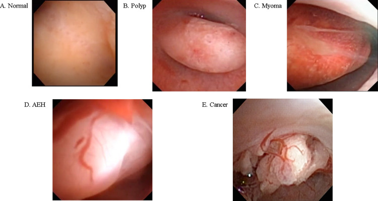
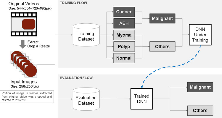
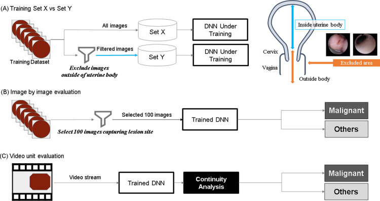
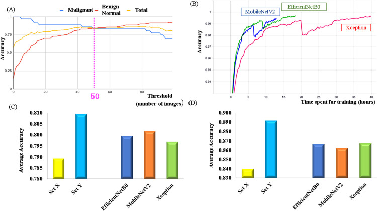
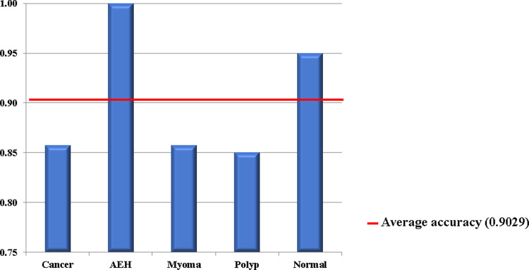
Endometrial cancer is a ubiquitous gynecological disease with increasing global incidence. Therefore, despite the lack of an established screening technique to date, early diagnosis of endometrial cancer assumes critical importance. This paper presents an artificial-intelligence-based system to detect the regions affected by endometrial cancer automatically from hysteroscopic images. In this study, 177 patients (60 with normal endometrium, 21 with uterine myoma, 60 with endometrial polyp, 15 with atypical endometrial hyperplasia, and 21 with endometrial cancer) with a history of hysteroscopy were recruited. Machine-learning techniques based on three popular deep neural network models were employed, and a continuity-analysis method was developed to enhance the accuracy of cancer diagnosis. Finally, we investigated if the accuracy could be improved by combining all the trained models. The results reveal that the diagnosis accuracy was approximately 80% (78.91-80.93%) when using the standard method, and it increased to 89% (83.94-89.13%) and exceeded 90% (i.e., 90.29%) when employing the proposed continuity analysis and combining the three neural networks, respectively. The corresponding sensitivity and specificity equaled 91.66% and 89.36%, respectively. These findings demonstrate the proposed method to be sufficient to facilitate timely diagnosis of endometrial cancer in the near future.
Conflict of interest statement
Kenbun Sone has a joint research agreement with Predicthy LLC. Katsuhiko Noda and Kaname Yoshida are members of Predicthy LLC. The other authors have no competing interests to disclose. This does not alter our adherence to PLOS ONE policies on sharing data and materials.
Figures
Similar articles
-
Hysteroscopy for asymptomatic postmenopausal women with sonographically thickened endometrium.Maturitas. 2009 Feb 20;62(2):176-8. doi: 10.1016/j.maturitas.2008.11.018. Epub 2009 Jan 3. Maturitas. 2009. PMID: 19121901
-
Hysteroscopy in women with abnormal uterine bleeding: a meta-analysis on four major endometrial pathologies.Arch Gynecol Obstet. 2015 Jun;291(6):1347-54. doi: 10.1007/s00404-014-3585-x. Epub 2014 Dec 19. Arch Gynecol Obstet. 2015. PMID: 25524536
-
Diagnostic hysteroscopy: a valuable diagnostic tool in the diagnosis of structural intra-cavital pathology and endometrial hyperplasia or carcinoma?. Six years of experience with non-clinical diagnostic hysteroscopy.Eur J Obstet Gynecol Reprod Biol. 2003 Sep 10;110(1):79-82. doi: 10.1016/s0301-2115(03)00165-9. Eur J Obstet Gynecol Reprod Biol. 2003. PMID: 12932877
-
Outpatient hysteroscopy and ultrasonography in the management of endometrial disease.Curr Opin Obstet Gynecol. 2004 Aug;16(4):305-11. doi: 10.1097/01.gco.0000136491.26463.c2. Curr Opin Obstet Gynecol. 2004. PMID: 15232484 Review.
-
Endometrial polyps. An evidence-based diagnosis and management guide.Eur J Obstet Gynecol Reprod Biol. 2021 May;260:70-77. doi: 10.1016/j.ejogrb.2021.03.017. Epub 2021 Mar 13. Eur J Obstet Gynecol Reprod Biol. 2021. PMID: 33756339 Review.
Cited by
-
Precision Medicine for Chronic Endometritis: Computer-Aided Diagnosis Using Deep Learning Model.Diagnostics (Basel). 2023 Mar 1;13(5):936. doi: 10.3390/diagnostics13050936. Diagnostics (Basel). 2023. PMID: 36900079 Free PMC article. Review.
-
Revolutionizing Women's Health: A Comprehensive Review of Artificial Intelligence Advancements in Gynecology.J Clin Med. 2024 Feb 13;13(4):1061. doi: 10.3390/jcm13041061. J Clin Med. 2024. PMID: 38398374 Free PMC article. Review.
-
Automated Detection of Endometrial Polyps from Hysteroscopic Videos Using Deep Learning.Diagnostics (Basel). 2023 Apr 13;13(8):1409. doi: 10.3390/diagnostics13081409. Diagnostics (Basel). 2023. PMID: 37189510 Free PMC article.
-
Clinical Prospects for Artificial Intelligence in Obstetrics and Gynecology.JMA J. 2025 Jan 15;8(1):113-120. doi: 10.31662/jmaj.2024-0197. Epub 2024 Dec 13. JMA J. 2025. PMID: 39926075 Free PMC article. Review.
-
Deep Learning-Based Classification of Uterine Cervical and Endometrial Cancer Subtypes from Whole-Slide Histopathology Images.Diagnostics (Basel). 2022 Oct 28;12(11):2623. doi: 10.3390/diagnostics12112623. Diagnostics (Basel). 2022. PMID: 36359467 Free PMC article.
References
Publication types
MeSH terms
LinkOut - more resources
Full Text Sources
Other Literature Sources
Medical
