. 2021 May 7;49(8):4643-4654.
doi: 10.1093/nar/gkab216.

Escherichia coli RNase E can efficiently replace RNase Y in Bacillus subtilis

Affiliations

Escherichia coli RNase E can efficiently replace RNase Y in Bacillus subtilis

Soumaya Laalami et al. Nucleic Acids Res. .

Abstract

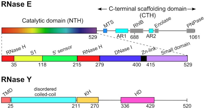
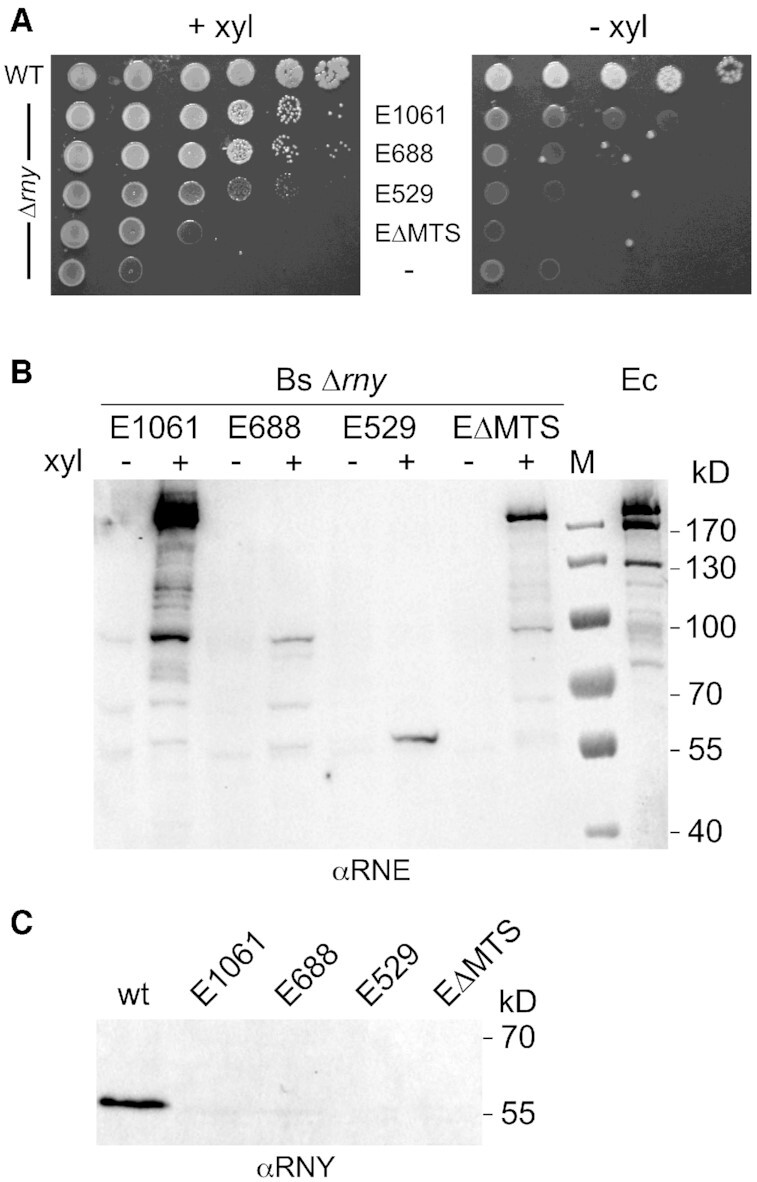
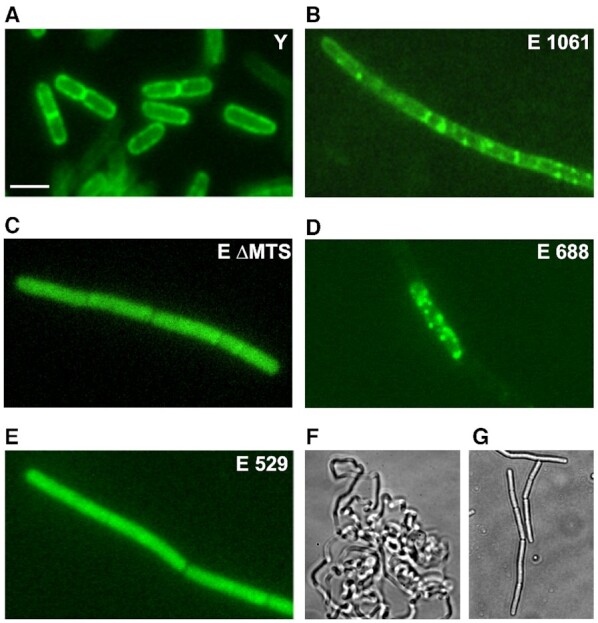
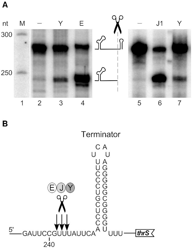
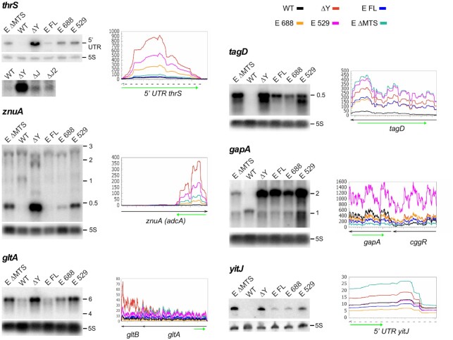
RNase Y and RNase E are disparate endoribonucleases that govern global mRNA turnover/processing in the two evolutionary distant bacteria Bacillus subtilis and Escherichia coli, respectively. The two enzymes share a similar in vitro cleavage specificity and subcellular localization. To evaluate the potential equivalence in biological function between the two enzymes in vivo we analyzed whether and to what extent RNase E is able to replace RNase Y in B. subtilis. Full-length RNase E almost completely restores wild type growth of the rny mutant. This is matched by a surprising reversal of transcript profiles both of individual genes and on a genome-wide scale. The single most important parameter to efficient complementation is the requirement for RNase E to localize to the inner membrane while truncation of the C-terminal sequences corresponding to the degradosome scaffold has only a minor effect. We also compared the in vitro cleavage activity for the major decay initiating ribonucleases Y, E and J and show that no conclusions can be drawn with respect to their activity in vivo. Our data confirm the notion that RNase Y and RNase E have evolved through convergent evolution towards a low specificity endonuclease activity universally important in bacteria.


Figures

Figure 1.
Figure 1.
Domain composition of E. coli RNase E (1061 aa) and B. subtilis RNase Y (520 aa) monomers. RNase E is composed of the N-terminal catalytic region (NTH, aa 1–529) and the C-terminal noncatalytic region (aa 530–1061). The catalytic region contains a large globular domain which is a composite of recurrent structural subdomains (50) and a small folded domain (aa 415–529). The C-terminal half (CTH) of the protein is predicted to be unfolded but contains microdomains that mediate interactions with the inner plasma membrane (MTS) and other components of the degradosome (the helicase RhlB, enolase, and PNPase). AR1 and AR2 are arginine-rich segments probably involved in RNA binding (51). B. subtilis RNase Y (520 aa) has a completely different domain structure including an N-terminal transmembrane domain (aa 1–25) anchoring the protein at the inner membrane, followed by a large region predicted to be disordered (aa ∼30–210), an RNA binding KH domain (aa 211–270) and a metal-chelating HD domain (aa 336–429) containing the conserved His/Asp motif required for RNase activity (1).
Figure 2.
Figure 2.
Functional complementation of B. subtilis Δrny by E. coli RNase E. (A) Growth of a B. subtilis Δrny mutant strain (SSB508) expressing different versions of E. coli RNase E. Cells from fresh colonies were resuspended and spotted in serial 10-fold dilutions on SMS medium agar plates containing 50 mM xylose to induce RNase E expression. Plates were incubated at 37°C for 48 h. (B) In vivo expression levels of RNase E variants in B. subtilis Δrny. A polyclonal antibody directed against E. coli RNase E was used to estimate expression of the various RNase E proteins from a xylose inducible promoter in a B. subtilis rny null mutant strain grown in LB medium with (+) or without xylose (−). E1061 : wild type full-length RNase E ; E688 : RNase E truncated after aa 688 ; E529 : RNase E truncated after aa 529; EΔMTS: full-length RNase E lacking the membrane tethering sequence (Δ567–582). The last lane (Ec) is a positive control that contains a total extract of E. coli strain JM109 grown in LB medium. (C) Western analysis of RNase Y expression in the B. subtilis wild type and the Δrny strain expressing the various forms of RNase E using an RNase Y specific antibody. The extracts analyzed were the same as those probed with anti-RNase E antibodies (panel B). M: marker proteins.
Figure 3.
Figure 3.
Localization of E. coli RNase E variants in B. subtilis by fluorescence microscopy. B. subtilis strains expressing GFP fusion proteins of RNase Y or various RNase E versions expressed from a xylose inducible promoter (panels A–E) were grown in LB medium to mid-log phase and imaged. Panels F and G show phase contrast images of the Δrny mutant (F) and the same mutant expressing wild-type RNase E without the GFP moiety (strain SSB500).
Figure 4.
Figure 4.
In vitro cleavage specificity of RNases Y, E and J. (A) Cleavage of 5′ end-labeled monophosphorylated thrS leader mRNA by purified B. subtilis RNase Y (Y), E. coli RNase E 529 (E) and B. subtilis RNase J1 (J1). The major cleavage site upstream of the leader terminator is indicated by a scissors symbol. Products were resolved on a 5% PAGE. M = marker. (B) Sequence context around the major cleavage site. Nucleotide numbering is with respect to the transcription start of the thrS gene. Arrows indicate the precise processing sites previously determined for RNase J1 (43).
Figure 5.
Figure 5.
Effect of RNase E expression on the transcriptome of the Δrny strain. (A) Browser view of RNA-Seq reads showing the integration and expression of the E. coli rne gene versions inserted at the amyE locus on the B. subtilis genome. Reads from a single replicate of each strain are shown as representative examples. The arrow represents the E. coli rne coding sequence and its orientation on the genome, the red rectangle indicates the position of the MTS domain. (B) Multidimensional scaling (MDS) plot of Δrny, Δrny + Pxyl-rne(1061), Δrny + Pxyl-rne(ΔMTS), Δrny + Pxyl-rne(688), Δrny + Pxyl-rne(529) and WT triplicate libraries. Distance between samples is based on the leading log2 fold-change (FC) of the top 500 most differentially regulated genes. (C) MA plots of differentially expressed genes (red dots) at FDR ≤ 0.05 identified in the mutant and complemented strains compared to WT. The ordinate represents log2-fold change.
Figure 6.
Figure 6.
Effect of E. coli RNase E expression on transcription profiles of coding and non-coding RNAs in a Δrny mutant. The left panels show a Northern analysis of total RNA isolated from WT, Δrny and Δrny strains expressing full-length E. coli RNase E (E FL), a cytoplasmic version of the full-length enzyme (E ΔMTS) and shorter RNase E proteins (E 688 and E 529). The blots were hybridized to specific riboprobes complementary to the RNA regions indicated by a green arrow below the corresponding RNAseq profiles (right panels). The curves representing the RNA-Seq coverage expressed as RPM of the various strains follow the color code presented on top of the first graph (thrS).

Similar articles

Cited by

References

    1. Laalami S., Zig L., Putzer H.. Initiation of mRNA decay in bacteria. Cell. Mol. Life Sci.: CMLS. 2014; 71:1799–1828. - PMC - PubMed
    1. Apirion D., Lassar A.B.. A conditional lethal mutant of Escherichia coli which affects processing of ribosomal RNA. J. Biol. Chem. 1978; 253:1738–1742. - PubMed
    1. Ono M., Kuwano M.. A conditional lethal mutation in an E. coli strain with a longer chemical lifetime of messenger RNA. J. Mol. Biol. 1979; 129:343–357. - PubMed
    1. Carpousis A.J., Van Houwe G., Ehretsmann C., Krisch H.M.. Copurification of E. coli RNAse E and PNPase: evidence for a specific association between two enzymes important in mRNA processing and degradaion. Cell. 1994; 76:889–900. - PubMed
    1. Py B., Higgins C.F., Krisch H.M., Carpousis A.J.. A DEAD-box RNA helicase in the Escherichia coli RNA degradosome. Nature. 1996; 381:169–172. - PubMed

Publication types

LinkOut - more resources