Review
. 2021 Nov 16;42(6):783-814.
doi: 10.1210/endrev/bnab009.

Normal and Premature Adrenarche

Affiliations
Review

Normal and Premature Adrenarche

Robert L Rosenfield. Endocr Rev. .

Abstract

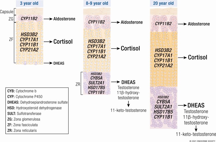
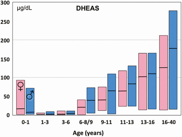
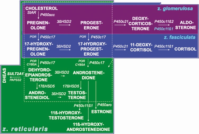
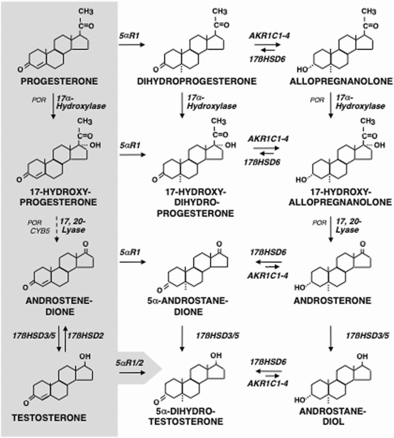
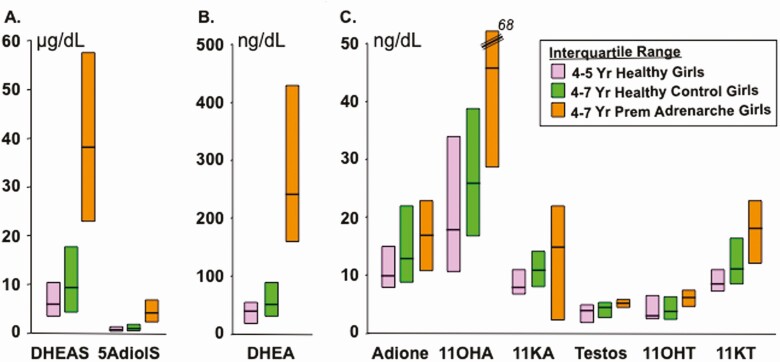
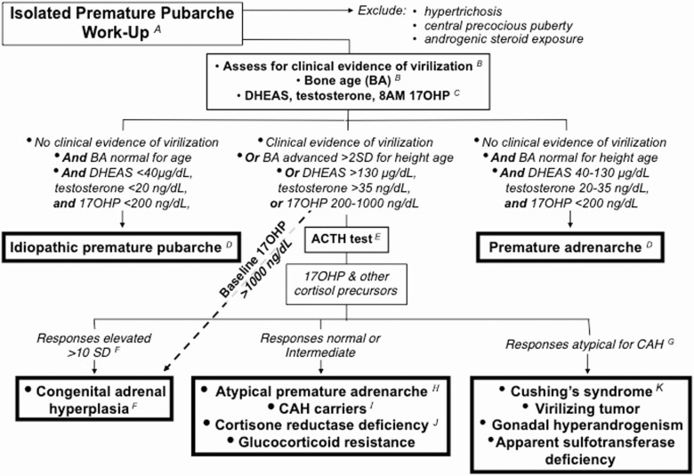
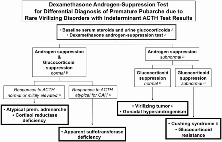
Adrenarche is the maturational increase in adrenal androgen production that normally begins in early childhood. It results from changes in the secretory response to adrenocorticotropin (ACTH) that are best indexed by dehydroepiandrosterone sulfate (DHEAS) rise. These changes are related to the development of the zona reticularis (ZR) and its unique gene/enzyme expression pattern of low 3ß-hydroxysteroid dehydrogenase type 2 with high cytochrome b5A, sulfotransferase 2A1, and 17ß-hydroxysteroid dehydrogenase type 5. Recently 11-ketotestosterone was identified as an important bioactive adrenarchal androgen. Birth weight, body growth, obesity, and prolactin are related to ZR development. Adrenarchal androgens normally contribute to the onset of sexual pubic hair (pubarche) and sebaceous and apocrine gland development. Premature adrenarche causes ≥90% of premature pubarche (PP). Its cause is unknown. Affected children have a significantly increased growth rate with proportionate bone age advancement that typically does not compromise growth potential. Serum DHEAS and testosterone levels increase to levels normal for early female puberty. It is associated with mildly increased risks for obesity, insulin resistance, and possibly mood disorder and polycystic ovary syndrome. Between 5% and 10% of PP is due to virilizing disorders, which are usually characterized by more rapid advancement of pubarche and compromise of adult height potential than premature adrenarche. Most cases are due to nonclassic congenital adrenal hyperplasia. Algorithms are presented for the differential diagnosis of PP. This review highlights recent advances in molecular genetic and developmental biologic understanding of ZR development and insights into adrenarche emanating from mass spectrometric steroid assays.

Keywords: adrenal androgens; adrenarche; polycystic ovary syndrome; pubarche; steroidogenic enzyme expression; zona reticularis.


Figures

Graphical Abstract
Graphical Abstract
Figure 1.
Figure 1.
Plasma DHEAS median and normal range during healthy childhood. DHEAS determined by LCMSMS. Concentrations fall during the neonatal period due to waning function of the fetal zone of the adrenal cortex. They begin to rise again starting at 3 to 6 years of age. Note: 6- to 8-year-old girls were studied (rather than 6- to 9-years-old, as in boys) to minimize the contribution of true puberty to the findings. Nevertheless, boys had significantly higher levels at most ages from 6 years onward, a trend consistent with most data. These results correlate closely with, but are lower than, those obtained by a standard direct assay of DHEAS in serum: DHEAS by radioimmunoassay = 1.8 (DHEAS by LCMSMS) – 8.4 (personal communication with AE Kulle, May 26, 2017). To convert DHEAS in µg/dL to µmol/L, multiply by 0.0271. Graphed from data of Kulle et al (2).
Figure 2.
Figure 2.
Changing rapid steroid secretory response to ACTH stimulation across adrenarche in females. Note increased responses of the Δ 5-3ß-hydroxysteroids 17-hydroxypregnenolone and DHEA through maturational stages, in parallel with baseline DHEAS. Serum androstenedione and its Δ 4-3-ketosteroid precursor 17-hydroxyprogesterone (17OHP) rise to a lesser extent. Cortisol responses remain unchanged. Basal levels were obtained early morning after overnight dexamethasone suppression. Post-ACTH levels obtained 30 min post-ACTH1-24 administered intravenously at 8 am. Prepubertal children were 2 to 12 years old; adrenarchal girls had PreAd; adults were normal volunteers in early follicular phase of menstrual cycle. For conversion factors to nmol/L, see Table 1. Data are from Rich et al (3). Figure adapted from Rosenfield RL, Cooke DW, Radovick S. Puberty and its disorders in the female. In: Sperling M, Majzoub JA, Menon RK, Stratakis CA, eds. Pediatric Endocrinology. 5th ed. Elsevier; 2021: 528-626. Copyright Elsevier 2021. Abbreviations: 11-deoxycortisol, Cmpd S; androstenedione, adione.
Figure 3.
Figure 3.
Major pathways of steroid hormone formation. Cholesterol carbon atoms are designated by conventional numbers and rings by conventional letters. The flow of steroidogenesis is generally downward and to the right. The top row is the pathway to progesterone and mineralocorticoids; the second row, the pathway to glucocorticoids; the third row, the 17-ketosteroids; the fourth row, 17ß-hydroxysteroids; and the bottom row, the 5α-reductase pathway to amplification and disposition of androgen. The dotted 17,20-lyase pathways are probably minor. The steroidogenic enzymes are italicized. Steroids before 3ßHSD action have the Δ 5-3ß-hydroxysteroid configuration, those formed by 3ßHSD are Δ 4-3-ketosteroids. Inset: 11ß-hydroxysteroid dehydrogenase interconversions of 11-oxysteroids, which occur in peripheral tissues. Abbreviations for enzymes are indicated in the side panel in approximate order of appearance. Modified from Rosenfield et al (10).
Figure 4.
Figure 4.
Organization of the adrenocortical zones. The area within the dotted square contains the core steroidogenic activities common to zona reticularis, ovarian theca cells, and testicular Leydig cells (although the latter express 17ßHSD type 3 rather than 17ßHSD5). The left column shows the Δ 5-3ß-hydroxysteroid pathway and the columns to its right shows the Δ 4-3-ketosteroid pathway. The adrenal zones are color-coded to facilitate visualizing overlapping and unique steps in the adrenal zones: the top row shows the zona glomerulosa pathway to mineralocorticoids culminating in aldosterone; the second row shows the zona fasciculata pathway to cortisol. The third row shows the zona reticularis steps to DHEAS and other 17-ketosteroids. The steroidogenic and accessory enzymes are italicized and abbreviated as in Figure 3 side panel. P450c11ß1 is expressed only in the zona fasciculata (P450c11ß1 blue lettering on mineralocorticoid pathway indicates the zona fasciculata formation of corticosterone, not shown) and zona reticularis. 3ßHSD2 expression is lower in the zona reticularis than other zones. Dotted P450c17 17,20-lyase pathways for Δ4-3-ketosteroids are relatively minor. Modified with permission from Rosenfield RL. Identifying children at risk of polycystic ovary syndrome. J Clin Endocrinol Metab. 2007;92:787-796.
Figure 5.
Figure 5.
Age-related changes in immunoreactivity per cell of 3ßHSD2, CYPB5, and SULT2A1 in each adrenocortical zone. (a) age-related changes in immunohistochemical staining optical density/cell, (b) immunohistochemistry at 8 to 9 years of age. 3ßHSD2 (left panel) immunoreactivity is strong in the cytoplasm of adrenocortical cells of the z. glomerulosa, the z. fasciculata, and the ZR from age 7 months to 3 years. However, after age 3, 3ßHSD2 expression begins to fall in the ZR, whereas its expression remains relatively constant in the z. glomerulosa and z. fasciculata. 3ßHSD2 immunohistochemical staining at 9 years of age is marked in the z. glomerulosa and fasciculata but is low in ZR. Immunoreactivity of CYB5 (middle panel) is weakly detected in cytoplasm of adrenocortical cells in the z. glomerulosa, z. fasciculata, and ZR from ages 7 months to 3 years; its immunoreactivity becomes more pronounced in the developing z. reticularis thereafter until it reaches a plateau after age 13, while CYB5 immunoreactivity remains relatively low in the other zones. Immunohistochemical staining at 8.4 years was strong in the zona reticularis, relatively weak in the other zones. SULT2A1 (right panel) immunoreactivity followed a similar pattern to CYB5 in the zona fasciculata and zona reticularis but was very low in zona glomerulosa. SULT2A1 immunohistochemical staining at 8.4 years was strong in the ZR. Key: z. glomerulosa, black triangles; z. fasciculata, blue circles; z. reticularis, red squares. Scale bar = 100 μm. Reprinted from Rainey et al (10) with permission from Elsevier.
Figure 6.
Figure 6.
Disposition pathways of steroid metabolism and the alternative pathway to DHT. Hepatic 5α-reduction, predominantly by the type 1 isoform, is a first step on the path towards excretion of Δ 4-3-ketosteroids as glucuronides and sulfates. The endocrine biosynthetic pathway from progesterone through testosterone (Fig. 3) is shown on the gray background to the left. The disposition pathways culminate in the formation of androsterone (and etiocholanolone via a parallel 5ß-reduced pathway) as water-soluble conjugates. Key fetal tissues express versions of this pathway that permit genital tissue to form DHT from androsterone, rather than testosterone; this constitutes the “backdoor” pathway to DHT. 17-Hydroxyallopregnanolone conversion to androsterone does not require CYB5, and both reductive and oxidative 3αHSD activities of AKR1C2/4 and 17ßHSD6 (retinol dehydrogenase/3α-hydroxysteroid epimerase) are required for this pathway. Based on Miller and Auchus (10), Flück et al (54), and Janner et al (167).
Figure 7.
Figure 7.
Peripheral serum concentrations (median and interquartile range) of adrenarchal C19 steroids in girls with and without PreAd. Samples obtained during clinic hours; subjects predominantly non-Hispanic White; PreAd group had Tanner pubic hair stage 2. Steroids all assayed by LCMSMS. All the differences between the steroids in PreAd and the age-matched control girls were statistically significant except for androstenedione. Note that each panel shows steroids on a different scale. Conversion multiplier for androstenediol sulfate (5AdioS) from µg/dL to µmol/L: 0.0270. Conversion multipliers from ng/dL to nmol/L for unconjugated steroids not listed in Table 1: 11β-hydroxyandrostenedione (11OHA) 0.0331; 11-ketotestosterone (11KT) 0.0331; 1-ketoandrostenedione (11KA) 0.0331; 11β-hydroxytestosterone (11OHT) 0.0328. For comparison, normal median (interquartile range) values for the 11-oxyandrogens in reproductive-age women are 11-hydroxyandrostenedione 117 (86-151), 11-ketoandrostenedione 15 (12-21), 11-hydroxytestosterone 11 (7-16), and 11-ketotestosterone 21 (16-32) ng/dL (99). The 11-oxyandrogen values of men this age are minimally higher (99). Graphed from data of Rege et al (54).
Figure 8.
Figure 8.
A work-up to screen for virilizing disorders as a cause of isolated premature pubarche. Premature adrenarche accounts for the great majority of isolated premature pubarche. Congenital adrenal hyperplasia due to 21-hydroxylase deficiency is the most common virilizing cause. (A) History and examination can usually exclude hypertrichosis, central precocious puberty, and exposure to androgenic/anabolic steroids. Hypertrichosis must be excluded by inspection to avoid fruitlessly pursing an endocrine cause for a cosmetic condition. Since pubarche may be the first sign of gonadotropin-dependent precocity in both girls and boys, in the absence of breast development or testicular enlargement, gonadotropin determination may be advisable (see text). Anabolic or androgenic steroid use by the child or a caretaker can usually be ruled out by a careful history; otherwise, special LCMSMS urine studies are required. (B) The earliest manifestations of virilization may be acceleration of height velocity followed by BA becoming disproportionately advanced for height. (C) A baseline serum DHEAS or testosterone much above the usual adrenarchal range (130 µg/dL and 35 ng/dL, respectively) or 8 am 17OHP ≥ 200 ng/mL (6.0 nmol/L) indicates increased risk for a virilizing disorder. (This 17OHP level has a ≥95% sensitivity and specificity for CAH/NCCAH among premature pubarche patients when obtained by 8:00 am). However, normal baseline levels do not necessarily exclude 21-hydroxylase deficiency or more rare forms of CAH. (D) Idiopathic premature pubarche is diagnosed when BA is normal for chronologic age and androgen and 17OHP levels are preadrenarchal with no clinical evidence of virilization. PreAd is characterized by a slightly advanced BA that is normal for height age and androgen and 17OHP levels appropriate for an early pubertal girl with no clinical evidence of virilization. (E) Cosyntropin (ACTH1-24) 0.250 mg is infused IV over 1 min intravenously after obtaining baseline steroids; peak serum steroid responses occur at 60 min. (F) The typical response in untreated CAH, whether classic or nonclassic, is that the steroid immediately prior to the enzyme block is extremely elevated (>10 SD above average), and steroids earlier in the biosynthetic pathway are successively less elevated the further removed they are from the block. For example, in 21-hydroxylase deficiency, 17OHP is >1000 ng/dL (30 nmol/L) and androstenedione and testosterone are successively less elevated; in 3ßHSD2 deficiency, 17OHP and DHEA responses are >4000 ng/dL (120 nmol/L) and 17OHP, androstenedione, and testosterone are mild-moderately elevated. 21-Hydroxylase deficiency is responsible for the vast majority of CAH, so many practitioners check only 17OHP responses, but we advise checking all the pertinent steroid intermediates from 17-hydroxypregnenolone to cortisol and to androstenedione through testosterone to potentially detect unusual disorders with a single ACTH test. (G) An example of a pattern that is atypical for any type of CAH would be androstenedione or testosterone levels greater than those of 17OHP. (H) Atypical premature adrenarche is the most common cause of baseline DHEAS and stimulated DHEA or 17-hydroxypregnenolone levels that approach those of 3ßHSD2 deficiency. It is a diagnosis of exclusion (see Fig. 9). (I) Carriers for 21-hydroxylase deficiency have 17OHP responses to ACTH that are >200 to 1000 ng/dL (6-30 nmol/L). Other forms of CAH may also have 17OHP responses in this range. (J) Normal or elevated cortisol levels may be found in endogenous Cushing’s syndrome, glucocorticoid resistance, and cortisone reductase deficiency (or apparent CRD). A DAST may be indicated for diagnosis (Fig. 9). (K) C19 steroid patterns atypical for CAH may arise from Cushing’s syndrome (androstenedione disproportionately elevated), virilizing tumors (very high DHEAS, etc.), male gonadal hyperandrogenism (testosterone elevated with pubertal steroid pattern), and apparent sulfotransferase deficiency (low DHEAS and mild DHEA hyperresponsiveness).
Figure 9.
Figure 9.
Dexamethasone androgen-suppression test for rare virilizing disorders causing premature pubarche with responses to ACTH that are indeterminate (normal, elevated, or atypical for CAH) (see Fig. 8). (A) First, an early morning blood sample for testosterone, cortisol, and DHEAS and any other suspect steroid and a` 24-h urine for glucocorticoids (ie, urine free cortisol, 17alpha-hydroxycorticosteroids and/or cortisol and cortisone metabolites for quantitative steroid profiling) are obtained. Then the DAST is begun: dexamethasone, 1 mg/m2/day is given in 3 to 4 divided doses daily for 4 days, and then serum cortisol and androgens are measured on the morning of the fifth day after a final dexamethasone dose. On days 2 and 4 another 24-hr urine for glucocorticoids may be collected. If Cushing’s disease is clinically suspect, one may then begin high-dose dexamethasone (2.5 mg/m2 every 6 h) on days 2 to 4. (B) Normal androgen and glucocorticoid suppression is indicated in young children when serum testosterone falls to <10 ng/dL, DHEAS to <40 ug/dL, and cortisol to <1 µg/dL (28 nmol/L). (C) Normal androgen and glucocorticoid suppression is found in atypical premature adrenarche and in cortisol reductase deficiency or apparent CRD. Among those with responses to ACTH atypical for CAH (Fig. 8), those with apparent sulfotransferase deficiency suppress normally as well. (D) Poor androgen suppression with normal glucocorticoid suppression is characteristic of virilizing tumors and gonadal hyperandrogenism. Testosterone-secreting tumors are usually distinguishable by an abnormal pattern of testosterone precursors. Male hyperandrogenism (eg, male-limited gonadotropin-independent sexual precocity) is characterized by a male pubertal steroid pattern. (E) The elevated androgens and glucocorticoids of endogenous Cushing’s syndrome and glucocorticoid resistance are not normally suppressible by low-dose dexamethasone (ie, DAST), but those of Cushing’s disease are suppressible by high-dose dexamethasone. Assay of serum dexamethasone can be added to assess the possibility of test noncompliance if cortisol suppression is subnormal.

References

    1. Ibáñez L, Dimartino-Nardi J, Potau N, Saenger P. Premature adrenarche: normal variant or forerunner of adult disease? Endocr Rev. 2000;21(6):671-696. - PubMed
    1. Kulle AE, Reinehr T, Simic-Schleicher G, Hornig NC, Holterhus PM. Determination of 17OHPreg and DHEAS by LC-MS/MS: impact of age, sex, pubertal stage, and BMI on the Δ5 steroid pathway. J Clin Endocrinol Metab. 2017;102(1):232-241. - PubMed
    1. Rich BH, Rosenfield RL, Lucky AW, Helke JC, Otto P. Adrenarche: changing adrenal response to adrenocorticotropin. J Clin Endocrinol Metab. 1981;52(6):1129-1134. - PubMed
    1. Dhom G. The prepuberal and puberal growth of the adrenal (adrenarche). Beitr Pathol. 1973;150(4):357-377. - PubMed
    1. Suzuki T, Sasano H, Takeyama J, et al. . Developmental changes in steroidogenic enzymes in human postnatal adrenal cortex: immunohistochemical studies. Clin Endocrinol (Oxf). 2000;53(6):739-747. - PubMed

Publication types