Review
. 2021 Mar 31;41(13):2796-2813.
doi: 10.1523/JNEUROSCI.2251-20.2021.

The Ubiquitinated Axon: Local Control of Axon Development and Function by Ubiquitin

Affiliations
Review

The Ubiquitinated Axon: Local Control of Axon Development and Function by Ubiquitin

Maria J Pinto et al. J Neurosci. .

Abstract

Ubiquitin tagging sets protein fate. With a wide range of possible patterns and reversibility, ubiquitination can assume many shapes to meet specific demands of a particular cell across time and space. In neurons, unique cells with functionally distinct axons and dendrites harboring dynamic synapses, the ubiquitin code is exploited at the height of its power. Indeed, wide expression of ubiquitination and proteasome machinery at synapses, a diverse brain ubiquitome, and the existence of ubiquitin-related neurodevelopmental diseases support a fundamental role of ubiquitin signaling in the developing and mature brain. While special attention has been given to dendritic ubiquitin-dependent control, how axonal biology is governed by this small but versatile molecule has been considerably less discussed. Herein, we set out to explore the ubiquitin-mediated spatiotemporal control of an axon's lifetime: from its differentiation and growth through presynaptic formation, function, and pruning.

Keywords: axons; neuronal disorders; presynaptic terminal; ubiquitin; ubiquitin-proteasome system.


Figures

Figure 1.
Figure 1.
Ubiquitin signaling. A, Attachment of ubiquitin moieties to naked substrates is promoted by a cascade of enzymes (E1 ubiquitin-activating enzyme, E2 ubiquitin-conjugating enzyme, and E3 ubiquitin-ligase) and counteracted by deubiquitinases (DUB) that trim bound ubiquitin. Several rounds of ubiquitination can lead to formation of polyubiquitin chains. B, Different topologies of ubiquitin signals on substrates. Substrates can be found in a monoubiquitinated state [single ubiquitin (monoUb) or multiple single ubiquitins (multi-monoUb)] or harboring polyubiquitin chains (polyUb). In homotypic chains, linkage between ubiquitin molecules occurs in the same lysine (K) residue, whereas heterotypic chains contain mixed linkages. For monoubiquitination and polyubiquitination, blue represents ubiquitin signals that may function as a tag for proteasome clearance. C, Proteins harboring a degradation tag are directed to the proteasome by shuttles, deubiquitinated, unfolded, and degraded within the 20S catalytic core.
Figure 2.
Figure 2.
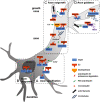
Molecular players in ubiquitin control of axon development. Local ubiquitin-mediated regulation occurs and supports all steps of axon morphogenesis, starting with the initial phase in which an axon gains its identity (A, axon specification), and continuing as the axon grows (B,C, axon outgrowth) and navigates its way to its target (D, axon guidance). Ubiquitin-targeted proteins (blue) and respective E3 ubiquitin ligases (orange) or deubiquitinases (yellow) are illustrated at their site of action. The type of ubiquitin chains involved is indicated. For complementary information, see Table 1. CHIP, C-terminus of Hsp70-interacting protein; DCC, deleted in colorectal cancer; EBAX-CRL, EBAX-type Cullin-RING; LIMK1, LIM kinase 1; RhoGEF, Rho guanine nucleotide exchange factor.
Figure 3.
Figure 3.
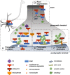
Ubiquitin-dependent signaling in the lifetime of presynaptic terminals. Following correct pathfinding, an axon establishes synaptic contacts (A, presynaptic formation), which mature into release-competent synaptic terminals (B, presynaptic function). Trimming of the axonal arbor (C, presynaptic elimination; and D, axon degeneration) can occur during development or adulthood. All these axonal events are locally controlled by ubiquitin and/or the proteasome. Ubiquitin-targeted proteins (blue) and respective E3 ubiquitin ligases (orange) or deubiquitinases (yellow) are illustrated at their site of action. The type of ubiquitin chains involved is indicated. For complementary information, see Table 1. ALK, Anaplastic lymphoma kinase; RIM1, Rab3-interacting molecule 1; UBC13, ubiquitin-conjugating enzyme 13; VAMP, vesicle-associated membrane protein; ZNRF1, zinc and ring finger 1.

References

    1. Aberle H, Haghighi AP, Fetter RD, McCabe BD, Magalhães TR, Goodman CS (2002) Wishful thinking encodes a BMP type II receptor that regulates synaptic growth in Drosophila. Neuron 33:545–558. 10.1016/s0896-6273(02)00589-5 - DOI - PubMed
    1. Anckar J, Bonni A (2015) Regulation of neuronal morphogenesis and positioning by ubiquitin-specific proteases in the cerebellum. PLoS One 10:e0117076. 10.1371/journal.pone.0117076 - DOI - PMC - PubMed
    1. Anderson C, Crimmins S, Wilson JA, Korbel GA, Ploegh HL, Wilson SM (2005) Loss of Usp14 results in reduced levels of ubiquitin in ataxia mice. J Neurochem 95:724–731. 10.1111/j.1471-4159.2005.03409.x - DOI - PubMed
    1. Angelman H (1965) 'Puppet' Children a report on three cases. Dev Med Child Neurol 7:681–688. - PubMed
    1. Arévalo JC, Waite J, Rajagopal R, Beyna M, Chen ZY, Lee FS, Chao MV (2006) Cell survival through Trk neurotrophin receptors is differentially regulated by ubiquitination. Neuron 50:549–559. 10.1016/j.neuron.2006.03.044 - DOI - PubMed

Publication types

LinkOut - more resources