. 2021 May 3;32(5):1097-1112.
doi: 10.1681/ASN.2020101459. Epub 2021 Mar 31.

The Rhesus Macaque Serves As a Model for Human Lateral Branch Nephrogenesis

Affiliations

The Rhesus Macaque Serves As a Model for Human Lateral Branch Nephrogenesis

Meredith P Schuh et al. J Am Soc Nephrol. .

Abstract

Background: Most nephrons are added in late gestation. Truncated extrauterine nephrogenesis in premature infants results in fewer nephrons and significantly increased risk for CKD in adulthood. To overcome the ethical and technical difficulties associated with studies of late-gestation human fetal kidney development, third-trimester rhesus macaques served as a model to understand lateral branch nephrogenesis (LBN) at the molecular level.

Methods: Immunostaining and 3D rendering assessed morphology. Single-cell (sc) and single-nucleus (sn) RNA-Seq were performed on four cortically enriched fetal rhesus kidneys of 129-131 days gestational age (GA). An integrative bioinformatics strategy was applied across single-cell modalities, species, and time. RNAScope validation studies were performed on human archival tissue.

Results: Third-trimester rhesus kidney undergoes human-like LBN. scRNA-Seq of 23,608 cells revealed 37 transcriptionally distinct cell populations, including naïve nephron progenitor cells (NPCs), with the prior noted marker genes CITED1, MEOX1, and EYA1 (c25). These same populations and markers were reflected in snRNA-Seq of 5972 nuclei. Late-gestation rhesus NPC markers resembled late-gestation murine NPC, whereas early second-trimester human NPC markers aligned to midgestation murine NPCs. New, age-specific rhesus NPCs (SHISA8) and ureteric buds (POU3F4 and TWIST) predicted markers were verified in late-gestation human archival samples.

Conclusions: Rhesus macaque is the first model of bona fide LBN, enabling molecular studies of late gestation, human-like nephrogenesis. These molecular findings support the hypothesis that aging nephron progenitors have a distinct molecular signature and align to their earlier human counterparts, with unique markers highlighting LBN-specific progenitor maturation.

Keywords: chronic kidney disease; lateral branch nephrogenesis; nephrogenesis; prematurity.


Figures

Figure 1.
Figure 1.
A 3D rendering of rhesus LBN. The 3D renderings of rhesus nephrogenesis from 126 to 136 DG (A1–A4 to C1–C4) and 32-WG human kidney (A5–C5) at a depth of 500 µm stained for SIX1/SIX2 (NPC), CDH1 (epithelia), and KRT8/18 (UB) are displayed from two perspectives: (A) cortical level and (B) lateral. (C) Two-dimensional immunofluorescence of representative images. White arrow denotes bulbous UB tip. Yellow arrow identifies the elongated ureteric branch after nephrogenesis ends. Note the variation in SIX2 signal in (C), caused by differences in fixation (samples in C2 and C3 were fixed in 4% paraformaldehyde, whereas samples C1, C4, C5, and C6 were fixed in formalin and incubated in Quadrol before staining). (D) Cross-sectional 3D renderings of a 32-WG human nephrogenic niche stained for KRT8/18, CDH1, and SIX2. KRT8/18 was absent from lateral branches. (E) The ureteric stalk in the rhesus compared with the human. (F) Visualization of rosette-like organization of niche tips in both human and rhesus. Scale bar, 100 µm.
Figure 2.
Figure 2.
Bifurcation rates, interniche distance, and tip per niche numbers during third-trimester rhesus development. (A) The 34 bifurcation identified within a 850 µm depth out of 797 ureteric stalks, examined from 126 to 138 GA rhesus. (B) The distance from the cortical surface to the bifurcating branch point at each age (average 482.6±166.3 µm). No bifurcating branch points were identified in 126 and 128 DG samples. Two out of 53 ureteric stalks in a 32-WG human kidney are shown for comparison. (C) The 3D rendering of recent bifurcating branch point (top, 129 DG, blue arrow) occurring at a depth of 198 µm and an older bifurcating branch point (bottom, 133 DG, blue arrow) occurring at a depth of 613 µm. Lateral branch identified by white arrow. (D) Tip to nearest-tip distances. White spheres indicate individual tip termini. (E) Minimum distance quantified for rhesus kidneys 124–136-DG and the 32-WG human kidney. See text for detail. (F) The number of niches per cluster as determined in 3D reconstructions. The cluster organization is shown by surface rendering and its corresponding z-stack section. Annotated colors represent the various number of tips per niche cluster (2, blue; 3, green; 4; yellow; 5, red; 6, white). (G) The number of tips per niche cluster quantified for rhesus kidneys 124–137-DG and the 32-WG human kidney. Scale bar, 100 µm.
Figure 3.
Figure 3.
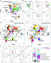
Comparative scRNA-Seq and snRNA-Seq analyses. (A) UMAP of 37 transcriptionally distinct cell populations predicted from the software ICGS2 from term rhesus kidney cortex scRNA-Seq in four biologic replicates. (B) Projection of animal-specific cell identifies on to the UMAP to assess possible batch effects (left), localization of cells from the predicted NPC cluster (c25, middle) and pseudotime trajectory predictions from the software SlingShot, with cluster 25 set as the origin. (C) Projected normalized gene expression (red = maximal expression, z-score normalized) for representative marker genes uniquely associated with cluster 25 (MarkerFinder algorithm). (D) UMAP of transcriptionally distinct cell-nuclei populations (snRNA-Seq) from a single kidney cortex, predicted by ICGS2. Associated labels were derived using prior defined single-cell marker genes, spanning 48 distinct adult and fetal body site locations, corresponding to over 2300 cell signatures. (E) Supervised classification of cell types on the basis of the predicted rhesus cell-type clusters from panel (A) in the snRNA-Seq. Unclassified cells are shown in gray. (F) snRNA-Seq predicted NPC marker genes (cluster 26) displayed similar to panel (C). (F) Projected normalized gene expression (red = maximal expression, z-score normalized) for CITED1, EYA1, SHISA8, BMPER, CACNA1E, and DPP6. (G) Comparison of the top 100 previously identified human fetal NPC cluster markers (16–17 weeks) to the top 100 mouse renal developmental dataset NPC markers by gene-set enrichment in the software GO-Elite.
Figure 4.
Figure 4.
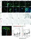
SHISA8 expression validated in the NPC of human archival material. (A) RNAScope was performed on human archival material from 16, 17 WG (“young” early second) and 26, 27 WG (“old” late second trimester) with RNA probes for SIX1 (green) and SHISA8 (red) to localize their transcripts, and antibodies to KRT8/18 (UB). SHISA8 colocalized with SIX1 at every gestational age. Images were acquired on the confocal microscope at ×20 magnification. (B) A ×40 magnification of the white box identified in (A). Separate channels show visualization of SIX1 (C) and SHISA8 (D) transcripts. (E) Absolute SHISA8 transcript counts and the ratio of SHISA8/SIX1 increase the further the NPC are from the cortical surface, marked NPC1 (green, cortical) to NPC3 (orange). (F) Quantification of SHISA8 and SIX1 transcripts performed using Bitlane Imaris spot detection algorithm. SHISA8 transcript counts (F1) were significantly increased between NPC1 versus NPC3 (P<0.0001) and NPC2 versus NPC3 (P=0.02) in the older gestation samples only. There was also a significant difference between NPC3 young and old samples (P=0.03). SHISA8/SIX1 ratio (F2) was significantly increased from NPC1 to NPC3 in both young (P=0.007) and old (P<0.0001) samples. There was a significant increase in SHISA1/SIX1 ratio in old NPC3 compared with young NPC3 (P=0.04). Scale bar 10 µm.
Figure 5.
Figure 5.
Transcriptional heterogeneity in the UB underlies inferred cell transitions. (A–C) Unsupervised ICGS2 clustering of UB clusters finds (A) six distinct cell states, with a possible progression of early ureteric tip to maturing stalk cells, (B and C) highlighted by the expression novel markers. (D and E) Visualization of ICGS2 clusters in (D) Monocle2 pseudotemporal ordered clusters. (E) Relative distribution of UB markers AQP2 and VSTM2A in (F) Monocle-ordered cells.
Figure 6.
Figure 6.
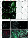
POU3F4 expression has a cortical bias. Confocal images of antibody stain and RNAScope in situ hybridization. RNA probes for SIX1 (green) and POU3F4 (red) and antibodies to KRT8/18 (collecting duct) were used to localize the transcripts in human archival material at (A) 16, (B) 26, and (C) 27 WG. KRT8/18 staining outlined in A3–A4, B3–B4, and C3–C4. POU3F4 colocalized KRT8/18 at every gestational age, located in both tip and stalk (A4 and B4), but absent from branch points (C4). (D and E) Representative widefield microscope images acquired at ×20 to determine POU3F4 distribution in the cortex, cortical-medullary junction, and medulla at 26 WG. POU3F4 is visualized in red, nonspecific, autofluorescence signal in light blue. Of note, there were some ureteric tips that showed tip-restricted staining (white arrow in box, E1 and D1).

Comment in

  • Monkeying about with Nephron Formation.
    Smyth IM. Smyth IM. J Am Soc Nephrol. 2021 May 3;32(5):1011-1013. doi: 10.1681/ASN.2021030320. Epub 2021 Apr 7. J Am Soc Nephrol. 2021. PMID: 33827903 Free PMC article. No abstract available.

References

    1. Hinchliffe SA, Sargent PH, Howard CV, Chan YF, van Velzen D: Human intrauterine renal growth expressed in absolute number of glomeruli assessed by the disector method and Cavalieri principle. Lab Invest 64: 777–784, 1991. - PubMed
    1. Nyengaard JR, Bendtsen TF: Glomerular number and size in relation to age, kidney weight, and body surface in normal man. Anat Rec 232: 194–201, 1992. - PubMed
    1. Hoy WE, Hughson MD, Bertram JF, Douglas-Denton R, Amann K: Nephron number, hypertension, renal disease, and renal failure. J Am Soc Nephrol 16: 2557–2564, 2005. - PubMed
    1. Bertram JF, Douglas-Denton RN, Diouf B, Hughson MD, Hoy WE: Human nephron number: Implications for health and disease. Pediatr Nephrol 26: 1529–1533, 2011. - PubMed
    1. Barker DJ: The intrauterine environment and adult cardiovascular disease. Ciba Found Symp 156: 3–10, discussion 10–16, 1991. - PubMed

Publication types

LinkOut - more resources