. 2021 Mar 31;11(1):7312.
doi: 10.1038/s41598-021-86470-1.

Age-dependent ataxia and neurodegeneration caused by an αII spectrin mutation with impaired regulation of its calpain sensitivity

Affiliations

Age-dependent ataxia and neurodegeneration caused by an αII spectrin mutation with impaired regulation of its calpain sensitivity

Arkadiusz Miazek et al. Sci Rep. .

Erratum in

Abstract

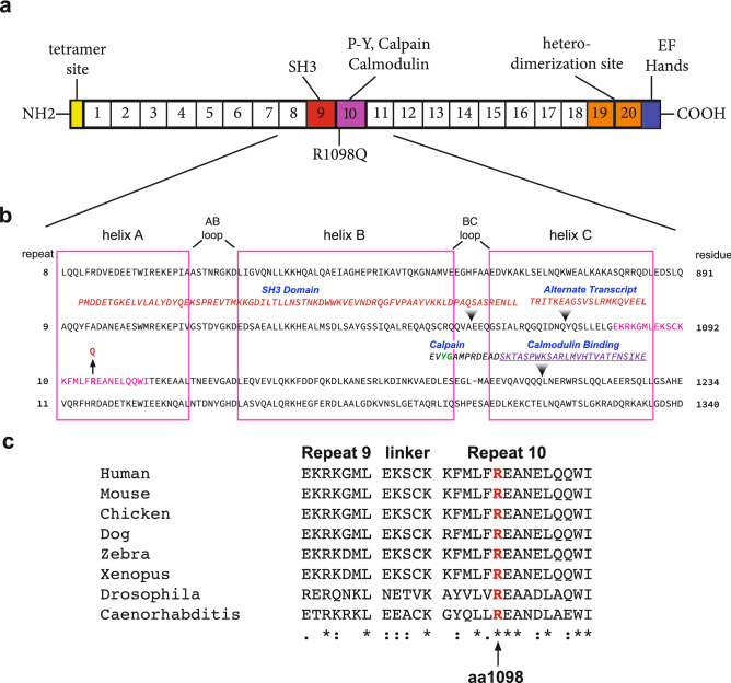
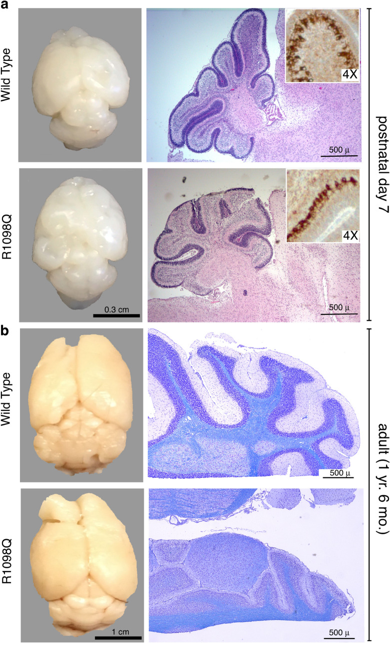
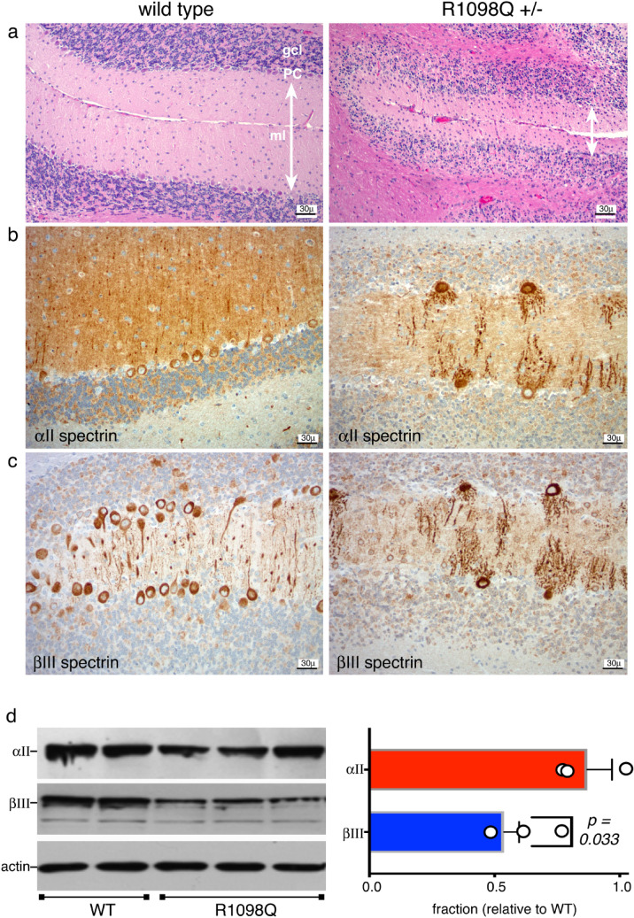
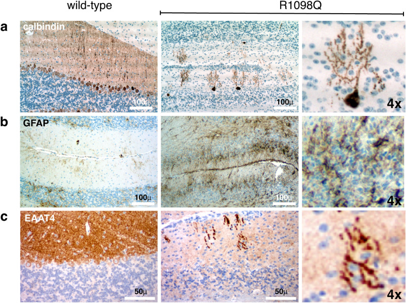
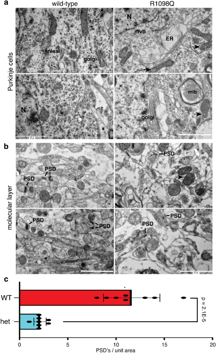
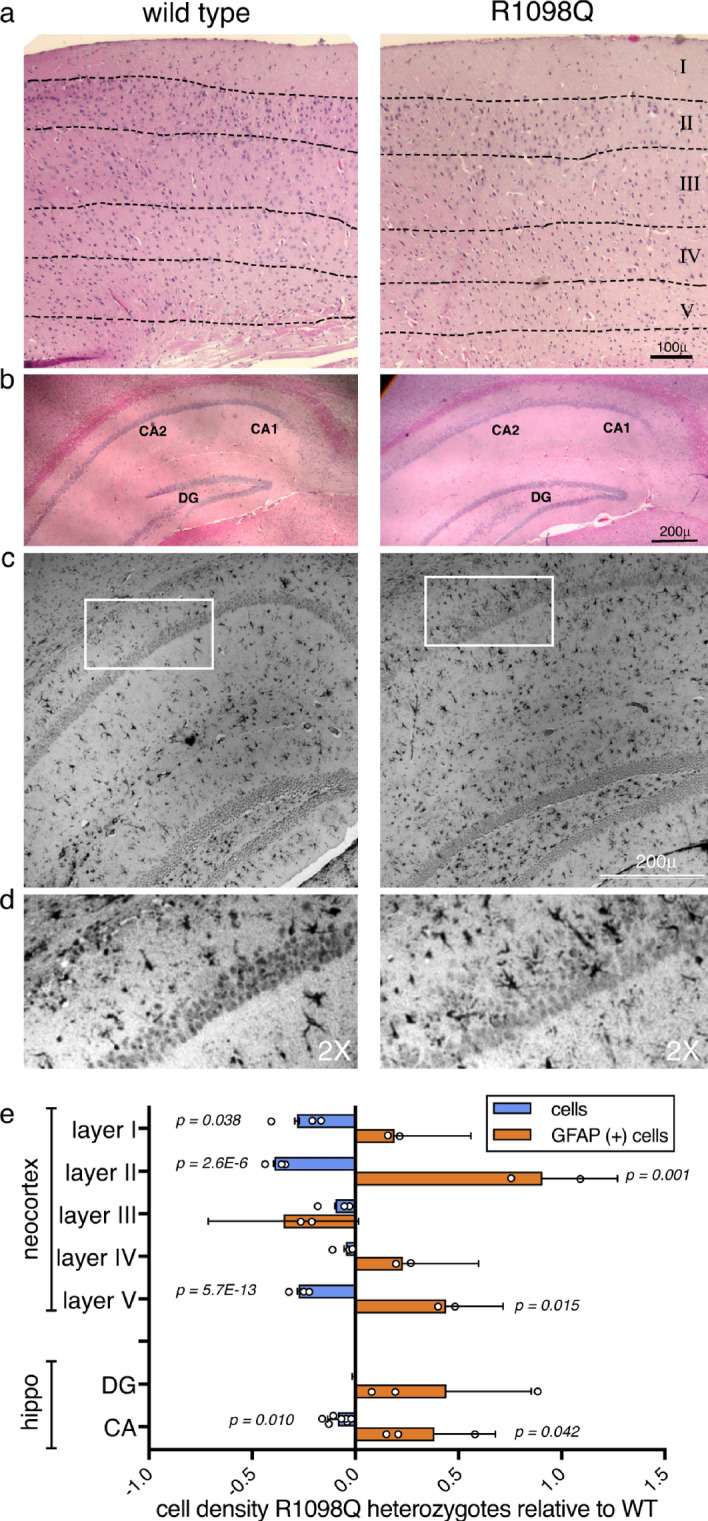
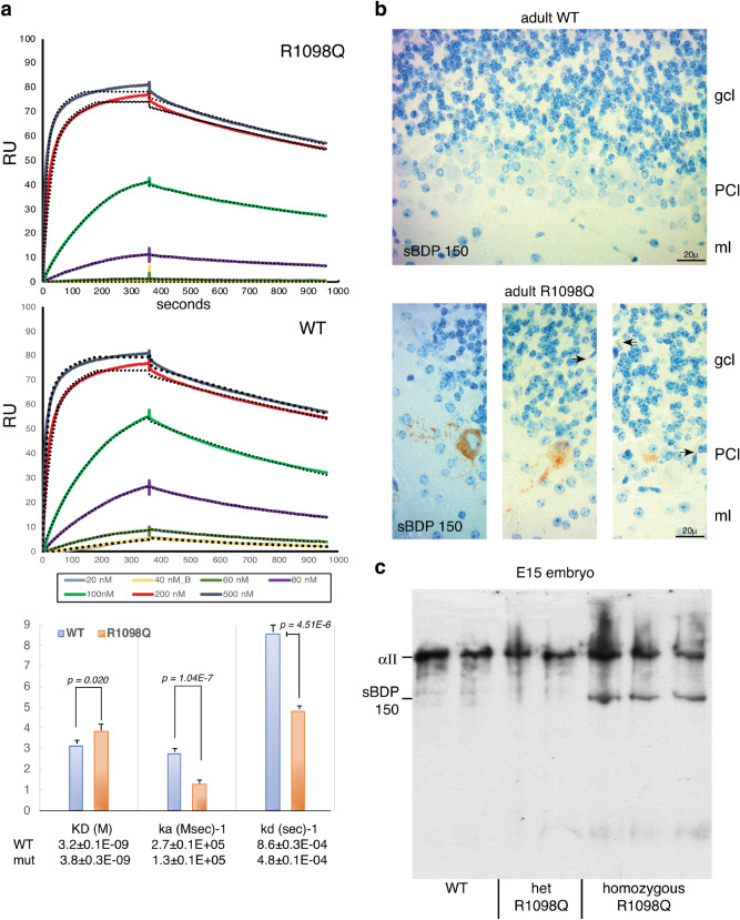
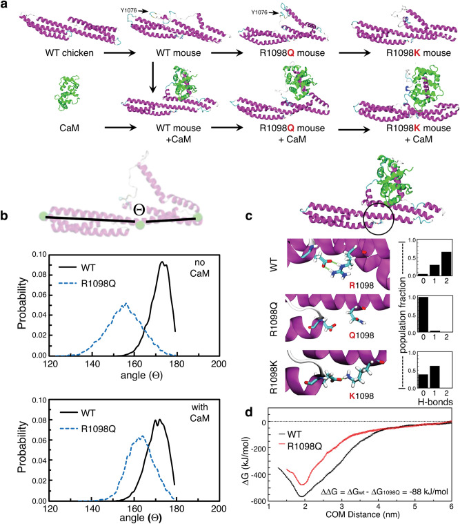
The neuronal membrane-associated periodic spectrin skeleton (MPS) contributes to neuronal development, remodeling, and organization. Post-translational modifications impinge on spectrin, the major component of the MPS, but their role remains poorly understood. One modification targeting spectrin is cleavage by calpains, a family of calcium-activated proteases. Spectrin cleavage is regulated by activated calpain, but also by the calcium-dependent binding of calmodulin (CaM) to spectrin. The physiologic significance of this balance between calpain activation and substrate-level regulation of spectrin cleavage is unknown. We report a strain of C57BL/6J mice harboring a single αII spectrin point mutation (Sptan1 c.3293G > A:p.R1098Q) with reduced CaM affinity and intrinsically enhanced sensitivity to calpain proteolysis. Homozygotes are embryonic lethal. Newborn heterozygotes of either gender appear normal, but soon develop a progressive ataxia characterized biochemically by accelerated calpain-mediated spectrin cleavage and morphologically by disruption of axonal and dendritic integrity and global neurodegeneration. Molecular modeling predicts unconstrained exposure of the mutant spectrin's calpain-cleavage site. These results reveal the critical importance of substrate-level regulation of spectrin cleavage for the maintenance of neuronal integrity. Given that excessive activation of calpain proteases is a common feature of neurodegenerative disease and traumatic encephalopathy, we propose that damage to the spectrin MPS may contribute to the neuropathology of many disorders.


Conflict of interest statement

The authors declare no competing interests.

Figures

Figure 1
Figure 1
Relationship of the R1098Q variant to αII spectrin structure. (a) Schematic representation of αII spectrin with 20 complete homologous repeats each ≈ 106 residues and the location of selected functional domains that interrupt the repeat motif. The 9th and 10th repeats (red and purple respectively) harbor an inserted SH3 domain, a variably appearing alternative transcript insertion, and specialized sequences encompassing the site of CaM binding and the site Y1176 and G1177 that is preferentially cleaved by calpain. (b) Sequence of repeats 8–11 aligned by homology. The boxes denote the boundaries of each of the three α-helices (A–C) of the canonical repeat unit; these are separated by AB and BC loops and inter-unit linkers. The sequences representing the SH3 domain and the alternative transcript inserted into repeat 9 and the calpain and CaM interaction sites in repeat 10 are indicated. The arrow marks the site of the R1098Q mutation near the start of the 10th repeat. The YG site of preferred calpain cleavage is colored green. (c) Clustal alignment of residues flanking the R1098Q mutation. This locus, and the flanking region, is almost perfectly conserved across diverse species. Residues fully conserved are denoted with asterisks (*); residues strongly conserved with a score greater than 0.5 on the PAM250 matrix are designated with a colon (:). Weakly similar residues are denoted with a period (.).
Figure 2
Figure 2
Whole brain images and cerebellum histology. (a) WT and heterozygous R1098Q mice brains are indistinguishable at birth. By P7, subtle gross and histologic abnormalities begin to emerge in the brains of the R1098Q heterozygotes. These include nearly imperceptible defects in their cerebellar folia and cerebral convolutions. However, at P7 Purkinje cells are abundant and their morphology is intact, excluding developmental failure due to the heterozygous mutation. This is most apparent when immunostained for βIII spectrin that highlights the PC soma and dendrites (insert, shown at 4 × magnification of the H&E stained section; also see supplemental Fig. S3 for enlarged image of the P7 cerebellum). (b) Older R1098Q heterozygote mice (1.5 years) exhibit profound atrophy of the cerebellum, correlating with near total collapse of the molecular layer and loss of Purkinje cells. Some cortical loss can also be perceived. These gradual morphologic changes correlate with the onset of the progressive ataxia apparent in the R1098Q mice, a phenotype that intensifies with age.
Figure 3
Figure 3
Cerebellar pathology in R1098Q heterozygous mice. (a) Hematoxylin and eosin (H&E) stained sections of cerebellum of 26 week old WT and heterozygous R1098Q mice. Most noticeable is the ≈ 50% loss in molecular layer width (double headed arrows) and extensive loss of cells in the Purkinje layer.. These changes are quantified in Supplemental Fig. S7. There may also be some loss of PC’s in the granular layer as well, but the disruption of layer organization in the hets made defining the boundaries of the granular layer problematic and rendered quantitation unreliable. (b) Immunostain for αII spectrin reveals its preservation in all cerebellar layers, and highlights the loss of PCs and the severe disruption and fragmentation of PC dendrites in the R1098Q mice. (c) Immunostain for βIII spectrin. This spectrin is primarily (although not exclusively) expressed in PCs in the cerebellum. Its pattern is the same in PC’s as that of αII spectrin, highlighting the major distortions and fragmentation of PC dendritic architecture. (d) Western blot of adult cerebellum for αII and βIII spectrin quantifying the global and selective PC loss in heterozygotes, expressed as a ratio relative to the WT levels. Results from two WT and three R1098Q heterozygotes are shown. While there is no significant reduction in αII spectrin levels (which is expressed in all cells), the βIII spectrin level is reduced to 64 ± 7% of the WT level (p = 0.033), reflecting the selective loss and vulnerability of PC’s to the R1098Q mutation. (Full length blots of the gels used for quantitation are presented in supplemental Fig. S6). Data analyzed by single tail, 2-sample homosedastic T-test. N = 2 for WT; 3 for het.
Figure 4
Figure 4
Evidence of cerebellar degeneration and gliosis. We used immunostains with markers of degeneration, reactive gliosis, and synaptic integrity to further characterize the cerebellar changes in 1.5 yo R1098Q heterozygous mice. Images in the right-most panel of each row are presented at 4 × magnification relative to the other images. (a) Calbindin, a marker of PCs, highlights their dramatic loss and their excessively branched, segmented, and coarse dendrites. (b) Glial fibrillary acidic protein (GFAP), a marker of reactive gliosis and an indirect marker of neuroinflammation, reveals a marked increase in both the molecular and granular cell layers. Astrocytes and their processes infiltrate all cerebellar layers. (c) EAAT4, a glutamate transporter that binds spectrin at Purkinje cell PSDs, is nearly absent in the R1098Q mice, and what remains is concentrated in the swollen dendritic shafts of PCs.
Figure 5
Figure 5
EM analysis reveals degenerative changes in R1098Q PCs and the molecular layer. (a) The PC’s of R1098Q 1.5 yo mice exhibit massively dilated organelle structures that appear to arise from Golgi and ER, along with dispersal of Nissl substance. Many multivesicular bodies (mvb) and multilamellar bodies (mlb) are present, presumably derived from autophagocytic vacuoles. Mitochondia (arrows) also frequently show internal vesiculation.(b) Within the cerebellar molecular layer of the heterozygous animals, dendrites are swollen and post synaptic densities (PSD) are attenuated and sparse. (c) The PSD’s identified in eight separate EM images of WT mouse and eight images from a heterozygous R1098Q mouse were counted. There is an approximately four-fold reduction in PSD’s in the heterozygous 1.5 yo mouse. The variation in the replicates comparing these two mice were evaluated by paired single tail T-test.
Figure 6
Figure 6
Degeneration of the neocortex and hippocampus in adult R1098Q mice. While not as dramatic as in the cerebellum, there is significant astrogliosis and neuronal loss in the cerebrum of 1.5 year old mice. (a) H&E stained neocortex exhibiting neuronal loss. When the cells per unit area and their GFAP staining were counted (below), losses and enhanced GFAP positive cells were significant in layers I, II and V. (b) H&E stained hippocampus. As in the neocortex, there is cell loss particularly in the CA1/CA2 regions. (c) GFAP immunostaining of the hippocampus exhibiting widespread astrogliosis. The region outlined is depicted in greater detail in panel (d), where the infiltration of astroglia is apparent throughout the CA1/2 layers in the R1098Q animals. (e) Comparison of changes in cell and astroglial (GFAP positive) density in R1098Q heterozygotes relative to the WT adult brains. The average density of cells in cortical layer I, layer II, and V are significantly reduced, while there is significantly increased numbers of GFAP cells per unit area in layers II and V. The CA1/2 region also exhibits less dramatic but still reduced cell counts, with marked increase in GFAP positive cells. There is no significant change in cell density in the dentate gyrus (DG), and the increase in GFAP positivity in this region also did not reach significance. For this analysis, images from comparable regions of two WT and two heterozygous mice were each divided into equal area units (18 to 46 units depending on the size of the region under consideration), and the number of cells in each unit area counted by three independent observers. All data for WT and het were then each pooled, and the average cell densities of each region calculated and compared (WT vs. het). Significance was evaluated by paired single-tailed T-test.
Figure 7
Figure 7
R1098Q spectrin exhibits reduced in vitro CaM affinity and enhanced in vivo calpain cleavage. (a) Surface plasmon resonance detected the binding of Ca2+/CaM to recombinant GST-spectrin peptides representing repeats 8–11 (as shown in Fig. 1). The concentration of CaM used for each curve is as indicated. The binding curves fit well to a 1:1 Langmuir binding model over the indicated range of ligand concentrations (dotted lines, generated by BIAevaluation 3.1, https://www.biacore.com/lifesciences/service/downloads/software_licenses/biaevaluation ). The derived rate constants for (ka) and (kd) and their confidence limits as reported by the BIAevaluation software were both reduced for the R1098Q peptide relative to the WT peptide. The derived KD for the mutant peptide of 3.8 ± 0.3E−09 represents a significant 19% reduction in its affinity for CaM. (b) To determine if spectrin was being excessively targeted by calpain in vivo, we examined the cerebellum of 26 week old mice with an antibody raised to the unique epitope created by calpain cleavage of spectrin at Y1176. This antibody only detects calpain-cleaved spectrin. No staining was detected in the cerebellum of either of two WT animals. Conversely, their respective R1098Q heterozygous littermates (two animals) at 26 weeks of age displayed calpain-cleaved spectrin in 5–18% of their PC soma and throughout the apical dendrite. Cleavage product was also detected in the granular cell layer (arrows); the origin of this staining in the granular layer (whether cells or synapses with sBDPs) is undetermined. (c) Embryos (14.5 d) were harvested in-utero and analysed by Western blotting with anti-αII spectrin antibodies to gauge the level of spectrin proteolysis. While break-down was minimal or absent in both the WT and heterozygous embryos, a single sBDP at ≈150 kDa was prominent only in the homozygotes. This sBDP is the major product generated by calpain. The characteristic cleavage fragment generated by caspase at ≈ 120 kDa during apoptosis was notably absent. These results indicate that the R1098Q spectrin is uniquely sensitive to calpain attack in the homozygous embryo (N = 2 WT; 2 het; and 3 homozygotes).
Figure 8
Figure 8
Molecular dynamics simulations predict enhanced flexibility of R1098Q spectrin with reduced calmodulin (CaM) binding and enhanced susceptibility to calpain cleavage. (a) Schematic of intermediate protein complexes simulated with molecular dynamics of spectrin repeats 9–10 with and without bound CaM. (Also see supplemental movie M2). (b) Convex bending of the spectrin scaffold (angle between adjacent monomers) is substantially shifted in R1098Q mutants, sampling both a more convex angle than WT spectrin and a broader distribution of values. (c) Hydrogen-bonding between D922 and either R1098, Q1098, or K1098 (a hypothetical variant) is markedly different between WT and variant spectrins. Salt-bridging between D922 and R1098 is strongest due to the presence of opposing charges and two hydrogen bond donors. Mutations that conserve native charge (e.g. R1098K) could still maintain intermediate hydrogen-bonding and scaffold flexibility, while pathological mutations such as R1098Q fully abrogate hydrogen-bonding and enhance spectrin flexibility. (d) The free energy of binding between CaM and spectrin is reduced by 88 kJ/mol in the R1098Q mutant, equivalent to a predicted 15% loss in binding affinity compared to WT complexes.

References

    1. Mahaman YAR, et al. Involvement of calpain in the neuropathogenesis of Alzheimer's disease. Med. Res. Rev. 2019;39:608–630. doi: 10.1002/med.21534. - DOI - PMC - PubMed
    1. Ahmad F, et al. Isoform-specific hyperactivation of calpain-2 occurs presymptomatically at the synapse in Alzheimer's disease mice and correlates with memory deficits in human subjects. Sci. Rep. 2018;8:13119. doi: 10.1038/s41598-018-31073-6. - DOI - PMC - PubMed
    1. Cheng SY, Wang SC, Lei M, Wang Z, Xiong K. Regulatory role of calpain in neuronal death. Neural Regen. Res. 2018;13:556–562. doi: 10.4103/1673-5374.228762. - DOI - PMC - PubMed
    1. Atherton J, et al. Calpain cleavage and inactivation of the sodium calcium exchanger-3 occur downstream of Abeta in Alzheimer's disease. Aging Cell. 2014;13:49–59. doi: 10.1111/acel.12148. - DOI - PMC - PubMed
    1. Hassen GW, et al. Effects of novel calpain inhibitors in transgenic animal model of Parkinson's disease/dementia with Lewy bodies. Sci. Rep. 2018;8:18083. doi: 10.1038/s41598-018-35729-1. - DOI - PMC - PubMed

Publication types

LinkOut - more resources