. 2021 May;593(7857):114-118.
doi: 10.1038/s41586-021-03413-6. Epub 2021 Mar 31.

An amygdala circuit that suppresses social engagement

Affiliations

An amygdala circuit that suppresses social engagement

Jeong-Tae Kwon et al. Nature. 2021 May.

Abstract

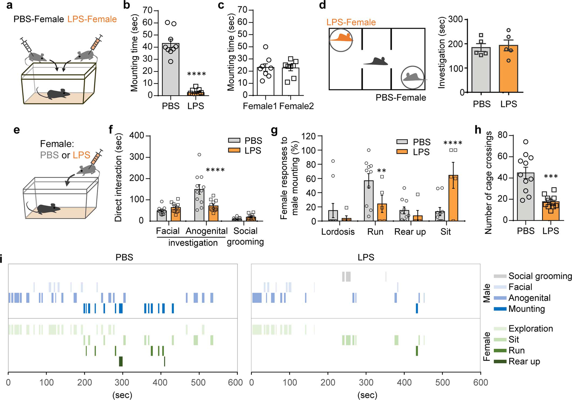
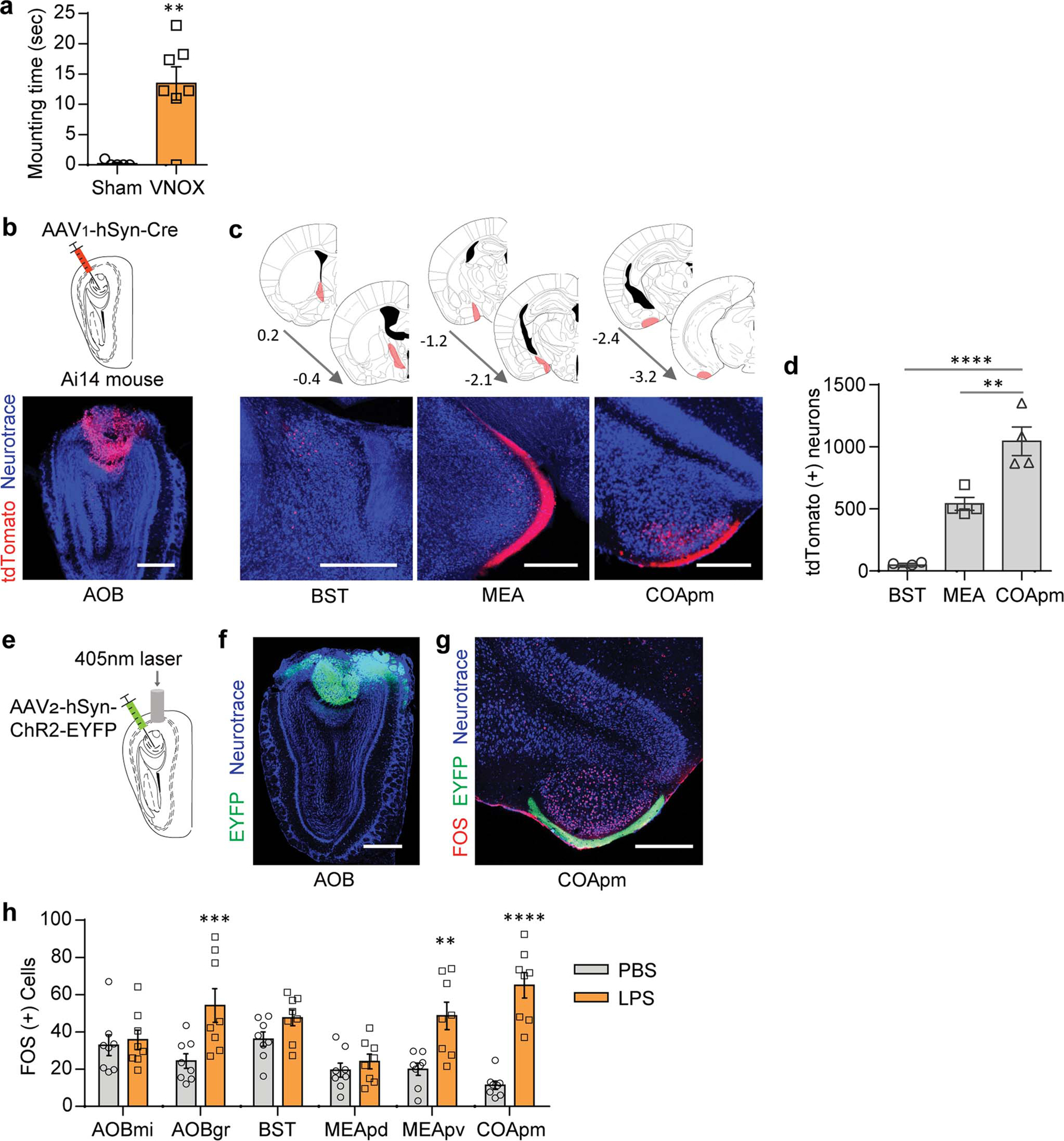
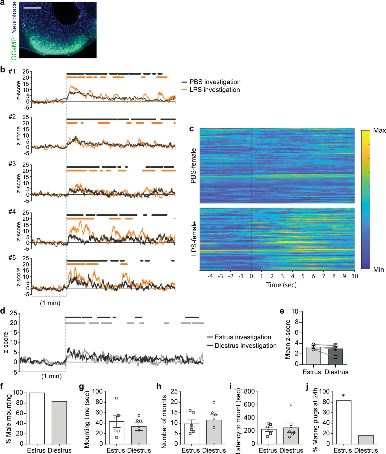
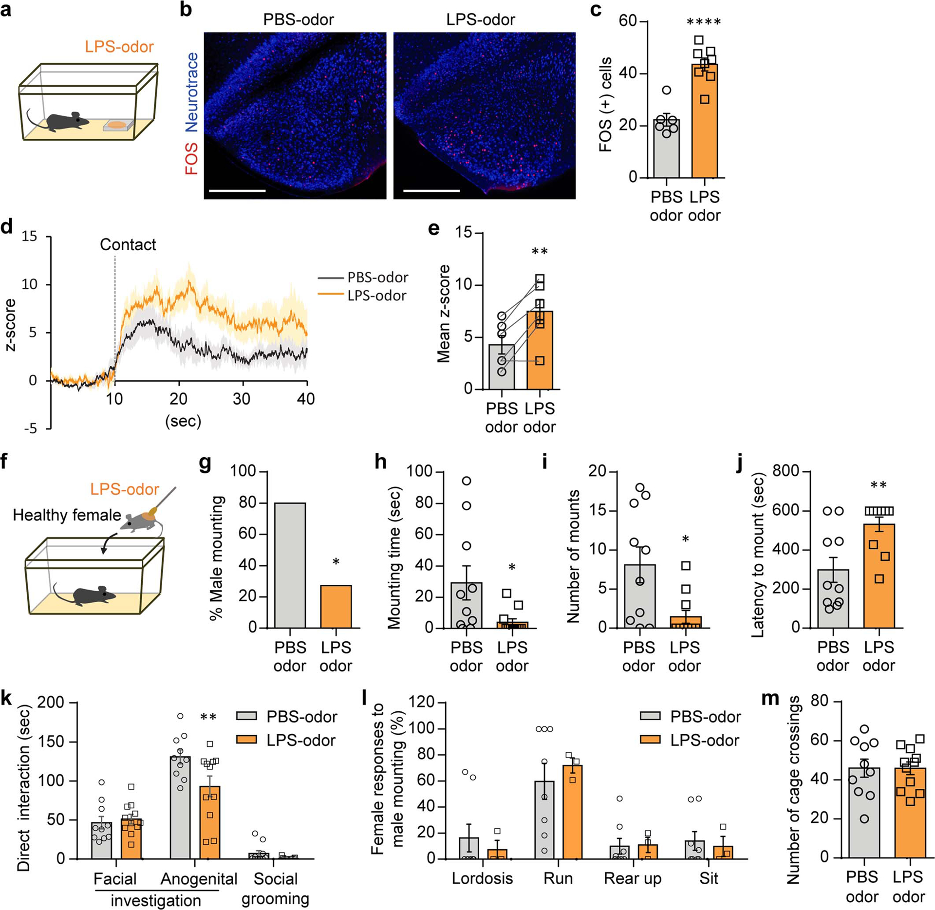
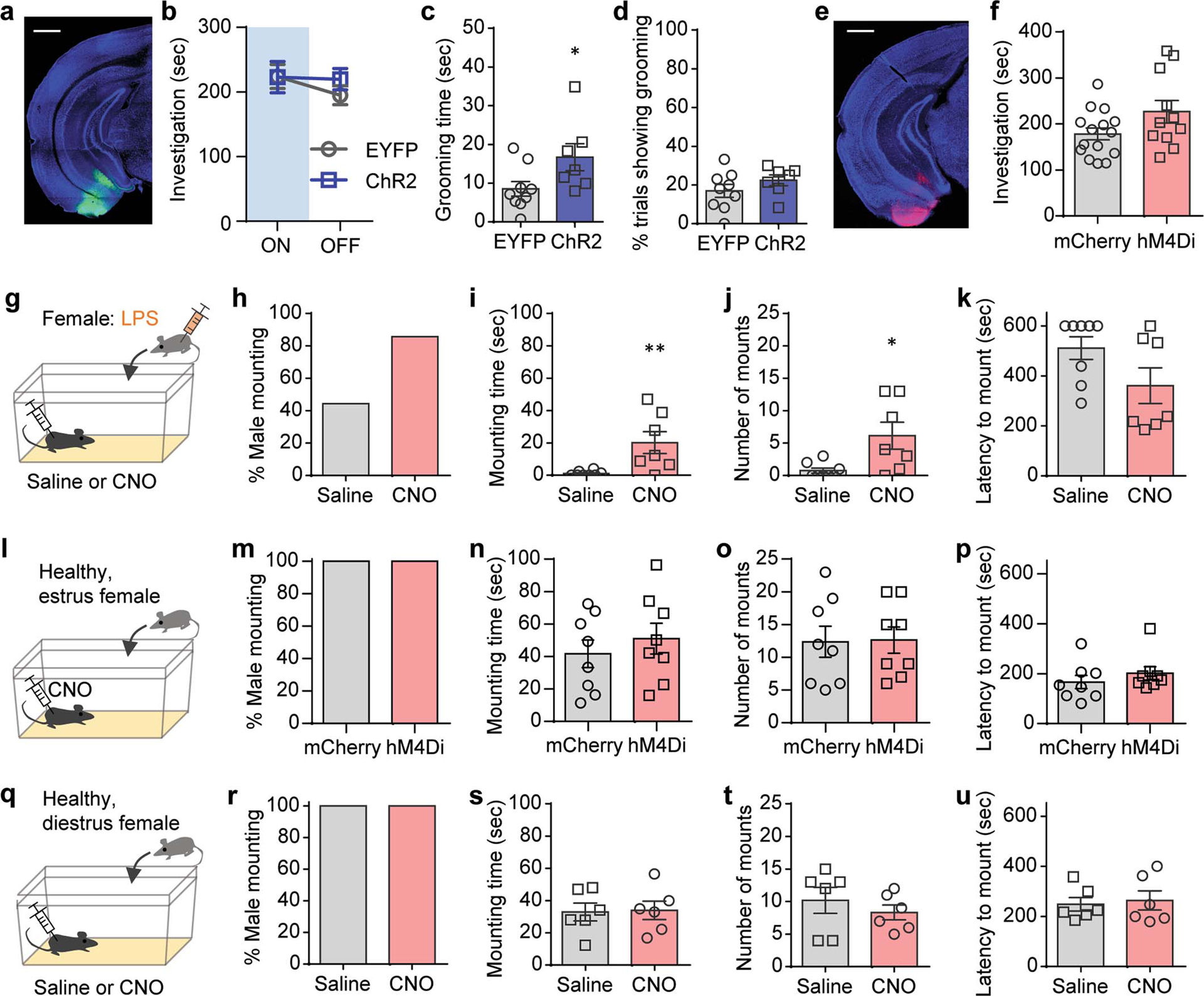
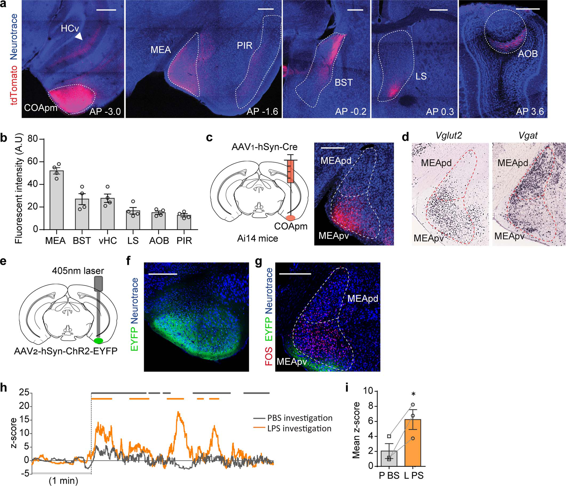
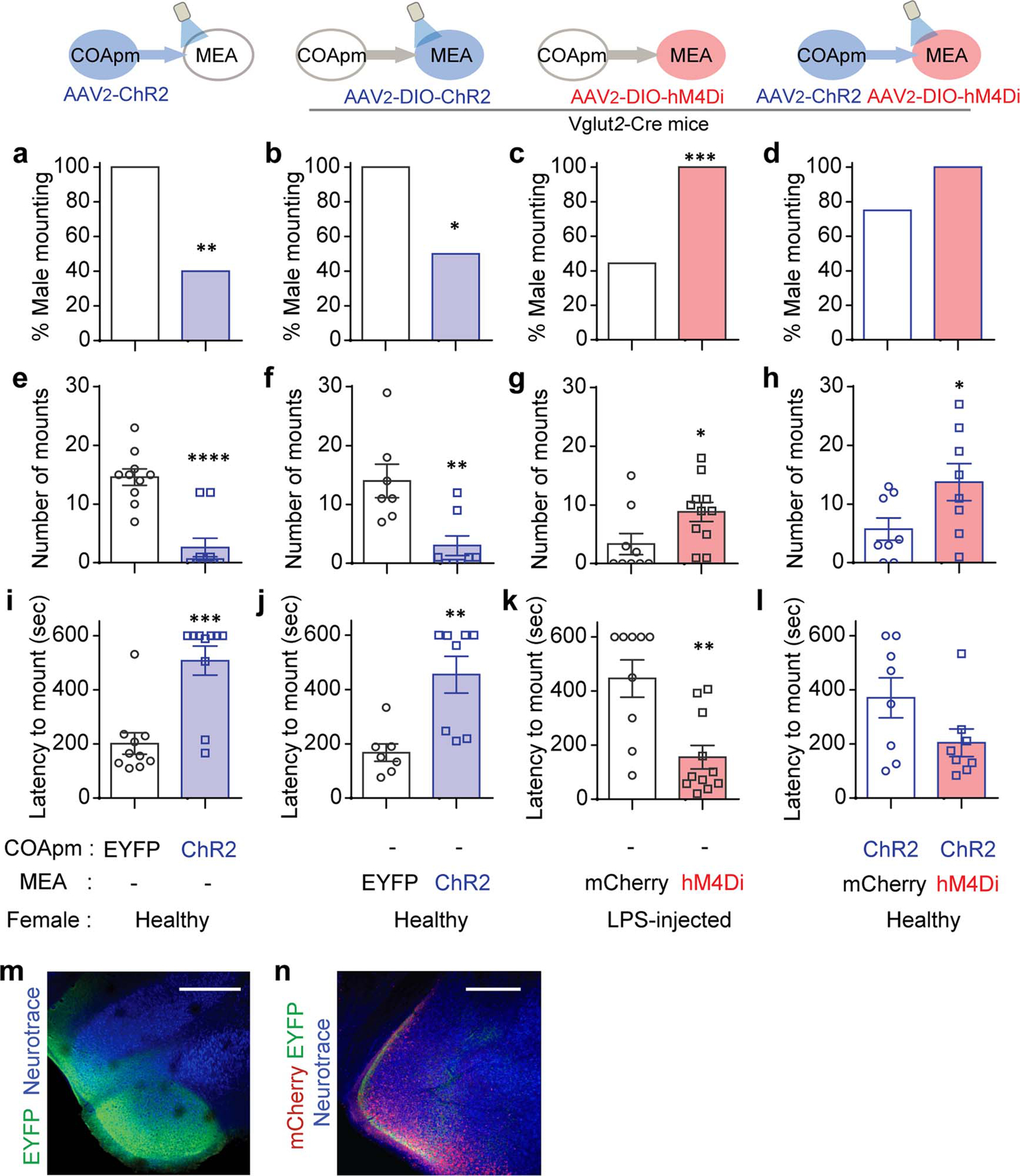
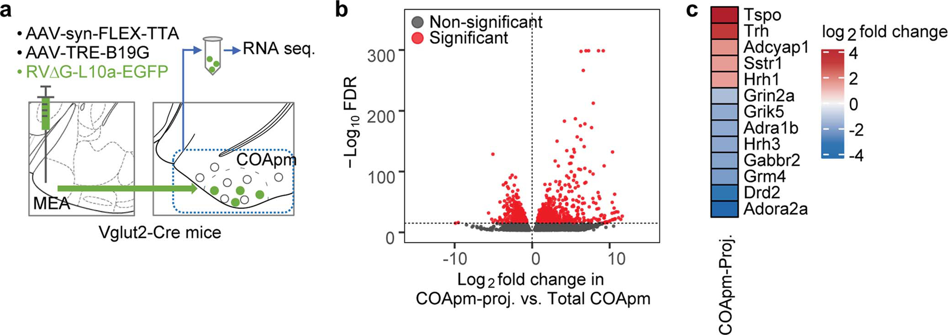
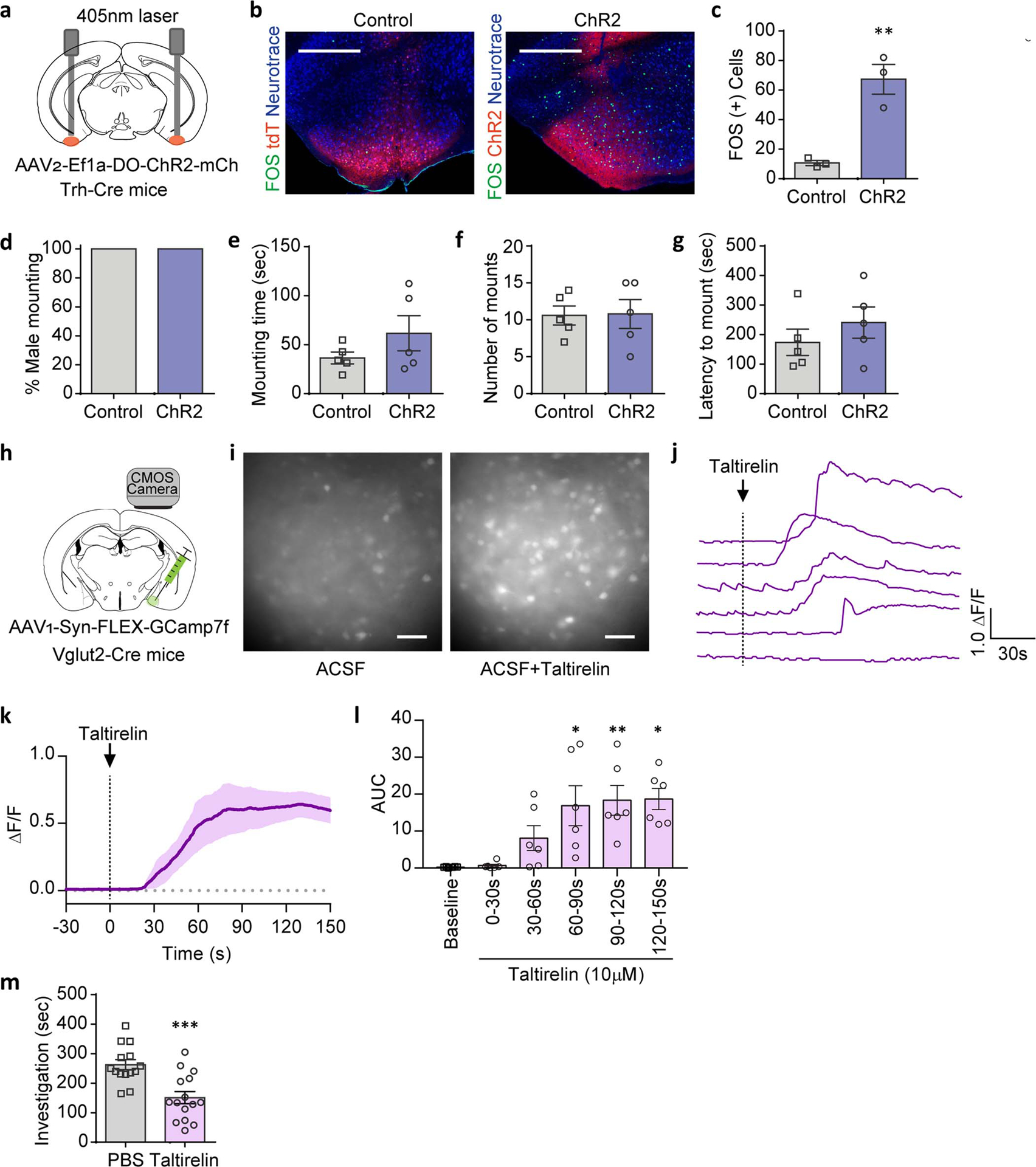
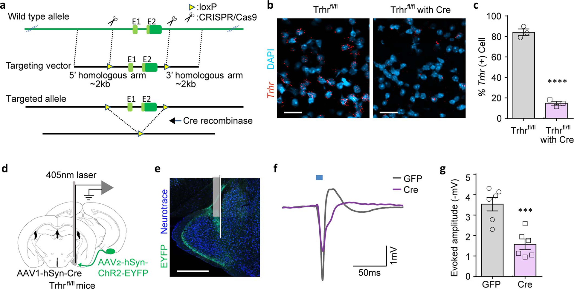
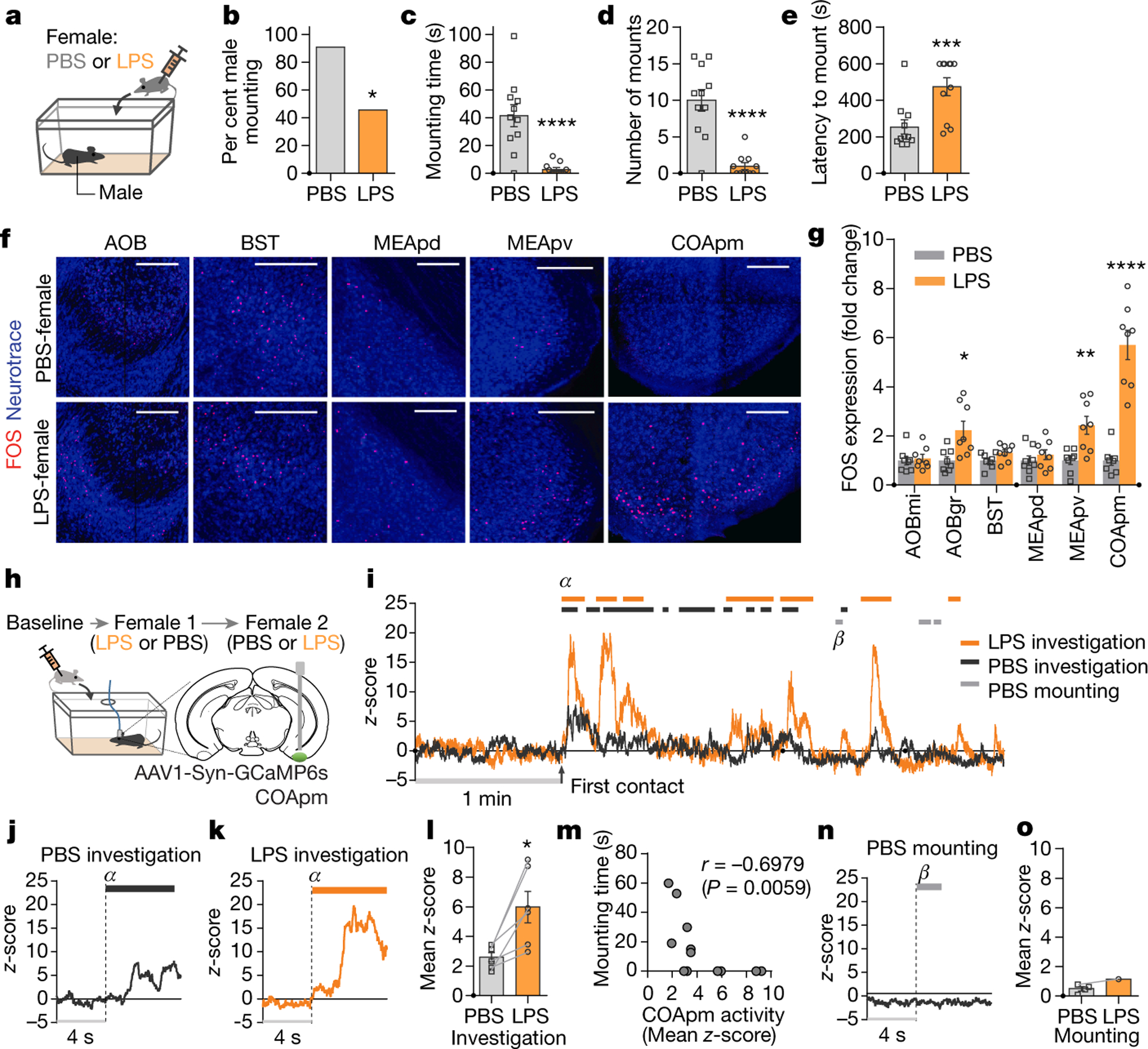
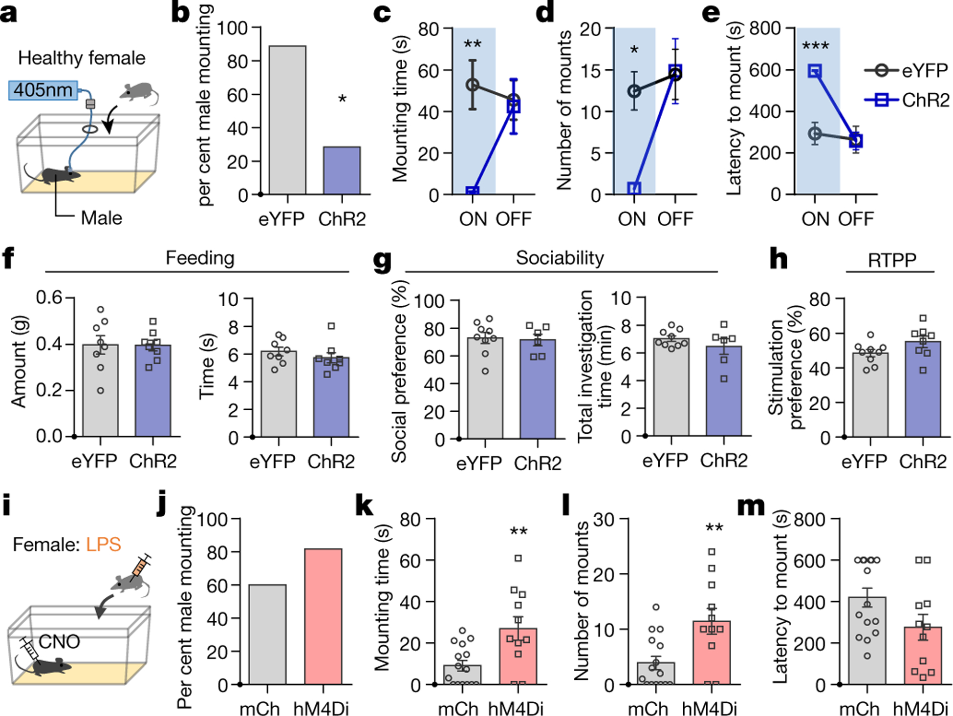
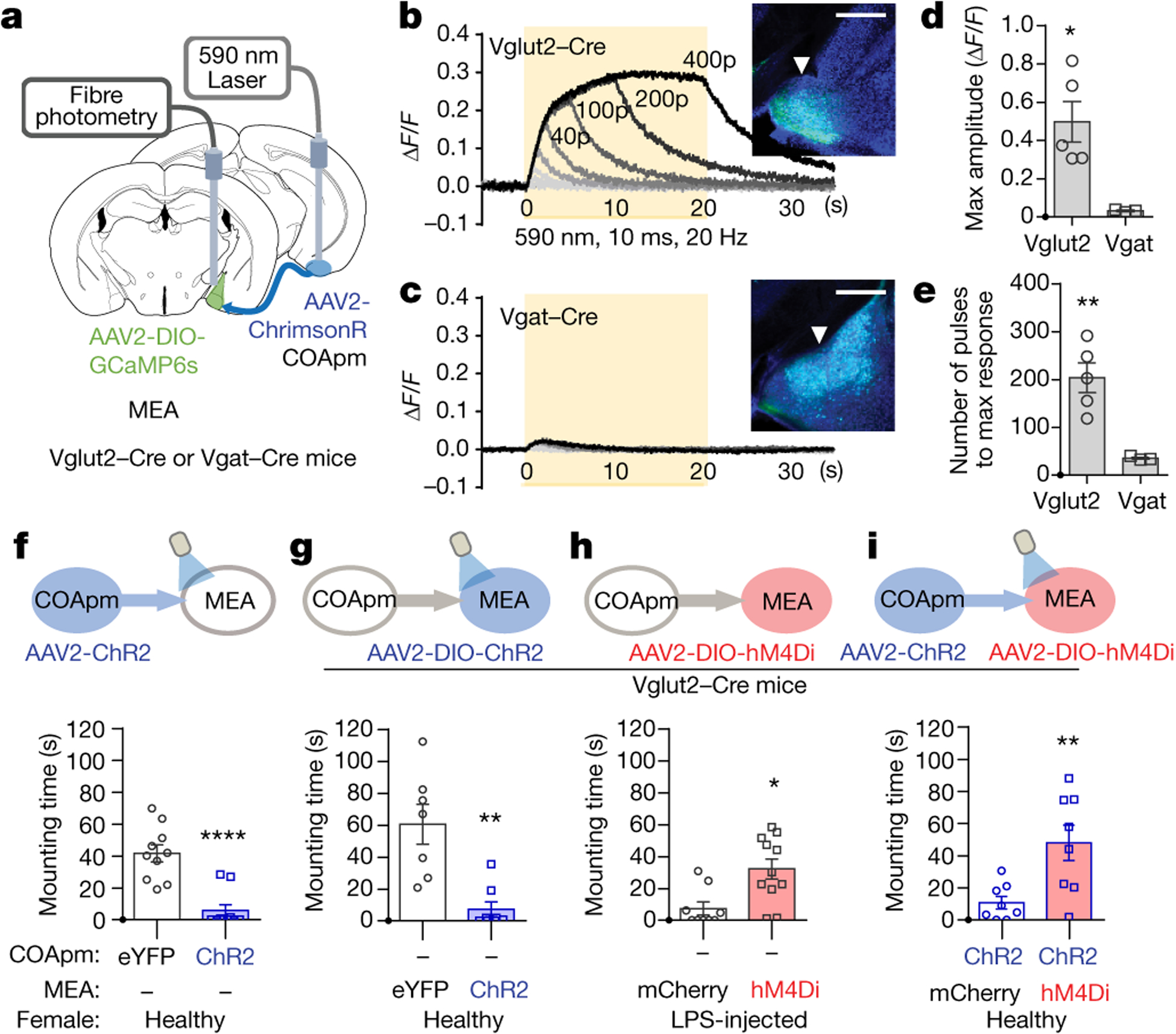
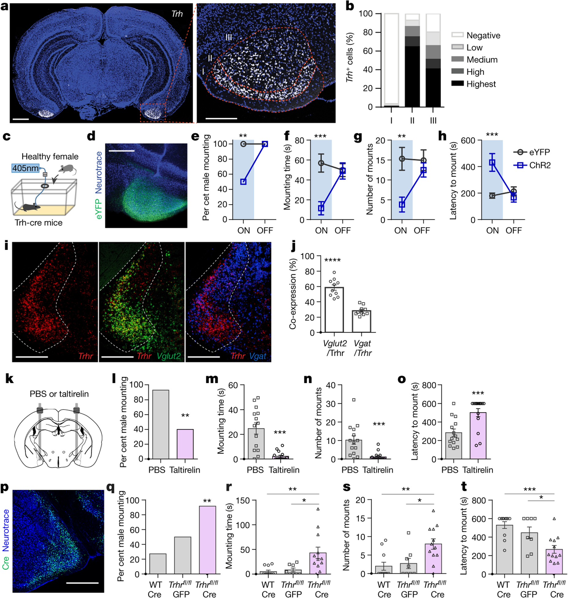
Innate social behaviours, such as mating and fighting, are fundamental to animal reproduction and survival1. However, social engagements can also put an individual at risk2. Little is known about the neural mechanisms that enable appropriate risk assessment and the suppression of hazardous social interactions. Here we identify the posteromedial nucleus of the cortical amygdala (COApm) as a locus required for the suppression of male mating when a female mouse is unhealthy. Using anatomical tracing, functional imaging and circuit-level epistatic analyses, we show that suppression of mating with an unhealthy female is mediated by the COApm projections onto the glutamatergic population of the medial amygdalar nucleus (MEA). We further show that the role of the COApm-to-MEA connection in regulating male mating behaviour relies on the neuromodulator thyrotropin-releasing hormone (TRH). TRH is expressed in the COApm, whereas the TRH receptor (TRHR) is found in the postsynaptic MEA glutamatergic neurons. Manipulating neural activity of TRH-expressing neurons in the COApm modulated male mating behaviour. In the MEA, activation of the TRHR pathway by ligand infusion inhibited mating even towards healthy female mice, whereas genetic ablation of TRHR facilitated mating with unhealthy individuals. In summary, we reveal a neural pathway that relies on the neuromodulator TRH to modulate social interactions according to the health status of the reciprocating individual. Individuals must balance the cost of social interactions relative to the benefit, as deficits in the ability to select healthy mates may lead to the spread of disease.


Conflict of interest statement

The authors declare no competing financial interests.

Figures

Extended Data Figure 1.
Extended Data Figure 1.. Male mice avoid mounting sick females.
a,b, Male mice were presented with a pair of estrus females each injected intraperitoneally with either PBS (PBS-female) or LPS (LPS-female) (a). Mounting time during a 10-min test (b) (n=9; from 2 independent experiments). c, Mounting time for male mice presented with two untreated, healthy females (n=8; from 2 independent experiments). d, Investigation time of PBS- and LPS-females during a 10-min three-chamber assay (n=5; from 2 independent experiments). e-i, These data are associated with Fig.1a–e. Duration of other typical male behaviors while engaged in direct interactions with a PBS- or LPS- female (e,f). Percentage of individual female behaviors during males’ mounting attempts (g) and the number of cage crossings (h). Representative traces of male and female behaviors during direct interactions (i). **P<0.01, ***P<0.001 and ****P<0.0001 calculated by paired two-tailed t-test (b), two-way ANOVA with Sidak’s post-hoc test (f,g) and unpaired two-tailed t-test (h). Graphs indicate mean ± s.e.m. p-values are described in the supplementary statistical information file.
Extended Data Figure 2.
Extended Data Figure 2.. Role of the vomeronasal pathway in mounting behavior.
a, Mounting time for males with a sham surgery (Sham) or the VNO removed (VNOX) towards LPS-females (Sham, n=5 and VNOX, n=7; from 2 independent experiments). b-d, Virus encoding the anterograde trans-synaptic tracer (AAV1-hSyn-Cre) was targeted to the accessory olfactory bulb (AOB) in Ai14 reporter mice that express tdTomato in a Cre-dependent manner (b). Representative images (c) and quantification (d) of trans-synaptically labeled tdTomato(+) neurons in BST, MEA, and COApm at the specified anterior-posterior axis (n=4; from 3 independent experiments). Scale bar=500μm. e-g, Virus encoding ChR2 (AAV2-hSyn-hChR2-EYFP) was targeted to the AOB (e,f). Representative image of FOS expression in the COApm upon photoactivation of the AOB, from n=3 animals (g). Scale bar=500μm. h, These data are associated with Fig.1g. Number of FOS-expressing neurons in the vomeronasal pathway after interaction with PBS- or LPS-females. **P<0.01, ***P<0.001 and ****P<0.0001 calculated by unpaired two-tailed t-test (a), one-way ANOVA with Bonferroni’s post-hoc test (d) and two-way ANOVA with Sidak’s post-hoc test (h). Graph indicates mean ± s.e.m. p-values are described in the supplementary statistical information file.
Extended Data Figure 3.
Extended Data Figure 3.. Investigation of LPS-females induces neural activity in the COApm of males.
a-c, These data are associated with Fig.1h–o. Representative image of GCaMP6s expression in the COApm (a). Individual traces of COApm bulk fluorescence signal during interactions with a PBS- or LPS-female (b). Heatmap of normalized COApm responses to PBS- or LPS-females. Each row represents a single investigation event. Investigation events were pooled from 6 animals from 3 independent experiments. Time=0 indicates initiation of investigation (c). Scale bar=300μm. d,e, Virus encoding GCaMP6s (AAV1-Syn-GCaMP6s) was targeted to the COApm for fiber photometry recordings. Male mice were sequentially presented with an estrus or diestrus female in counterbalanced sessions. Representative traces of COApm bulk fluorescence signal (d) and the mean z-score of the fluorescence during direct investigation of the estrus or diestrus female (e) (Estrus, n=5 and Diestrus, n=5; from 3 independent experiments). f-j, Male mounting behaviors towards an estrus or diestrus, healthy female. Percent male mounting (f), mounting time (g), number of mounts (h), latency to mount (i), and percent female partners with mating plugs (j) (Estrus, n=6 and Diestrus, n=6; from 2 independent experiments). *P<0.05 calculated by Chi-Square Test of Independence (j). Graph indicates mean ± s.e.m. p-values are described in the supplementary statistical information file.
Extended Data Figure 4.
Extended Data Figure 4.. LPS-odor suppresses male mating behaviors.
a-c, Male mice were presented with PBS- or LPS-odor (a). Representative images (b) and quantification (c) of FOS expression in the COApm of males after exposure to PBS- or LPS-odor (PBS-odor, n=6 and LPS-odor, n=8; from 2 independent experiments). Scale bar=500μm. d,e, Virus encoding GCaMP6s (AAV1-Syn-GCaMP6s) was targeted to the COApm for fiber photometry recordings. Male mice were sequentially presented with a PBS- or LPS-odor in counterbalanced sessions. Traces of COApm bulk fluorescence signal (solid line=average, shaded area=s.e.m.) (d) and the mean z-score of the fluorescence during the first 20 sec of direct investigation of the odor (e) (n=6; from 3 independent experiments). f-m, Male mice were presented with a healthy female painted with PBS- or LPS-odor (f). Percent male mounting (g), mounting time (h), number of mounts (i), latency to mount (j), and duration of additional male behaviors while engaged in direct interactions with the female (k). Percentage of female behaviors in response to males’ mounting attempts (l) and the number of cage crossings (m) (PBS-odor, n=10 and LPS-odor, n=11; from 2 independent experiments). *P<0.05, **P<0.01 and ****P<0.0001 calculated by unpaired two-tailed t-test (c,h-j), paired two-tailed t-test (e), Chi-Square Test of Independence (g) and two-way ANOVA with Sidak’s post-hoc test (k). Graphs indicate mean ± s.e.m. p-values are described in the supplementary statistical information file.
Extended Data Figure 5.
Extended Data Figure 5.. COApm mediates suppression of mating behaviors towards unhealthy females.
a-d, These data are associated with Fig.2a–e. Representative image of ChR2 expression in the COApm (a). Total direct investigation time of healthy females in the presence (ON) and absence (OFF) of COApm photoactivation (b). Duration of self grooming (c) and percentage of photoactivation trials with self grooming (d). Scale bar=1mm. e,f, These data are associated with Fig.2i–m. Representative image of hM4Di expression in the COApm (e) and total direct investigation time of LPS-females (f). Scale bar=1mm. g-k, Additional control experiments for data in Fig.2i–m. Male mice expressing inhibitory DREADD (hM4Di, AAV2-hSyn-hM4D(Gi)-mCherry) in COApm were injected with either saline or CNO and tested for mounting behavior towards LPS-females (g). Percent male mounting (h), mounting time (i), number of mounts (j), and latency to mount (k) (Saline, n=8 and CNO, n=7; from 2 independent experiments). l-p, Male mice expressing mCherry (AAV2-hSyn-mCherry) or inhibitory DREADD (hM4Di, AAV2-hSyn-hM4D(Gi)-mCherry) in COApm were injected with CNO and tested for mounting behavior towards untreated, healthy estrus females (l). Percent male mounting (m), mounting time (n), number of mounts (o), and latency to mount (p) (mCherry, n=8 and hM4Di, n=8; from 2 independent experiments). q-u, Male mice expressing inhibitory DREADD (hM4Di, AAV2-hSyn-hM4D(Gi)-mCherry) in COApm were injected with saline or CNO and tested for mounting behavior toward untreated, healthy diestrus females (q). Percent male mounting (r), mounting time (s), number of mounts (t), and latency to mount (u) (Saline, n=6 and CNO, n=6; from 2 independent experiments). *P<0.05, **P<0.01 calculated by unpaired two-tailed t-test (c,i,j). Graphs indicate mean ± s.e.m. p-values are described in the supplementary statistical information file.
Extended Data Figure 6.
Extended Data Figure 6.. COApm neurons preferentially project to MEA-Vglut2(+) neurons.
a-b, Virus encoding the anterograde tracer (AAV2-hSyn-tdTomato) was targeted to the COApm. Total fluorescence intensity was measured in sub-regions receiving COApm axonal projections: MEA, BST, ventral hippocampus (HCv), lateral septum (LS), AOB and piriform cortex (PIR) (n=4 mice; from 2 independent experiments). Scale bar=500μm. c, Virus encoding the anterograde trans-synaptic tracer (AAV1-hSyn-Cre) was targeted to the COApm of Ai14 reporter mice that express tdTomato in a Cre-dependent manner. Representative image of MEA post-synaptic neurons at anterior-posterior (AP) −1.7 mm, from 3 independent experiments. Scale bar=300μm. d, mRNA expression of Vglut2 and Vgat in MEA (Image credit: Allen Institute). e-g, ChR2 (AAV2-hSyn-ChR2-EYFP) was targeted to the COApm (e). Representative images of ChR2 expression in COApm (f) and FOS expression in MEA upon photoactivation of COApm (g) (n=3; from 2 independent experiments). Scale bar=300μm. h,i, Virus encoding GCaMP6s (AAV1-Syn-GCaMP6s) was targeted to the MEA of Vglut2-Cre mice for fiber photometry recordings. Male mice were sequentially presented with a PBS- or LPS-female in counterbalanced sessions. Representative traces of MEA-vGlut2(+) bulk fluorescence signal (h) and the mean z-score of the fluorescence during direct investigation of the PBS- or LPS-female (i) (n=3; from 2 independent experiments). *P<0.05 calculated by paired two-tailed t-test (i). Graphs indicate mean ± s.e.m. p-values are described in the supplementary statistical information file.
Extended Data Figure 7.
Extended Data Figure 7.. COApm suppresses male mating behaviors by engaging MEA-Vglut2(+) neurons.
These data are associated with Fig.3f–i. Percent male mounting (a-d), number of mounts (e-h), latency to mount (i-l). Representative images of ChR2 expression in COApm (m) and hM4Di expression in MEA (n) of Vglut2-Cre mice with concurrent photoactivation of COApm-MEA projections and hM4Di-inhibition of MEA-Vglut2(+) neurons. Scale bar=500μm. *P<0.05, **P<0.01, ***P<0.001 and ****P<0.0001 calculated by Chi-Square Test of Independence (a-c) and unpaired two-tailed t-test (e-k). Graphs indicate mean ± s.e.m. p-values are described in the supplementary statistical information file.
Extended Data Figure 8.
Extended Data Figure 8.. Summary of gene expression profiling in COApm neurons projecting to MEA-Vglut2(+) neurons.
a, A combination of AAV1-syn-FLEX-TTA and AAV-TRE-B19G, and RVΔG-L10a-EGFP were sequentially injected into the MEA of Vglut2-Cre mice to label COApm neurons projecting to MEA-Vglut2(+) neurons (COApm-proj). COApm tissue was harvested and immediately used for TRAP analyses. Control mice were injected with AAV.PHP.eB-Syn-L10a-EGFP via retro-orbital injection in order to label COApm neurons with L10a-EGFP independently of their efferent projections (Total COApm). b, Volcano plot showing log2-fold change plotted against −log10 FDR for the labeled COApm neurons projecting to the MEA-Vglut2(+) population (COApm-Proj.) compared to the Total COApm. Differentially expressed genes that pass the threshold for the FDR are highlighted in red. c, Heatmap showing COApm-differentially expressed genes that belong to the KEGG neuroactive ligand-receptor interaction pathway. p-values are described in the supplementary statistical information file.
Extended Data Figure 9.
Extended Data Figure 9.. COApm-TRH(+) neurons mediate the suppression of male mating towards unhealthy females.
a-g, Trh-Cre male mice expressing tdTomato (AAV1/2-Ef1a-DO-DIO-tdTomato(tdT)-EGFP, Control) or ChR2 (AAV1/2-Ef1a-DO-ChR2-mCherry, ChR2) in Cre(−) neurons were tested for mating behaviors towards healthy females with COApm photoactivation (a). Representative images (b) and quantification (c) of FOS expression in COApm. Percent male mounting (d), mounting time (e), number of mounts (f), and latency to mount (g) with photoactivation of COApm-TRH(−) cells (Control, n=5 and ChR2, n=5; from 2 independent experiments). Scale bar=500μm. h-l, Calcium imaging of MEA brain slices from Vglut2-Cre mice expressing GCaMP7f (AAV1-hSyn-FLEX-GCaMP7f) in MEA-Vglut2(+) neurons upon taltirelin (10 μM) application (h). Representative images of MEA slices before (−15s) and after (+35s) taltirelin application (i). Example traces of fluorescence signal from individual neurons (j) and the average of the fluorescence signal (solid line=average, shaded area=s.e.m.) (k) upon taltirelin application. Area under the curve (AUC) of the average fluorescence signal from individual MEA slices binned every 30 sec (l) (n=6 slices, from 3 mice). Scale=50μm. m, These data are associated with Fig.4k–o. Total duration of direct investigation following microinjection of the TRH analog taltirelin into MEA. *P<0.05, **P<0.01 and ***P<0.001 calculated by unpaired two-tailed t-test (c,m) and Friedman test with Dunn’s multiple comparisons test (l). Graphs indicate mean ± s.e.m. p-values are described in the supplementary statistical information file.
Extended Data Figure 10.
Extended Data Figure 10.. Suppression of mating engages TRH-TRHR signaling in the COApm-MEA projection.
a, Schematic depicting the targeting construct used to generate the Trhr conditional knock-out mouse line. b,c Representative images (AP −1.8 mm) (b) and quantification (c) of Trhr mRNA expression in the MEApv of Trhrfl/fl mice with or without Cre expression (AAV1-hSyn-Cre) (Trhrfl/fl, n=3 and Trhrfl/fl with Cre, n=3; from 2 independent experiments). Scale bar=20μm. d-g, In vivo recordings of MEA responses to COApm inputs in Trhr conditional knock-out mice. Trhrfl/fl mice were injected with AAV2-hSyn-ChR2-EYFP in COApm and either AAV1-hSyn-GFP (GFP) or AAV1-hSyn-Cre (Cre) in MEApv. Local field potentials evoked by a 10-msec photoactivation were recorded from MEApv in anesthetized mice using an optrode (d). Representative image of electrode localization (e). Representative waveforms (f) and amplitudes (baseline-to-negative peak) of MEApv responses evoked by photoactivation of COApm inputs (g) (GFP, n=6 and Cre, n=6; from 6 independent experiments). Scale bar=300μm. ***P<0.001 and ****P<0.0001 calculated by unpaired two-tailed t-test (c,g). Graphs indicate mean ± s.e.m. p-values are described in the supplementary statistical information file.
Figure 1.
Figure 1.. COApm is activated by LPS-treated females.
a-e, Male mounting behaviors towards a PBS- or LPS-treated female. Percentage of males engaged in mounting behavior (percent male mounting) (b), total duration of mounting bouts (mounting time) (c), number of mounting attempts (number of mounts) (d) and latency to the first mounting attempt (latency to mount) (e) during a 10-min test (PBS, n=11 and LPS, n=11; from 2 independent experiments). f,g Representative images of FOS expression in the vomeronasal pathway after interaction with PBS- or LPS-females (f) and fold change in the number of FOS-expressing neurons normalized to the mean of the PBS-female group for each region (g) (PBS, n=8 and LPS, n=8; from 2 independent experiments). Scale bar=200μm. AOBmi: mitral layer of the accessory olfactory bulb, AOBgr: granular layer of the accessory olfactory bulb, BST: bed nucleus of the stria terminalis, MEApd: posterodorsal part of the medial amygdalar nucleus, MEApv: posteroventral part of the medial amygdalar nucleus. h-o, Virus encoding GCaMP6s (AAV1-Syn-GCaMP6s) was targeted to the COApm for fiber photometry recordings. Male mice were sequentially presented with a PBS- or LPS-female in counterbalanced sessions (h). Representative trace of COApm bulk fluorescence signal during interactions with the PBS- (black) or LPS-female (orange) (i). Representative traces (marked as α in (i)) (j, k) and the average mean z-score of the fluorescence during direct investigation of the PBS- or LPS-female (l). Spearman’s correlation between mean z-score during investigation and mounting time. Data are pulled from the sessions with PBS- and LPS-females (m). Representative trace (marked as β in (i)) (n) and the average mean z-score of the fluorescence during mounting of the PBS- or LPS-female (o) (n=6 from 3 independent experiments). *P<0.05, **P<0.01, ***P<0.001 and ****P<0.0001 calculated by Chi-Square Test of Independence (b), unpaired two-tailed t-test (c-e), two-way ANOVA with Sidak’s post-hoc test (g) and paired two-tailed t-test (l). Graphs indicate mean ± s.e.m. p-values are described in the supplementary statistical information file.
Figure 2.
Figure 2.. COApm mediates suppression of mating behaviors towards unhealthy females.
a-e, Male mice expressing EYFP (AAV2-hSyn-EYFP) or channelrhodopsin (ChR2, AAV2-hSyn-hChR2-EYFP) in COApm were tested for mating behaviors towards healthy females in the presence (ON) or absence (OFF) of COApm photoactivation. Percent male mounting (b), mounting time (c), number of mounts (d), and latency to mount (e) (EYFP, n=9 and ChR2, n=7; from 2 independent experiments). f, Total amount of food eaten and time spent eating used to assess feeding behavior during COApm photoactivation (EYFP, n=8 and ChR2, n=8; from 2 independent experiments). g, Social preference (time spent investigating a social object/total time spent investigating social and inanimate objects) and total investigation time measured during a sociability test with COApm photoactivation (EYFP, n=9 and ChR2, n=6; from 2 independent experiments). h, Stimulation preference (time spent in the compartment where photoactivation occurs/total testing time) during a real-time place preference (RTPP) test (EYFP, n=9 and ChR2, n=8; from 2 independent experiments). i-m, Male mice expressing mCherry (AAV2-hSyn-mCherry) or inhibitory DREADD (hM4Di, AAV2-hSyn-hM4D(Gi)-mCherry) in COApm were injected with Clozapine-N-Oxide (CNO) and tested for mating behaviors towards LPS-females (i). Percent male mounting (j), mounting time (k), number of mounts (l), and latency to mount (m) (mCherry, n=15 and hM4Di, n=11; from 3 independent experiments). *P<0.05, **P<0.01 and ***P<0.001 calculated by Chi-Square Test of Independence (b), two-way repeated measures ANOVA with Bonferroni’s post-hoc test (c-e) and unpaired two-tailed t-test (k, l). Graphs indicate mean ± s.e.m. p-values are described in the supplementary statistical information file.
Figure 3.
Figure 3.. COApm projections to MEA-Vglut2(+) neurons mediate suppression of mating towards LPS-females.
a-e, Red-shifted opsin ChrimsonR (AAV2-hSyn-ChrimsonR-tdTomato) was expressed in COApm, while GCaMP6s was expressed in MEA for fiber photometry analyses. Bulk fluorescence signal upon COApm photoactivation was measured in either MEA-Vglut2(+) or MEA-Vgat(+) neurons. Representative traces of changes in fluorescence signal in MEA upon photoactivation of COApm using light trains with 1, 5, 10, 20, 40, 100, 200 and 400 pulses, respectively. Inset images show GCaMP6s expression in MEA-Vglut2(+) or MEA-Vgat(+) neurons. Arrowheads indicate the placement of the optic fiber for fiber photometry (b,c). Maximum amplitude evoked by photoactivation of COApm with 400 pulses of light (d) and number of light pulses to reach max amplitude (e) (Vglut2-Cre, n=5 and Vgat-Cre, n=3; from 3 independent experiments). Scale bar=500μm. f-i, Quantification of male mounting behaviors during concurrent manipulation of COApm-MEA axonal projections and MEA-Vglut2(+) neurons. Male subjects were wild type for (f) or Vglut2-Cre mice for (g, h and i). Female subjects were healthy for (f, g and i) or LPS-injected for (h). f, Mounting time with photoactivation of COApm-MEA projections (EYFP, n=10 and ChR2, n=10; from 2 independent experiments). g, Mounting time with photoactivation of MEA-Vglut2(+) neurons (EYFP, n=7 and ChR2, n=8; from 2 independent experiments). h, Mounting time towards LPS-females with hM4Di-inhibition of MEA-Vglut2(+) neurons (mCherry, n=9 and hM4Di, n=11; from 2 independent experiments). i, Mounting time with concurrent photoactivation of COApm-MEA projections and hM4Di-inhibition of MEA-Vglut2(+) neurons (mCherry, n=8 and hM4Di, n=8; from 2 independent experiments). *P<0.05, **P<0.01 and ****P<0.0001 calculated by unpaired two-tailed t-test. Graphs indicate mean ± s.e.m. p-values are described in the supplementary statistical information file.
Figure 4.
Figure 4.. Suppression of social behaviors engages COApm-TRH(+) neurons.
a,b, Representative image (a) and quantification (b) of Trh mRNA expression in wild-type mice at anterior-posterior (AP) −2.9 mm according to cortical layers (n=13 sections from 4 mice; from 2 independent experiments). Dotted line defines cortical layers I, II, and III. Scale bar=1mm (left) and 300μm (right). c-h, Trh-Cre male mice expressing EYFP (AAV2-hSyn-DIO-EYFP) or ChR2 (AAV2-hSyn-DIO-hChR2-EYFP) in COApm-TRH(+) neurons were tested for mating behaviors towards healthy females in the presence (ON) or absence (OFF) of photoactivation (c). Representative image of ChR2 expression in COApm (d). Percent male mounting (e), mounting time (f), number of mounts (g), and latency to mount (h) (EYFP, n=13 and ChR2, n=10; from 2 independent experiments). Scale bar=500μm. i,j, Representative images (i) and quantification (j) of the overlap between cells expressing Trhr (red), Vglut2 (green) and Vgat (blue) mRNA in MEA of wild-type mice at AP −1.8 mm (n=10 MEA sections from total 5 mice; from 4 independent experiments). Dotted line indicates MEA. Scale bar=500μm. k-o, TRH analog taltirelin was bilaterally injected into MEA (k). Percent male mounting (l), mounting time (m), number of mounts (n), and latency to mount (o) (PBS, n=14 and Taltirelin, n=15; from 2 independent experiments). p-t, Wild-type (WT) or TRHRfl/fl male mice expressing GFP (AAV1-hSyn-GFP) or Cre (AAV1-Syn-Cre) in the MEA were tested for mating behaviors towards LPS-females. Representative image of Cre expression in MEA (p). Percent male mounting (q), mounting time (r), number of mounts (s), and latency to mount (t) (WT:Cre, n=11; TRHRfl/fl:EYFP, n=8 and TRHRfl/fl:Cre, n=12; from 2 independent experiments). Scale bar= 500μm. *P<0.05, **P<0.01, ***P<0.001 and ****P<0.0001 calculated by Chi-Square Test of Independence (e,l,q), two-way repeated measures ANOVA with Bonferroni’s post-hoc test (f-h) and paired two-tailed t-test (j), unpaired two-tailed t-test (m-o) and one-way ANOVA with Bonferroni’s post-hoc test (r-t). Graphs indicate mean ± s.e.m. p-values are described in the supplementary statistical information file.

References

    1. Tinbergen N The study of instinct. New York, NY: Claredon Press/Oxford University Press; (1951).
    1. Altizer S et al. Social Organization and Parasite Risk in Mammals: Integrating Theory and Empirical Studies. Annual Review of Ecology, Evolution, and Systematics 34, 517–547 (2003).
    1. Hart BL Behavioral adaptations to pathogens and parasites: five strategies. Neuroscience and biobehavioral reviews 14, 273–294, doi:10.1016/s0149-7634(05)80038-7 (1990). - DOI - PubMed
    1. Ehman KD & Scott ME Female mice mate preferentially with non-parasitized males. Parasitology 125, 461–466, doi:10.1017/s003118200200224x (2002). - DOI - PubMed
    1. Chen P & Hong W Neural Circuit Mechanisms of Social Behavior. Neuron 98, 16–30, doi:10.1016/j.neuron.2018.02.026 (2018). - DOI - PMC - PubMed

Publication types