. 2021 Mar 15:12:628777.
doi: 10.3389/fphys.2021.628777. eCollection 2021.

Inhibiting Mitochondrial Cytochrome c Oxidase Downregulates Gene Transcription After Traumatic Brain Injury in Drosophila

Affiliations

Inhibiting Mitochondrial Cytochrome c Oxidase Downregulates Gene Transcription After Traumatic Brain Injury in Drosophila

Ekta J Shah et al. Front Physiol. .

Abstract

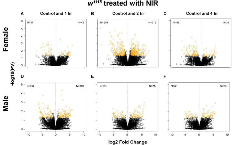
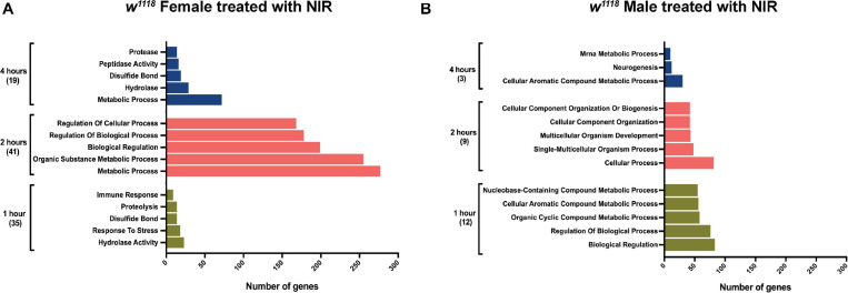
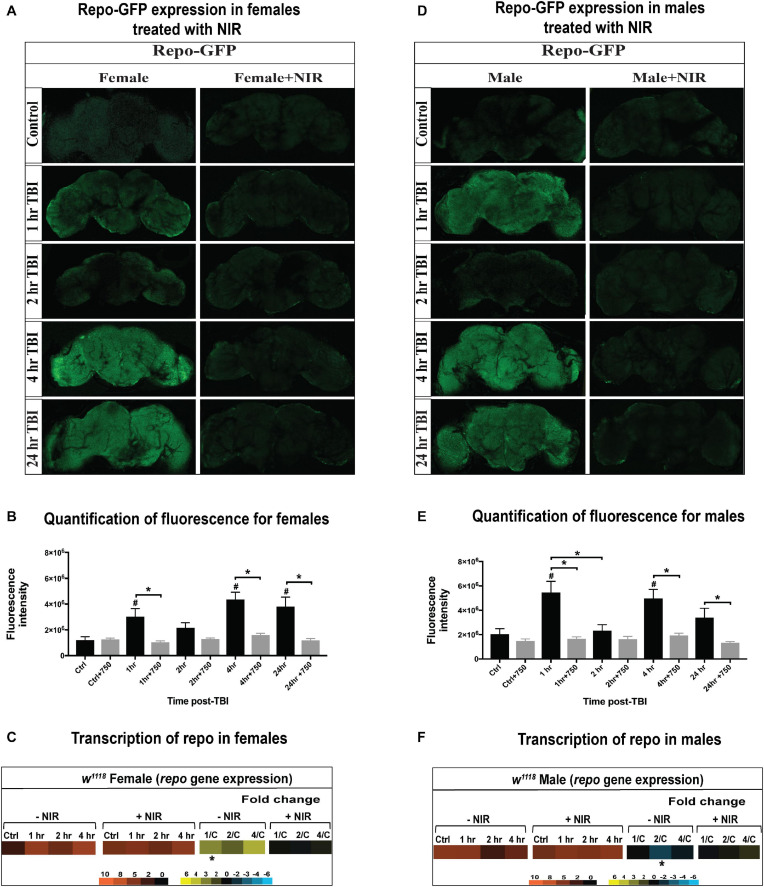
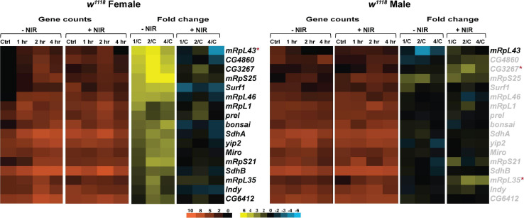
Traumatic brain injuries (TBIs) caused by a sudden impact to the head alter behavior and impair physical and cognitive function. Besides the severity, type and area of the brain affected, the outcome of TBI is also influenced by the patient's biological sex. Previous studies reporting mitochondrial dysfunction mainly focused on exponential reactive oxygen species (ROS) generation, increased mitochondrial membrane potential, and altered mitochondrial dynamics as a key player in the outcome to brain injury. In this study, we evaluated the effect of a near-infrared (NIR) light exposure on gene expression in a Drosophila TBI model. NIR interacts with cytochrome c oxidase (COX) of the electron transport chain to reduce mitochondrial membrane potential hyperpolarization, attenuate ROS generation, and apoptosis. We subjected w 1118 male and female flies to TBI using a high-impact trauma (HIT) device and subsequently exposed the isolated fly brains to a COX-inhibitory wavelength of 750 nm for 2 hours (hr). Genome-wide 3'-mRNA-sequencing of fly brains revealed that injured w 1118 females exhibit greater changes in transcription compared to males at 1, 2, and 4 hours (hr) after TBI. Inhibiting COX by exposure to NIR downregulates gene expression in injured females but has minimal effect in injured males. Our results suggest that mitochondrial COX modulation with NIR alters gene expression in Drosophila following TBI and the response to injury and NIR exposure varies by biological sex.

Keywords: gene expression; mitochondria; near-infrared light; sex-differences; traumatic brain injury.


Conflict of interest statement

MH and TS are co-founders of Mitovation Inc., that develops infrared light therapy for ischemia/reperfusion injury applications. The remaining authors declare that the research was conducted in the absence of any commercial or financial relationships that could be construed as a potential conflict of interest.

Figures

FIGURE 1
FIGURE 1
Plot depicting transcriptional changes after NIR exposure in w1118 injured male and female flies. Volcano plots depicting log2 fold change and –log10(PV:p-value) of differentially expressed genes at 1, 2, and 4 hr after injury and subsequent NIR exposure compared to control for females (A–C) and males (D–F). The number of significantly upregulated and downregulated gene changes are indicated in each plot (| log2FC| > 1; p-value < 0.05). Injured w1118 females show more transcriptional changes in response to NIR exposure than males.
FIGURE 2
FIGURE 2
Gene ontology (GO) enrichment in w1118 injured flies exposed to NIR. Significant genes (| log2FC| > 1; p-value < 0.05) identified from sequencing were classified for their biological functions using RDAVID. The plot shows top five biological processes enriched in injured females (A) and males (B) after NIR exposure at each time-point. The total number of GO terms differentially regulated at each time-point is indicated in parenthesis.
FIGURE 3
FIGURE 3
Immune response gene expression is downregulated in injured w1118 flies with post-NIR exposure. Heatmaps depicting immune response gene expression changes in w1118 females and males at control, 1, 2, and 4 hr after injury and NIR exposure. The orange scale represents average normalized counts for three replicates in each of the indicated groups. Yellow-blue scale shows fold change for each gene at 1-, 2-, and 4-hr post-injury compared to control. The genes indicated in black font are significantly changed in injured flies not exposed to NIR whereas red asterisk (*) indicates significant alteration in injured flies exposed to NIR (| log2FC| > 1, p-value < 0.05). 1/C: Fold change at 1 hr compared to control; 2/C: Fold change at 2 hr compared to control and 4/C: Fold change at 4 hr compared to control. (–NIR: not exposed to NIR and +NIR: exposed to NIR).
FIGURE 4
FIGURE 4
GFP-tagged repo expression in w1118 male and female flies. Confocal images showing change in GFP expression in Repo-GFP flies at control, 1, 2, 4, and 24 hr after TBI in females (A) and males (D) with and without exposure to NIR. About 10–15 brains were imaged for each condition and representative images from each group are shown here. Average fluorescence intensity for each time-point (10–15 brains) was assessed using ImageJ marking whole brain area as region of interest (B,E). A significant increase (One-way ANOVA, Dunnett test, p-value < 0.05) in GFP expression was observed for both sexes at 1, 4, and 24 hr after injury. GFP expression was unchanged in both sexes inflicted with TBI after NIR exposure. Repo transcription was significantly upregulated (| log2FC| > 1, p-value < 0.05) in females 1 hr after injury (C) and downregulated in males (F) 2 hr after injury. NIR exposure had no change in repo transcription in both sexes.
FIGURE 5
FIGURE 5
NIR exposure downregulates mitochondrial gene transcription in w1118 flies subjected to TBI. Heatmaps depicting mitochondrial gene expression changes in w1118 females and males at control, 1, 2, and 4 hr after injury and NIR exposure. The orange scale represents average normalized counts for three replicates in each of the indicated groups. Yellow-blue scale shows fold change for each gene at 1-, 2-, and 4-hr post-injury compared to control. The genes indicated in black font are significantly changed in injured flies not exposed to NIR whereas red asterisk (*) indicates significant alteration in injured flies exposed to NIR (| log2FC| > 1, p-value < 0.05). 1/C: Fold change at 1 hr compared to control; 2/C: Fold change at 2 hr compared to control and 4/C: Fold change at 4 hr compared to control. (–NIR: not exposed to NIR and +NIR: exposed to NIR).
FIGURE 6
FIGURE 6
Cytoskeletal gene transcription is downregulated after NIR exposure. Heatmaps depicting cytoskeletal gene expression changes in w1118 females and males at control, 1, 2, and 4 hr after injury and NIR exposure. The orange scale represents average normalized counts for three replicates in each of the indicated groups. Yellow-blue scale shows fold change for each gene at 1-, 2-, and 4-hr post-injury compared to control. The genes indicated in black font are significantly changed in injured flies not exposed to NIR whereas red asterisk (*) indicates significant alteration in injured flies exposed to NIR (| log2FC| > 1, p-value < 0.05). 1/C: Fold change at 1 hr compared to control; 2/C: Fold change at 2 hr compared to control and 4/C: Fold change at 4 hr compared to control. (–NIR: not exposed to NIR and +NIR: exposed to NIR).

Similar articles

Cited by

References

    1. Allen N. J., Barres B. A. (2009). Neuroscience: glia - more than just brain glue. Nature 457 675–677. 10.1038/457675a - DOI - PubMed
    1. Anders S., Pyl P. T., Huber W. (2015). HTSeq–a Python framework to work with high-throughput sequencing data. Bioinformatics 31 166–169. 10.1093/bioinformatics/btu638 - DOI - PMC - PubMed
    1. Anesti V., Scorrano L. (2006). The relationship between mitochondrial shape and function and the cytoskeleton. Biochim. Biophys. Acta 1757 692–699. 10.1016/j.bbabio.2006.04.013 - DOI - PubMed
    1. Bartolak-Suki E., Imsirovic J., Nishibori Y., Krishnan R., Suki B. (2017). Regulation of mitochondrial structure and dynamics by the cytoskeleton and mechanical factors. Int. J. Mol. Sci. 18:1812. 10.3390/ijms18081812 - DOI - PMC - PubMed
    1. Begum R., Calaza K., Kam J. H., Salt T. E., Hogg C., Jeffery G. (2015). Near-infrared light increases ATP, extends lifespan and improves mobility in aged Drosophila melanogaster. Biol. Lett. 11:20150073. 10.1098/rsbl.2015.0073 - DOI - PMC - PubMed