Review
. 2021 Mar 15:12:591535.
doi: 10.3389/fmicb.2021.591535. eCollection 2021.

Emergence of Bat-Related Betacoronaviruses: Hazard and Risks

Affiliations
Review

Emergence of Bat-Related Betacoronaviruses: Hazard and Risks

Roger Frutos et al. Front Microbiol. .

Abstract

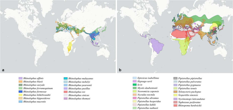
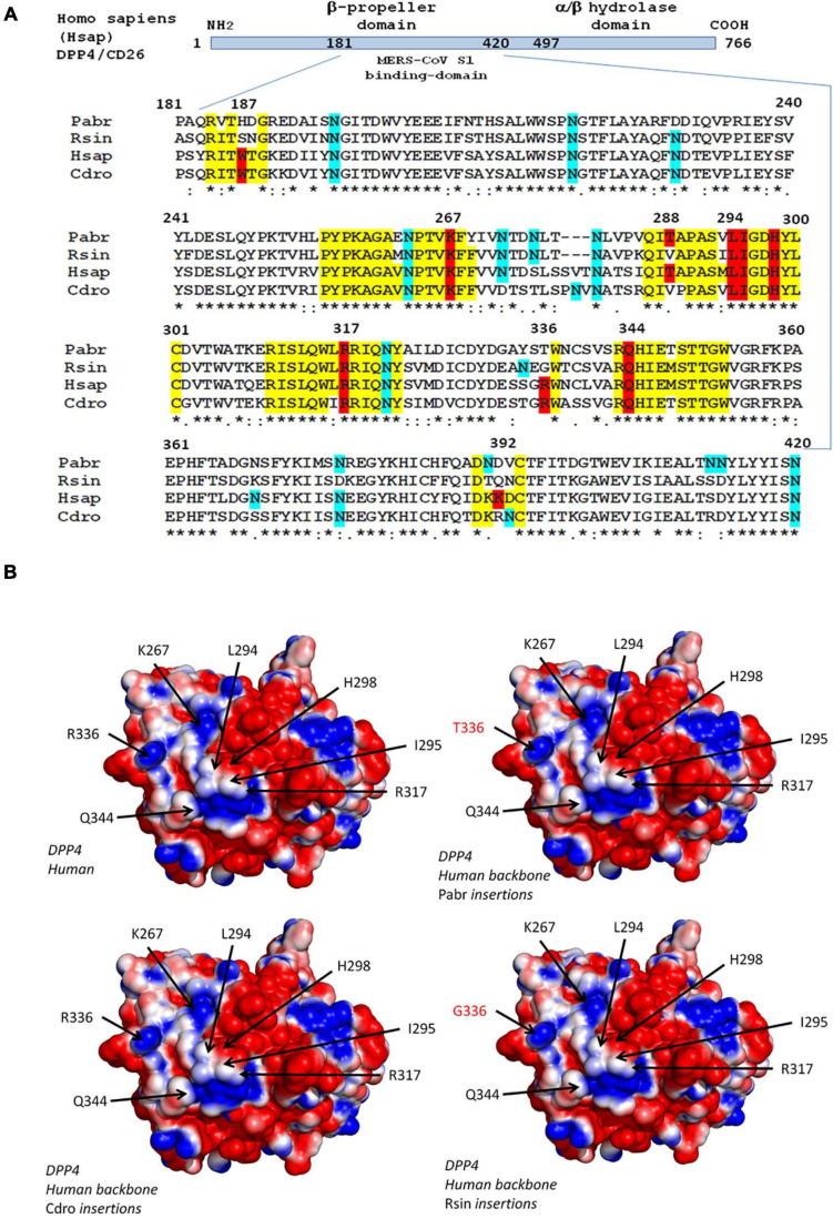
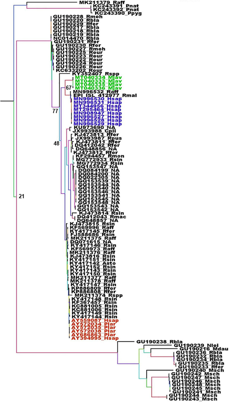
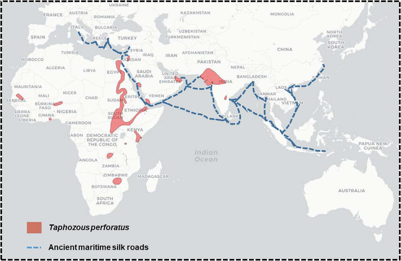
The current Coronavirus Disease 2019 (COVID-19) pandemic, with more than 111 million reported cases and 2,500,000 deaths worldwide (mortality rate currently estimated at 2.2%), is a stark reminder that coronaviruses (CoV)-induced diseases remain a major threat to humanity. COVID-19 is only the latest case of betacoronavirus (β-CoV) epidemics/pandemics. In the last 20 years, two deadly CoV epidemics, Severe Acute Respiratory Syndrome (SARS; fatality rate 9.6%) and Middle East Respiratory Syndrome (MERS; fatality rate 34.7%), plus the emergence of HCoV-HKU1 which causes the winter common cold (fatality rate 0.5%), were already a source of public health concern. Betacoronaviruses can also be a threat for livestock, as evidenced by the Swine Acute Diarrhea Syndrome (SADS) epizootic in pigs. These repeated outbreaks of β-CoV-induced diseases raise the question of the dynamic of propagation of this group of viruses in wildlife and human ecosystems. SARS-CoV, SARS-CoV-2, and HCoV-HKU1 emerged in Asia, strongly suggesting the existence of a regional hot spot for emergence. However, there might be other regional hot spots, as seen with MERS-CoV, which emerged in the Arabian Peninsula. β-CoVs responsible for human respiratory infections are closely related to bat-borne viruses. Bats are present worldwide and their level of infection with CoVs is very high on all continents. However, there is as yet no evidence of direct bat-to-human coronavirus infection. Transmission of β-CoV to humans is considered to occur accidentally through contact with susceptible intermediate animal species. This zoonotic emergence is a complex process involving not only bats, wildlife and natural ecosystems, but also many anthropogenic and societal aspects. Here, we try to understand why only few hot spots of β-CoV emergence have been identified despite worldwide bats and bat-borne β-CoV distribution. In this work, we analyze and compare the natural and anthropogenic environments associated with the emergence of β-CoV and outline conserved features likely to create favorable conditions for a new epidemic. We suggest monitoring South and East Africa as well as South America as these regions bring together many of the conditions that could make them future hot spots.

Keywords: COVID-19; MERS; SARS; coronavirus; hazard and risks assessment.


Conflict of interest statement

The authors declare that the research was conducted in the absence of any commercial or financial relationships that could be construed as a potential conflict of interest.

Figures

FIGURE 1
FIGURE 1
Distribution of bat species according to the group of coronavirus. (a) Distribution of bat species displaying an ACE2 receptor and associated with Sarbecoviruses. (b) Distribution of bat species displaying a DPP4 receptor and associated with Merbecoviruses.
FIGURE 2
FIGURE 2
Polymorphism of the DPP4 receptor. (A) DPP4 multiple sequence alignments. Schematic representation of the DPP4 protein organization (upper panel). The MERS-CoV S1 spike binding site is located in a region of DPP4 that extends from amino acid position 181 to amino acid 420. A clustal omega multiple sequence alignment (EMBL-EBI bioinformatic tool; Copyright EMBL 2020), spanning the 181-420 region of DPP4 from Pipistrella abramus (Pabr), Rhinolophus sinicus (Rsin), Homo sapiens (Hsap), and Camelus dromedarius (Cdro) is shown (lower panel). All DPP4 sequences were downloaded from Genbank (NCBI): Homo sapiens (GenBank: AAH13329.2); Camelus dromedarius (GenBank: AIG55259.1); Rhinolophus sinicus (GenBank: AZO92863.1); Pipistrellus abramus (GenBank: AZO922861.1). Within the amino acid sequences of DPP4 important for MERS-CoV spike binding, the conserved amino acids are highlighted in yellow, those critical for MERS-CoV-DPP4 binding are highlighted in red and the potential N-glycosylation sites are highlighted in blue. (B) 3-D analysis and electrostatic potential surface potential of the DPP4 receptor. The 3-D structure of DPP4 was retrieved according to the published data (PDB : 6M1D and 4L72). Critical amino acid sequences required to allow coronaviruses spike binding to human DPP4 were substituted by the corresponding sequence from Rhinolophus sinicus, Pipistrella abramus and Camelus dromedarius into a human DPP4 backbone sequence to determine whether or not these substitutions may change the 3-D structure of the receptors. Protein modeling for these chimeric sequences was performed using the Phyre2 server (Kelley et al., 2015). The PyMOL 1.8.0 software (https://sourceforge. net/projects/pymol/files/pymol/1.8/) and the Adaptive Poisson-Boltzmann Solver (APBS) tools plugin (https://pymolwiki.org/index.php/APBS) was used to generate electrostatic potential surfaces of the human receptors and their modified chimeric versions. The red color indicates an excess of negative charges near the surface and the blue color arises from a positively charged surface, while white is neutral.
FIGURE 3
FIGURE 3
Phylogenetic analysis of Sarbecoviruses RdRp genes. The alignment of the full RdRp genes was performed with MUSCLE from the SeaView package (Gouy et al., 2010). The tree was built using the maximum likelihood method under the GTR model with 500 repeats. The tree was rooted using the RdRp sequence of a MERS-CoV from Camelus dromedarius (KT368883) as outgroup. Violet: RdRp sequences from human SARS-CoV-2. Green: RdRp sequences from pangolins’ Sarbecoviruses. Red: RdRp sequences from SARS-CoV. Sample names are built with the GenBank accession number followed by a four-letter code identifying the species. The species codes are as follows: Asto, Aselliscus stoliczkanus; Cpli, Chaerephon plicata; Hsap, Homo sapiens; Mdau, Myotis daubentonii; Mjav, Manis javanica; Msch, Miniopterus schreibersii; Nlei, Nyctalus leisleri; Plar, Paguma larvata; Pnat, Pipistrellus nathusii; Ppyg, Pipistrellus pygmaeus; Raff, Rhinolophus affinis; Rbla, Rhinolophus blasii; Reur, Rhinolophus euryale; Rfer, Rhinolophus ferrumequinum; Rmac, Rhinophilus maculatus; Rmal, Rhinolophus malayanus; Rmeh, Rhinolophus mehelyi; Rmon, Rhinophilus monoceros; Rpus, Rhinolophus pusillus; Rsin, Rhinolophus sinensis; Rspp, Rhinolophus unidentified species; NA, Not available.
FIGURE 4
FIGURE 4
Phylogenetic analysis of Merbecoviruses RdRp genes. The alignment of the full RdRp genes was performed with MUSCLE from the SeaView package (Gouy et al., 2010). The tree was built using the maximum likelihood method under the GTR model with 500 repeats. The tree was rooted using the RdRp sequence of a Sarbecovirus from Manis javanica (MT040333) as outgroup. Deep blue: RdRp sequences from human MERS-CoV. Red: RdRp sequences from dromedaries MERS-CoV. Sample names are built with the GenBank accession number followed by a four-letter code identifying the species. The species codes are as follows: Cdro, Camelus dromedarius; Eeur, Erinaceus europaeus; Hpul, Hypsugo pulveratus; Hsap, Homo sapiens; Hsav, Hypsugo savii; Ncap, Neoromicia capensis; Pabr, Pipistrellus abramus; Pkuh, Pipistrellus kuhlii; Pspp, Pipistrellus unidentified species; Tpac, Tylonycteris pachypus; Tper, Taphozous perforatus; Tspp, Tylonycteris unidentified species; Vsup, Vespertilio superans; NA, Not available.
FIGURE 5
FIGURE 5
Comparison of the distribution of Taphozous perforatus and the ancient silk roads.

References

    1. Afelt A., Frutos R., Devaux C. A. (2018). Bats, coronaviruses, and deforestation: toward 631 the emergence of novel infectious diseases? Front. Microbiol. 9:702. 10.3389/fmicb.2018.00702 - DOI - PMC - PubMed
    1. Afelt A., Lacroix A., Zawadzka-Pawlewska U., Pokojski W., Buchy P., Frutos R. (2017). Distribution of bat-borne viruses and environment patterns. Infect. Genetics Evolut. 58 181–191. 10.1016/j.meegid.2017.12.009 - DOI - PMC - PubMed
    1. Alagaili A. N., Briese T., Mishra N., Kapoor V., Sameroff S. C., de Wit E., et al. (2014). Middle East respiratory syndrome coronavirus infection in dromedary camels in Saudi Arabia. mBio 5:e00884-14. 10.1128/mBio.01002-14 - DOI - PMC - PubMed
    1. Andersen K. G., Rambaut A., Lipkin W. I., Holmes E. C., Garry R. F. (2020). The proximal origin of SARS-CoV-2. Nat. Med. 26 450–452. 10.1038/s41591-020-0820-9 - DOI - PMC - PubMed
    1. Annan A., Baldwin H. J., Corman V. M., Klose S. M., Owusu M., Nkrumah E. E., et al. (2013). Human Betacoronavirus 2c EMC/2012–related viruses in bats. Ghana and Europe. Emerg. Infect. Dis. 19 456–456. 10.3201/eid1903.121503 - DOI - PMC - PubMed