

Invasive Pneumococcal Disease in a Patient With COVID-19: A Case Report
- PMID: 33791177
- PMCID: PMC8004547
- DOI: 10.7759/cureus.13559
Invasive Pneumococcal Disease in a Patient With COVID-19: A Case Report
Abstract
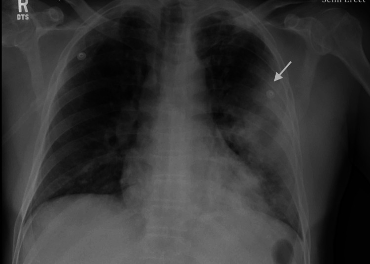
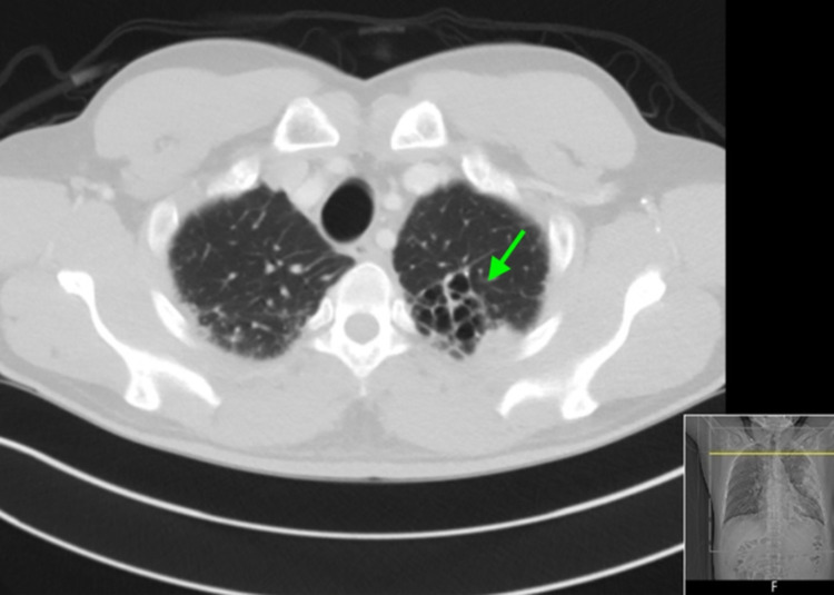
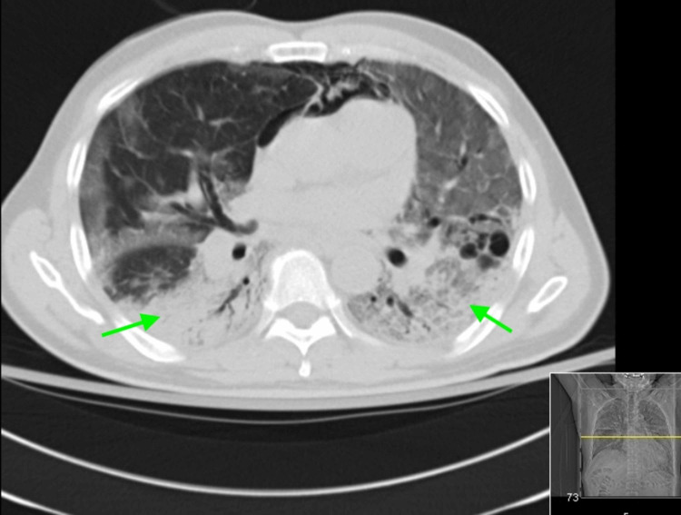
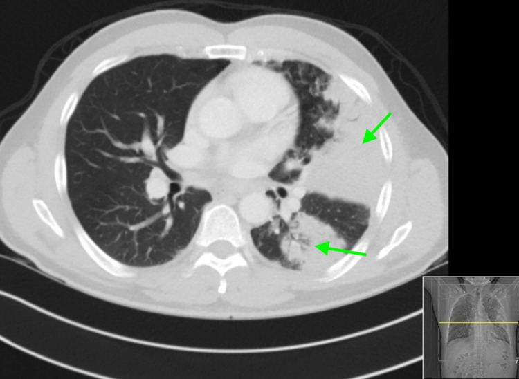
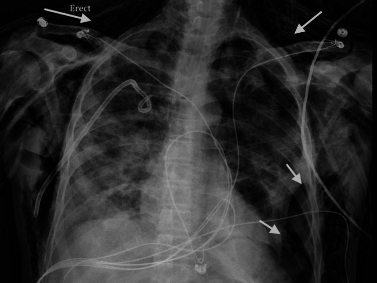
The spread of the new severe acute respiratory syndrome coronavirus 2 (SARS-CoV-2), which causes coronavirus disease 2019 (COVID-19), has resulted in a global health pandemic and caused profound morbidity and mortality worldwide. The virus is known to cause severe hypoxemic respiratory failure and has been associated with extrapulmonary manifestations and end-organ dysfunction in the setting of extensive inflammatory response. Recently, the association between COVID-19 and pneumococcal pneumonia co-infection or superinfections has gained increasing interest. In this report, we present the case of a 58-year-old man with a past medical history significant for pulmonary tuberculosis, diagnosed over two decades ago, who presented with pleuritic chest pain, myalgia, intermittent fevers, chills, and productive cough and was found to have invasive pneumococcal disease and COVID-19. To our knowledge, this is the first reported case of invasive pneumococcal infection in a patient with COVID-19.
Keywords: covid-19; invasive pneumococcal disease; sars cov 2; streptococcus pneumoniae.
Copyright © 2021, Ayad et al.
Conflict of interest statement
The authors have declared that no competing interests exist.
Figures
References
-
- WHO coronavirus disease (COVID-19) dashboard. [Feb;2021 ];https://covid19.who.int/ WHO. 2021
Publication types
LinkOut - more resources
Full Text Sources
Other Literature Sources
Miscellaneous