. 2021 Mar 15:9:643522.
doi: 10.3389/fcell.2021.643522. eCollection 2021.

LRH1 Acts as an Oncogenic Driver in Human Osteosarcoma and Pan-Cancer

Affiliations

LRH1 Acts as an Oncogenic Driver in Human Osteosarcoma and Pan-Cancer

Yang Song et al. Front Cell Dev Biol. .

Abstract

Osteosarcoma (OS) that mainly occurs during childhood and adolescence is a devastating disease with poor prognosis presented by extreme metastases. Recent studies have revealed that liver receptor homolog 1 (LRH-1) plays a vital role in the metastasis of several human cancers, but its role is unknown in the metastasis of OS. In this study, Gene Ontology (GO) enrichment analyses based on high-throughput RNA-seq data revealed that LRH-1 acted a pivotal part in the positive regulation of cell migration, motility, and angiogenesis. Consistently, LRH-1 knockdown inhibited the migration of human OS cells, which was concurrent with the downregulation of mesenchymal markers and the upregulation of epithelial markers. In addition, short hairpin RNAs (shRNAs) targeting LRH-1 inactivated transforming growth factor beta (TGF-β) signaling pathway. LRH-1 knockdown inhibited human umbilical vein endothelial cell (HUVEC) proliferation, migration, and tube formation. Vascular endothelial growth factor A (VEGFA) expression was also downregulated after LRH-1 knockdown. Immunohistochemistry (IHC) revealed that the expression of LRH-1 protein was significantly higher in tumor tissues than in normal bone tissues. We found that high LRH-1 expression was associated with poor differentiation and advanced TNM stage in OS patients using IHC. Based on The Cancer Genome Atlas (TCGA) database, high LRH-1 expression predicts poor survival in lung squamous cell carcinoma (LUSC), kidney renal papillary cell carcinoma (KIRP), and pancreatic adenocarcinoma (PAAD). The downregulation of LRH-1 significantly hindered the migration and motility of LUSC cells. Using multi-omic bioinformatics, the positive correlation between LRH-1- and EMT-related genes was found across these three cancer types. GO analysis indicated that LRH-1 played a vital role in "blood vessel morphogenesis" or "vasculogenesis" in KIRP. Our results indicated that LRH-1 plays a tumor-promoting role in human OS, could predict the early metastatic potential, and may serve as a potential target for cancer therapy.

Keywords: LRH1; angiogenesis; epithelial-mesenchymal transition; metastasis; osteosarcoma; pan-cancer.


Conflict of interest statement

The authors declare that the research was conducted in the absence of any commercial or financial relationships that could be construed as a potential conflict of interest.

Figures

FIGURE 1
FIGURE 1
Elevated LRH1 expression correlates with advanced TNM stage of osteosarcoma (OS) patients. (A) Representative immunohistochemistry (IHC) images showing the expression of LRH1 in OS and nontumoural bone tissues (non-tumour). Original magnification, ×400; Scale bar = 25 μm. A histogram shows the percentage of LRH1 expression in tumour and nontumour tissues. (B) Representative IHC staining images are shown of high expression (left) and low expression (right) of LRH1 in OS tissues. Original magnification, ×100; Scale bar = 100 μm. (C,D) The percentages of patients with different TNM stages (C) and different differentiation (D) were assigned according to the expression level of LRH1 in OS. *P < 0.05, **P < 0.01.
FIGURE 2
FIGURE 2
LRH1 is a metastatic-promoting regulator in OS cells. (A) RT-PCR and (B) western blot analysis results are shown of LRH1 in 143B and SJSA-1 cells untreated (UT) or transfected with control shRNA (Ctrl) or two shRNAs (LRH1-sh1 and LRH1-sh2) against LRH1. Mean ± SD, n = 3, ***P < 0.001. (C) A volcano plot indicates upregulated (red dots) and downregulated (gray dots) genes in the shLRH1 group compared with the control group. Black dotted lines represent a cut-off range of 1.0-fold and P < 0.05. (D) The top 20 enriched pathways identified by GO (BP) terms based on differentially expressed genes are shown.
FIGURE 3
FIGURE 3
LRH1 depletion suppresses the motility and migration of OS cells. (A) Wound healing assays indicate the motility and migration of OS cells in shLRH1 group and control group. (B) Transwell assays revealed the migration of Ctrl and shLRH1 groups. Representative images from 3 experiments are shown in (A) and (B). Migrated cell numbers are quantified in (A) and (B), respectively (mean ± SEM, n = 3). *P < 0.05 and **P < 0.01 versus Ctrl, by 2-way ANOVA with Tukey’s t test.
FIGURE 4
FIGURE 4
Effect of LRH1 loss on EMT and the TGFβ signalling pathway. (A) Heatmaps of GO (BP) categories show the differential expression of epithelial to mesenchymal transition pathway gene signatures in control and shLRH1 OS cells. Red and green indicate high and low mRNA expression levels, respectively. (B) The GSEA results show a correlation between LRH1 levels and the gene set “ONDER CDH1 TARGETS 3 DN.” (C) The top 10 enriched pathways identified by KEGG (Kyoto Encyclopedia of Genes and Genomes) pathway analysis based on differentially expressed genes. Rich factor = number of enriched genes/number of background genes in the pathway. (D) western blot analysis of E-cadherin, N-cadherin and TGFβ signalling pathway was conducted in Ctrl OS cells and shLRH1 OS cells. β-actin served as a loading control.
FIGURE 5
FIGURE 5
Decreased LRH1 inhibits HUVEC proliferation and tube formation. (A,B) HUVECs were treated with CM from OS cells transfected with shLRH1 or Ctrl. (A) A CCK-8 kit was used to evaluate the proliferation ability of HUVECs. (B) Tube formation tests were performed. Tube length and branch points were analyzed to evaluate angiogenic activity. (C) The expression of VEGFA in OS cells transfected with shLRH1 or Ctrl was determined by ELISA. The values are represented as the means of three independent experiments. *P < 0.05 and **P < 0.01 versus Ctrl.
FIGURE 6
FIGURE 6
A pan-cancer analysis of the oncogenic role of LRH1. (A) Kaplan–Meier curves show the overall survival for KIRP, LUSC and PAAD patients with high or low LRH1 expression from the TCGA database. (B) A volcano plot shows genes (red dots) that positively correlate with LRH1 and genes (green dots) that positively correlate with LRH1 in KIRP. (C) Heat maps show genes positively and negatively correlated with LRH1 mRNA expression in KIRP (top 50). (D) A correlation map displays Pearson correlation values for each pair of genes in three types of cancer. The bar on the left of the map indicates the color legend of the Pearson correlationvalues calculated for each couple of genes. (E) An interactive network of the top 20 enriched terms is colored by cluster ID in KIRP and LUSC. Each color represents one enrichment pathway.
FIGURE 7
FIGURE 7
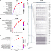
KEGG pathway analysis of related genes and DNA methylation of the LRH1 promoter. (A–C) KEGG pathway enrichment analysis of the DEGs in KIRP (A), LUSC (B) and PAAD (C) was performed. (D) The correlation of LRH1 expression and DNA methylation of theLRH1 promoter region from MEXPRESS, including 223 samples in pancreatic adenocarcinoma from TCGA datasets is shown. The statistics [correlation coefficient (r) and P-value] on the right show the relationship between expression and DNA methylation of the promoter. *P < 0.05, **P < 0.01, and ***P < 0.001.

Similar articles

Cited by

References

    1. Bianco S., Brunelle M., Jangal M., Magnani L., Gévry N. (2014). LRH-1 governs vital transcriptional programs in endocrine-sensitive and -resistant breast cancer cells. Cancer Res. 74 2015–2025. 10.1158/0008-5472.Can-13-2351 - DOI - PubMed
    1. Bianco S., Jangal M., Garneau D., Gévry N. (2015). LRH-1 controls proliferation in breast tumor cells by regulating CDKN1A gene expression. Oncogene 34 4509–4518. 10.1038/onc.2014.382 - DOI - PubMed
    1. Botter S. M., Neri D., Fuchs B. (2014). Recent advances in osteosarcoma. Curr. Opin. Pharmacol. 16 15–23. 10.1016/j.coph.2014.02.002 - DOI - PubMed
    1. Cai H., Liu X., Zheng J., Xue Y., Ma J., Li Z., et al. (2017). Long non-coding RNA taurine upregulated 1 enhances tumor-induced angiogenesis through inhibiting microRNA-299 in human glioblastoma. Oncogene 36 318–331. 10.1038/onc.2016.212 - DOI - PubMed
    1. Cheng D. D., Li S. J., Zhu B., Zhou S. M., Yang Q. C. (2018). EEF1D overexpression promotes osteosarcoma cell proliferation by facilitating Akt-mTOR and Akt-bad signaling. J. Exp. Clin. Cancer Res. 37 50. 10.1186/s13046-018-0715-5 - DOI - PMC - PubMed

LinkOut - more resources