. 2021 Mar 15:8:639541.
doi: 10.3389/fcvm.2021.639541. eCollection 2021.

Donepezil Ameliorates Pulmonary Arterial Hypertension by Inhibiting M2-Macrophage Activation

Affiliations

Donepezil Ameliorates Pulmonary Arterial Hypertension by Inhibiting M2-Macrophage Activation

Haihua Qiu et al. Front Cardiovasc Med. .

Abstract

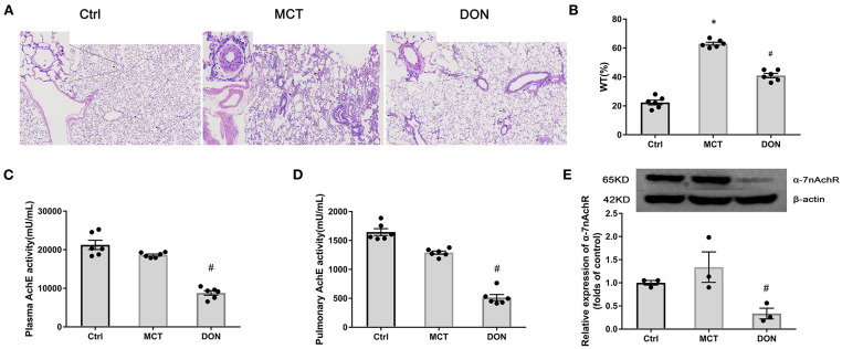
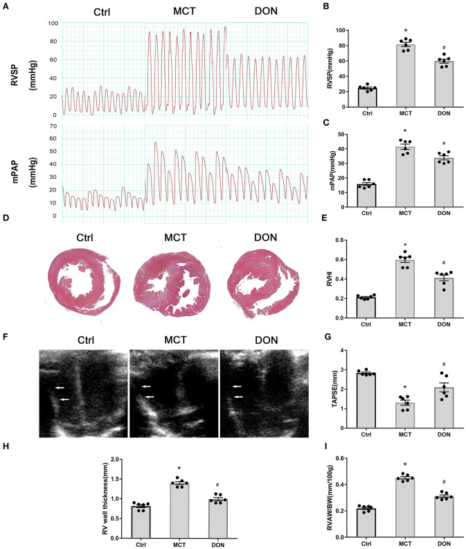
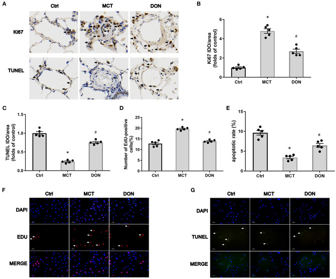
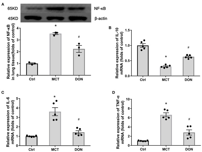
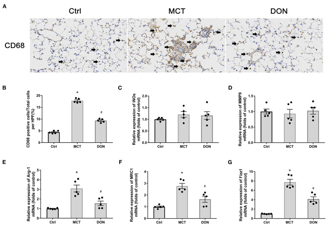
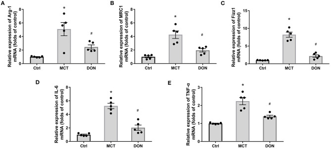
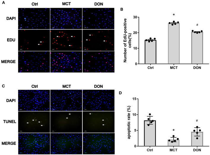
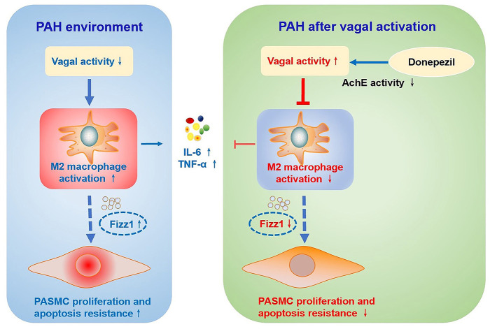
Background: The beneficial effects of parasympathetic stimulation in pulmonary arterial hypertension (PAH) have been reported. However, the specific mechanism has not been completely clarified. Donepezil, an oral cholinesterase inhibitor, enhances parasympathetic activity by inhibiting acetylcholinesterase, whose therapeutic effects in PAH and its mechanism deserve to be investigated. Methods: The PAH model was established by a single intraperitoneal injection of monocrotaline (MCT, 50 mg/kg) in adult male Sprague-Dawley rats. Donepezil was administered via intraperitoneal injection daily after 1 week of MCT administration. At the end of the study, PAH status was confirmed by echocardiography and hemodynamic measurement. Testing for acetylcholinesterase activity and cholinergic receptor expression was used to evaluate parasympathetic activity. Indicators of pulmonary arterial remodeling and right ventricular (RV) dysfunction were assayed. The proliferative and apoptotic ability of pulmonary arterial smooth muscle cells (PASMCs), inflammatory reaction, macrophage infiltration in the lung, and activation of bone marrow-derived macrophages (BMDMs) were also tested. PASMCs from the MCT-treated rats were co-cultured with the supernatant of BMDMs treated with donepezil, and then, the proliferation and apoptosis of PASMCs were evaluated. Results: Donepezil treatment effectively enhanced parasympathetic activity. Furthermore, it markedly reduced mean pulmonary arterial pressure and RV systolic pressure in the MCT-treated rats, as well as reversed pulmonary arterial remodeling and RV dysfunction. Donepezil also reduced the proliferation and promoted the apoptosis of PASMCs in the MCT-treated rats. In addition, it suppressed the inflammatory response and macrophage activation in both lung tissue and BMDMs in the model rats. More importantly, donepezil reduced the proliferation and promoted the apoptosis of PASMCs by suppressing M2-macrophage activation. Conclusion: Donepezil could prevent pulmonary vascular and RV remodeling, thereby reversing PAH progression. Moreover, enhancement of the parasympathetic activity could reduce the proliferation and promote the apoptosis of PASMCs in PAH by suppressing M2-macrophage activation.

Keywords: M2-macrophage; cholinesterase inhibitor; donepezil; pulmonary arterial hypertension; pulmonary arterial smooth muscle cells.


Conflict of interest statement

The authors declare that the research was conducted in the absence of any commercial or financial relationships that could be construed as a potential conflict of interest.

Figures

Figure 1
Figure 1
DON enhances parasympathetic activity and ameliorates pulmonary arterial remodeling. Lung tissue of MCT-induced rats after DON treatment were fixed and stained with HE at the end of the study. The pathological sections of lung tissue were scanned and analyzed by DSAssistantLite software (Motic, China; ×100 magnification, image size: 2,860 × 1,631 μm). Pulmonary arteriole from 5 random fields were analyzed. AchE from plasma and lung tissue were tested by colorimetry. Relative protein expression of α-7nAchR from lung tissue was assayed by western blot. (A) Representative images of HE-stained lung sections under light microscope in each group, the black arrow points to pulmonary vessels, and the enlarged vessel is presented in the upper left corner. (B) Statistical analysis of pulmonary arterial WT%. Pulmonary vessels with external diameters smaller than 200 μm were selected in each field (n = 6). (C) Statistical analysis of AchE activity in plasma (n = 6). (D) Statistical analysis of AchE activity in lung tissue (n = 6). (E) Relative protein expression of α-7nAchR in lung tissue (n = 3). AchE, acetylcholinesterase; α-7nAchR, nicotinic acetylcholine receptor alpha 7; Ctrl, control; DON, donepezil; HE, hematoxylin and eosin; MCT, monocrotaline; WT, wall thickness. *P < 0.05, vs. Ctrl group, #P < 0.05, vs. MCT group.
Figure 2
Figure 2
DON improves hemodynamics and RV dysfunction in MCT-induced PAH rats. At the end of the study, RVSP and mPAP were measured with closed-chest technique to show the hemodynamic changes of MCT-induced rats after DON treated. The heart of rats was fixed and stained with HE to show the RV expanding and remodeling. RVHI indicates the ratio of RV weight to LV + S weight. Transthoracic echocardiography at the apical 4-chamber view to show the indicators of RV remodeling and dysfunction. (A) RVSP and mPAP measurement in experimental rats. (B) Statistical analysis of RVSP. (C) Statistical analysis of mPAP. (D) Representative HE staining of heart samples. (E) Quantitative analysis of RVHI. (F) Transthoracic echocardiography at the apical 4-chamber view. (G) Statistical analysis of TAPSE. (H) Statistical analysis of RV wall thickness. (I) Statistical analysis of the ratio of RVAW to BW. BW, body weight; Ctrl, control; DON, donepezil, HE, hematoxylin and eosin; LV, left ventricle; MCT, monocrotaline; mPAP, mean pulmonary arterial pressure; RVSP, right ventricular systolic pressure; RV, right ventricle; RVHI, RV hypertrophy index; RVAW, right ventricular anterior wall; S, septum; TAPSE, tricuspid annular plane systolic excursion. *P < 0.05, vs. Ctrl group, #P < 0.05, vs. MCT group (n = 6).
Figure 3
Figure 3
DON reverses PASMC proliferation and apoptosis resistance in MCT-induced PAH rats. Immunohistochemical staining for Ki67 and TUNEL in lung tissue to show the effect of DON on PASMC proliferation and apoptosis in MCT-induced rats in vivo (×400 magnification). Relative positive cells of the pulmonary artery were counted. To further test PASMC proliferative ability in vitro, PASMCs in the Ctrl, MCT, DON groups were isolated and cultured. The proliferative and apoptotic abilities of PASMCs were, respectively, assayed by immunofluorescence staining with EdU and TUNEL. The ratio of EdU and TUNEL positive cells to total cell numbers per high power field (×200 magnification) were calculated. (A) Representative immunohistochemical staining for Ki67 and TUNEL of pulmonary artery samples. (B) Quantitative analysis of Ki67 positive rate. (C) Quantitative analysis of TUNEL positive rate. (D) Statistical analysis of the EdU+ cell rate. (E) Statistical analysis of the apoptosis rate of PASMCs. (F) Representative immunofluorescent staining for EdU of PASMCs. (G) Representative immunofluorescent staining for TUNEL of PASMCs. Ctrl, control; DON, donepezil; DAPI, 4',6-diamidino-2-phenylindole; EdU, 5-ethynyl-20-deoxyuridine; MCT, monocrotaline; OD, optical density; PASMCs, pulmonary artery smooth muscle cells; TUNEL, terminal deoxynucleotidyl transferase dUTP nick end labeling. *P < 0.05, vs. Ctrl group, #P < 0.05, vs. MCT group (n = 5).
Figure 4
Figure 4
DON reduced pulmonary inflammation. Relative protein expression of NF-κB in lung tissue in MCT-induced rats treated with DON were tested by western blot. Relative expression of inflammatory mRNA in lung tissue were detected by real time-PCR. (A) Relative protein expression of NF-κB (n = 3). (B) Quantitative analysis of relative mRNA expression of IL-10 (n = 5). (C) Quantitative analysis of relative mRNA expression of IL-6 (n = 5). (D) Quantitative analysis of relative mRNA expression of TNF-α (n = 5). Ctrl, control; DON, donepezil; IL-6, interleukin-6; IL-10, interleukin-10; MCT, monocrotaline; NF-κB, nuclear factor-κB; TNF-α, tumor necrosis factor-α. *P < 0.05, vs. Ctrl group, #P < 0.05, vs. MCT group.
Figure 5
Figure 5
DON reduced macrophage activation in the lung. Immunohistochemical staining for CD68 antibody in lung tissue to show the effect of DON on macrophages infiltration in MCT-induced rats, which were scanned and analyzed by DSAssistantLite software (Motic, China; ×400 magnification, image size: 715 × 408 μm). The positive staining rate are counted with the mean of 5 random fields. The biomarkers of M1- and M2-macrophage in lung tissue were tested by real time-PCR. (A) Representative immunohistochemical staining for CD68. (B) Quantitative analysis of the CD68 positive cell rate. (C) Quantitative analysis of relative mRNA expression of iNOS. (D) Quantitative analysis of relative mRNA expression of MMP9. (E) Quantitative analysis of relative mRNA expression of Arg-1. (F) Quantitative analysis of relative mRNA expression of MRC1. (G) Quantitative analysis of relative mRNA expression of Fizz1. Arg-1, argirase-1; Ctrl, control; DON, donepezil; Fizz1, found in inflammatory zone 1; iNOs, inducible nitric oxide synthase; MCT, monocrotaline; MMP9, matrix metalloproteinase 9; MRC1, mannose receptor C1. *P < 0.05, vs. Ctrl group, #P < 0.05, vs. MCT group (n = 5).
Figure 6
Figure 6
DON reduced M2-macrophage phenotype in BMDMs. BMDMs from the three groups were isolated. After purified and cultured, the biomarkers of M2-macrophage and inflammatory factors of BMDMs were identified by real time-PCR. (A) Quantitative analysis of relative mRNA expression of Arg-1. (B) Quantitative analysis of relative mRNA expression of MRC1. (C) Quantitative analysis of relative mRNA expression of Fizz1. (D) Quantitative analysis of relative mRNA expression of IL-6. (E) Quantitative analysis of relative mRNA expression of TNF-α. Arg-1, argirase-1; BMDMs, bone marrow-derived macrophages; Ctrl, control; DON, donepezil; Fizz1, found in inflammatory zone 1; IL-6, interleukin-6; MCT, monocrotaline; MRC1, mannose receptor-1; TNF-α, tumor necrosis factor-α. *P < 0.05, vs. Ctrl group, #P < 0.05, vs. MCT group (n = 5).
Figure 7
Figure 7
M2-macrophages inhibited PASMC proliferation and promoted its apoptosis. PASMCs in MCT-induced rats were isolated and cultured, which were co-cultured with the supernatant of BMDMs isolated from the Ctrl, MCT and DON group, respectively. After 24 h cultivation, the proliferative and apoptotic abilities of PASMCs were, respectively, assayed by EdU and TUNEL Assay Kit. The ratio of EdU and TUNEL positive cells to total cell numbers per high power field (×200 magnification) were calculated. (A) EdU stain to test proliferation of PASMCs. (B) Statistical analysis of EdU+ cell rate. (C) TUNEL assay to detect apoptosis of PASMCs. (D) Statistical analysis of apoptosis rate. BMDMs, bone marrow-derived macrophages; Ctrl, control; DON, donepezil; DAPI, 4',6-diamidino-2-phenylindole; EdU, 5-ethynyl-20-deoxyuridine; MCT, monocrotaline; PASMCs, pulmonary artery smooth muscle cells; TUNEL, Terminal deoxynucleotidyl transferase dUTP nick end labeling. *P < 0.05, vs. Ctrl group, #P < 0.05, vs. MCT group (n = 5).
Figure 8
Figure 8
Schematic illustration of the effect of DON on ameliorating MCT-induced PAH by regulating M2 macrophage activation. In the MCT-induced PAH rat model, parasympathetic activity is reduced, inflammatory response and M2-macrophages are activated, and the expression of pro-inflammatory factors and pro-proliferation factor Fizz1 is increased, and subsequently increase PASMC proliferation and apoptosis resistance. However, DON enhances parasympathetic nerve activity by reducing AchE activity, and suppresses the inflammatory response and M2-macrophage activation, thereby inhibiting PASMC proliferation and promoting its apoptosis to reverse PAH, which is speculated to be related to decreased expression of Fizz1. AchE, acetylcholinesterase; Fizz1, found in inflammatory zone 1; IL-6, interleukin-6; MCT, monocrotaline; PAH, pulmonary arterial hypertension; PASMCs, pulmonary artery smooth muscle cells; TNF-α, tumor necrosis factor-α.

Similar articles

Cited by

References

    1. Voelkel NF, Tamosiuniene R, Nicolls MR. Challenges and opportunities in treating inflammation associated with pulmonary hypertension. Expert Rev Cardiovasc Ther. (2016) 14:939–51. 10.1080/14779072.2016.1180976 - DOI - PMC - PubMed
    1. Thenappan T, Ormiston ML, Ryan JJ, Archer SL. Pulmonary arterial hypertension: pathogenesis and clinical management. BMJ. (2018) 360:j5492. 10.1136/bmj.j5492 - DOI - PMC - PubMed
    1. Vonk Noordegraaf A, Groeneveldt JA, Bogaard HJ. Pulmonary hypertension. Eur Respir Rev. (2016) 25:4–11. 10.1183/16000617.0096-2015 - DOI - PMC - PubMed
    1. Qiu H, He Y, Ouyang F, Jiang P, Guo S, Guo Y. The role of regulatory T Cells in pulmonary arterial hypertension. J Am Heart Assis. (2019) 8:e014201. 10.1161/JAHA.119.014201 - DOI - PMC - PubMed
    1. Frid MG, Thurman JM, Hansen KC, Maron BA, Stenmark KR. Inflammation, immunity, and vascular remodeling in pulmonary hypertension; evidence for complement involvement? Glob Cardiol Sci Pract. (2020) 2020:e202001.10.21542/gcsp.2020.1 - DOI - PMC - PubMed

LinkOut - more resources