. 2020 Jul 29;2(1):obaa021.
doi: 10.1093/iob/obaa021. eCollection 2020.

Bifunctional Role of the Sternohyoideus Muscle During Suction Feeding in Striped Surfperch, Embiotoca lateralis

Affiliations

Bifunctional Role of the Sternohyoideus Muscle During Suction Feeding in Striped Surfperch, Embiotoca lateralis

J J Lomax et al. Integr Org Biol. .

Abstract

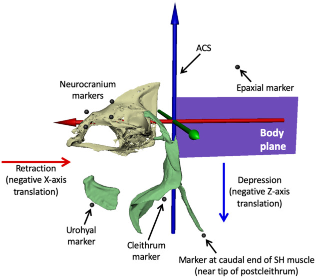
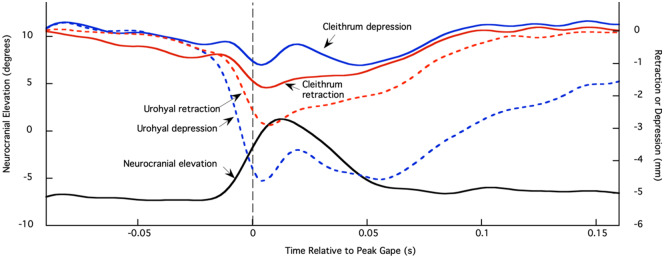
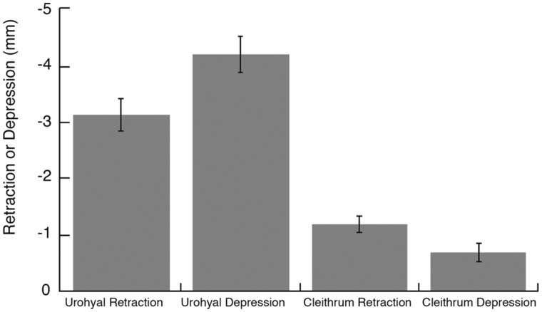
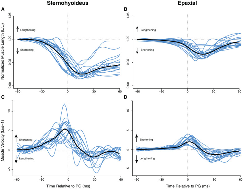
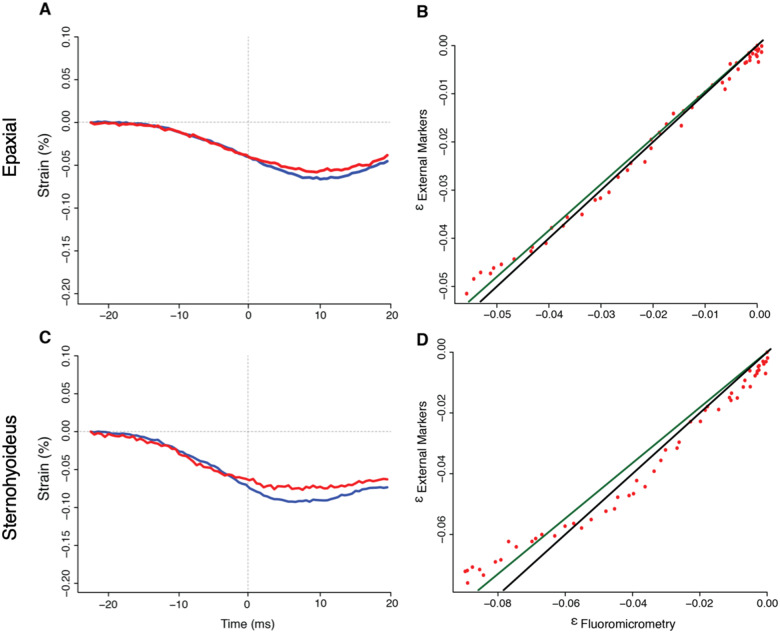
In ray-finned fishes, the sternohyoideus (SH) is among the largest muscles in the head region and, based on its size, can potentially contribute to the overall power required for suction feeding. However, the function of the SH varies interspecifically. In largemouth bass (Micropterus salmoides) and several clariid catfishes, the SH functions similarly to a stiff ligament. In these species, the SH remains isometric and transmitts power from the hypaxial musculature to the hyoid apparatus during suction feeding. Alternatively, the SH can shorten and contribute muscle power during suction feeding, a condition observed in the bluegill sunfish (Lepomis macrochirus) and one clariid catfish. An emerging hypothesis centers on SH muscle size as a predictor of function: in fishes with a large SH, the SH shortens during suction feeding, whereas in fish with a smaller SH, the muscle may remain isometric. Here, we studied striped surfperch (Embiotoca lateralis), a species in which the SH is relatively large at 8.8% of axial muscle mass compared with 4.0% for L. macrochirus and 1.7% for M. salmoides, to determine whether the SH shortens during suction feeding and is, therefore, bifunctional-both transmitting and generating power-or remains isometric and only transmits power. We measured skeletal kinematics of the neurocranium, urohyal, and cleithrum with Video Reconstruction of Moving Morphology, along with muscle strain and shortening velocity in the SH and epaxial muscles, using a new method of 3D external marker tracking. We found mean SH shortening during suction feeding strikes (n = 22 strikes from four individual E. lateralis) was 7.2 ± 0.55% (±SEM) of initial muscle length. Mean peak speed of shortening was 4.9 ± 0.65 lengths s-1, and maximum shortening speed occurred right around peak gape when peak power is generated in suction feeding. The cleithrum of E. lateralis retracts and depresses but the urohyal retracts and depresses even more, a strong indicator of a bifunctional SH capable of not only generating its own power but also transmitting hypaxial power to the hyoid. While power production in E. lateralis is still likely dominated by the axial musculature, since even the relatively large SH of E. lateralis is only 8.8% of axial muscle mass, the SH may contribute a meaningful amount of power given its continual shortening just prior to peak gape across all strikes. These results support the finding from other groups of fishes that a large SH muscle, relative to axial muscle mass, is likely to both generate and transmit power during suction feeding.


Figures

Fig. 1
Fig. 1
Anatomy of the SH muscle in the striped surfperch (E. lateralis) in lateral view. (A) Photograph of superficial dissection of a fresh specimen. (B) Drawing from the photograph demonstrating the length and height of the SH muscle. The approximate location of the postcleithrum is provided as a landmark to connect this drawing with Fig. 3.
Fig. 2
Fig. 2
VROMM animation of suction feeding in a striped surfperch. Three camera views with neurocranium, cleithrum, tracked markers (animated as white spheres), and body plane (dark blue) animated from body markers. See Supplementary Movie S1.
Fig. 3
Fig. 3
ACS and external marker locations. We animated the body plane from at least five beads attached to the outside of the body (see Fig. 2) and parented the motion of the ACS to the body plane, with the blue Z-axis pointing dorsally, the green Y-axis pointing laterally to the left, and the red X-axis pointing rostrally. Depression of the urohyal and cleithrum was measured as negative translation of their associated markers along the Z-axis, and retraction was measured as negative translation along the X-axis. SH muscle strain was measured as the change in distance between the urohyal marker and the marker at the caudal end of SH muscle (near the tip of postcleithrum), and epaxial strain from the most caudal neurocranium marker to the most cranial epaxial marker. Mesh models of the urohyal, cleithrum, and postcleithrum are shown for reference. These bones were not animated; their motions were measured from translations of their attached external markers.
Fig. 4
Fig. 4
Neurocranial elevation (degrees) and retraction and depression of the urohyal and cleithrum (millimeter) markers from one feeding event. Left axis is neurocranial rotation in degrees relative to the body plane and right axis is retraction or depression (in millimeter) of the urohyal and cleithrum markers (see Fig. 3 for marker placements and ACS). Neurocranium, black; urohyal retraction, red dashed; urohyal depression, blue dashed; cleithrum retraction, red solid; cleithrum depression, blue solid. Time zero and gray dashed line correspond to peak gape.
Fig. 5
Fig. 5
Urohyal and cleithrum kinematics. Mean retraction and depression (±SEM) of the urohyal and cleithrum for 19 strikes from three individual fish. Results from individual fish were not significantly different for all four variables (P > 0.05), so results were pooled.
Fig. 6
Fig. 6
Normalized muscle length and velocity during suction feeding in E. lateralis. Time is relative to peak gape. Traces from individual strikes (blue lines) are shown (n = 22 strikes from four individuals), with the mean for all strikes in black. (A, B) Change in muscle length over time normalized by mean initial muscle length (Li) for epaxial and SH, respectively. Decreasing values indicate shortening. (C, D) Instantaneous velocity for epaxial and SH. For velocity, positive values indicate shortening.
Fig. 7
Fig. 7
Evaluation of external muscle marker tracking method relative to fluoromicrometry for SH and epaxial muscle length in a bluegill sunfish, L. macrochirus, for a representative trial. Muscle strain was measured both by fluoromicrometry (blue) and external marker tracking (red) for (A) epaxial and (C) SH muscles. Regression plots comparing the generated strain values from both methods are presented for the (B) epaxial and (D) SH. Strain measurements were compared following methods in Camp et al. (2016). External marker strain is plotted as a function of fluoromicrometry measured strain and fit with a linear regression line (dark green), the slope of which was compared to the 1:1 ratio of the ideal line (black). Red points represent the true strain calculations when plotting εexternal as a function of εfluoromicrometry. ε is a SI unit for the unit-less strain.

References

    1. Aerts P. 1991. Hyoid morphology and movements relative to abducting forces during feeding in Astatotilapia elegans (Teleostei: Cichlidae). J Morph 208:323–45. - PubMed
    1. Askew GN, Marsh RL. 1998. Optimal shortening velocity (V/Vmax) of skeletal muscle during cyclical contractions: length-force effects and velocity-dependent activation and deactivation. J Exp Biol 201:1527–40. - PubMed
    1. Bishop KL, Wainwright PC, Holzman R. 2008. Anterior-to-posterior wave of buccal expansion in suction feeding fishes is critical for optimizing fluid flow velocity profile. J R Soc Interface 5:1309–16. - PMC - PubMed
    1. Brainerd EL, Blob RW, Hedrick TL, Creamer AT, Müller UK. 2017. Data management rubric for video data in organismal biology. Integr Comp Biol 57:33–47. - PMC - PubMed
    1. Brown G, Wellings S. 1970. Electron microscopy of the skin of the teleost, Hippoglossoides elassodon. Z Zellforsch Mikrosk Anat 103:149–69. - PubMed

LinkOut - more resources