Review
. 2021 Apr 27;15(4):6008-6029.
doi: 10.1021/acsnano.0c10756. Epub 2021 Apr 1.

Antimicrobial Nano-Agents: The Copper Age

Affiliations
Review

Antimicrobial Nano-Agents: The Copper Age

Maria Laura Ermini et al. ACS Nano. .

Abstract

The constant advent of major health threats such as antibacterial resistance or highly communicable viruses, together with a declining antimicrobial discovery, urgently requires the exploration of innovative therapeutic approaches. Nowadays, strategies based on metal nanoparticle technology have demonstrated interesting outcomes due to their intrinsic features. In this scenario, there is an emerging and growing interest in copper-based nanoparticles (CuNPs). Indeed, in their pure metallic form, as oxides, or in combination with sulfur, CuNPs have peculiar behaviors that result in effective antimicrobial activity associated with the stimulation of essential body functions. Here, we present a critical review on the state of the art regarding the in vitro and in vivo evaluations of the antimicrobial activity of CuNPs together with absorption, distribution, metabolism, excretion, and toxicity (ADMET) assessments. Considering the potentiality of CuNPs in antimicrobial treatments, within this Review we encounter the need to summarize the behaviors of CuNPs and provide the expected perspectives on their contributions to infectious and communicable disease management.

Keywords: ADMET; antimicrobials; antiviral; bacteria; biodistribution; communicable diseases; copper; nanoparticles; virus; wound healing.


Conflict of interest statement

The authors declare no competing financial interest.

Figures

Figure 1
Figure 1
Scheme of the several reactions in which copper plays a role in wound healing. Reprinted with permission from ref (20). Copyright 2016 Elsevier.
Figure 2
Figure 2
Left: SEM images showing the morphology of substrates with different amount of CuNPs at two magnifications: (a, b) outer eggshell membrane (ESM), (c, d) 0Cu-BG/ESM, (e, f) 2Cu-BG/ESM, and (g, h) 5Cu-BG/ESM. Right: Detection of increased vessel by immunofluorescence of CD31 (green) at day 7. Nuclei are stained with DAPI (blue). Vascularized areas are indicated by pink arrows. Scale bar = 100 μm. Reprinted with permission from ref (68). Copyright 2016 Elsevier.
Figure 3
Figure 3
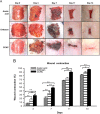
(A) Photographs of wounds at days 0, 3, 7, 11, and 14 after treatment with acetic acid, chitosan, and CCNC. (B) Wound contraction (%) at different days. Reprinted with permission from ref (53). Copyright 2014 Elsevier.
Figure 4
Figure 4
Upper graphs: Ratio between the weight of swollen hydrogel at time t and the weight of swollen hydrogel at equilibrium state. Lower graphs: Antibacterial activity of the hydrogels against E. coli, C. albicans, and S. aureus (itaconic acid concentration is varied). Reprinted with permission from ref (74). Copyright 2015 Elsevier.
Figure 5
Figure 5
Live–dead staining at laser scanning confocal microscopy are in picture 1, 2, and 3 (red cells = dead, green cells = dead, fibers = red for autofluorescence). SEMs of copper–cotton substrates are in picture 4, 5, and 6. Graph A: Antimicrobial activity against A. baumannii at different times. Graph B: A direct comparison among Cu- and Ag-coated cotton substrates and a commercial silver wound dressing, Acticoat. Graph C: Plot showing about 3-log kill for Cu-cotton samples in the presence of A. baumannii. Reprinted with permission from ref (77). Copyright 2011 John Wiley and Sons.
Figure 6
Figure 6
(A) Steps of healing of infected wounds in mice on days 0, 2, 4, 8, and 14. (B) Wound area closure (%) at different times points. (C) Bacteria from the wound tissues on LB agar plates (a: control; b: hydrogel; c: hydrogel + laser; d: Cu-NP-embedded hydrogel; e: Cu-NP-embedded hydrogel + laser). (D) Log of total bacterial CFU on the LB agar plates. Reprinted with permission from ref (54). Copyright 2013 Royal Society of Chemistry.
Figure 7
Figure 7
Upper panel: Photos and relative survival rates of ESBL E. coli (up) and MRSA E. coli (down). In the graphs The amount of bacteria is reported vs the concentration of NPs. Down panel: TEM images of ESBL E. coli and MRSA for CuS NPs, CuS NDs with and without laser irradiation (2.5 W/cm2, 10 min). Reprinted with permission from ref (91). Copyright 2019 American Chemical Society, with Creative Commons Attribution (CC BY) license.
Figure 8
Figure 8
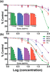
Cells viability normalized on the control vs concentration (logarithmic) of CuNPs at (a) 4 h and (b) 24 h. Lines and colors refer to different surface ligands on NPs (8-mercaptooctanoic acid (MOA), 12-mercaptododecanoic acid (MDA), and 16-mercaptohexa-decanoic acid (MHA)). Reprinted with permission from ref (103). Copyright 2009 Royal Society of Chemistry.
Figure 9
Figure 9
(A) Copper in the urine of mice at different times post injection. (B) Biodistribution of Cu(I)-GSH and Cu(I) complex (Cu(II)-GSSG). (C) Liver to urine ratios of the two complexes at 24 h. (D) Fluorescence intensity of the complexes (black is Cu(I) complex, red the Cu(II) complex). (E) Time dependence of copper distribution in kidneys and bladder. Reprinted with permission from ref (109). Copyright 2017 The Authors, published open access by MDPI under Creative Commons Attribution (CC BY) license.
Figure 10
Figure 10
Upper panels: Renal clearance and biodistribution studies of GSH-CuS NDs. (A) Absorption spectra of urine samples before and 1 h after the injection. (B) Amount of Cu excreted in urine. (C) Biodistribution of GSH-CuS NDs at 24 h post-injection. D) MR images of GSH-CuS NDs enhanced (from a to j: pre-injection and 30 s, 1 min, 2.5 min, 5.5 min, 10.5 min, 30.5 min, 1 h, 2 h, and 24 h post-injection signal enhancement in heart (B) and bladder (C) caused by GSH-CuS NDs. Reprinted with permission from ref (111). Copyright 2013 Royal Society of Chemistry.
Figure 11
Figure 11
Fluorescence images in vivo of mice treat before and after treatment with NCM (400 μM, 200 μL) and Cu2–xSe–NCM NPs. Reprinted with permission from ref (114). Copyright 2019 Royal Society of Chemistry.

References

    1. Luo G.; Gao S. Global Health Concerns Stirred by Emerging Viral Infections. J. Med. Virol. 2020, 92 (4), 399–400. 10.1002/jmv.25683. - DOI - PMC - PubMed
    1. Fisher R. A.; Gollan B.; Helaine S. Persistent Bacterial Infections and Persister Cells. Nat. Rev. Microbiol. 2017, 15, 453–464. 10.1038/nrmicro.2017.42. - DOI - PubMed
    1. Akhavan O.; Ghaderi E. Cu and CuO Nanoparticles Immobilized by Silica Thin Films as Antibacterial Materials and Photocatalysts. Surf. Coat. Technol. 2010, 205 (1), 219–223. 10.1016/j.surfcoat.2010.06.036. - DOI
    1. Labruère R.; Sona A. J.; Turos E. Anti-Methicillin-Resistant Staphylococcus aureus Nanoantibiotics. Front. Pharmacol. 2019, 10 (October), 1121.10.3389/fphar.2019.01121. - DOI - PMC - PubMed
    1. Merlin C. Reducing the Consumption of Antibiotics: Would That Be Enough to Slow Down the Dissemination of Resistances in the Downstream Environment?. Front. Microbiol. 2020, 11 (January), 33.10.3389/fmicb.2020.00033. - DOI - PMC - PubMed

Publication types

MeSH terms

Substances

LinkOut - more resources