. 2021 Apr 1;131(7):e140300.
doi: 10.1172/JCI140300.

Hypoxia-inducible factor-1α-dependent induction of miR122 enhances hepatic ischemia tolerance

Affiliations

Hypoxia-inducible factor-1α-dependent induction of miR122 enhances hepatic ischemia tolerance

Cynthia Ju et al. J Clin Invest. .

Abstract

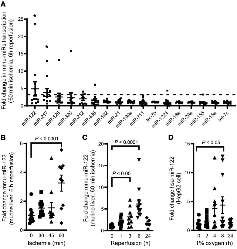
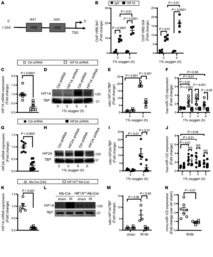
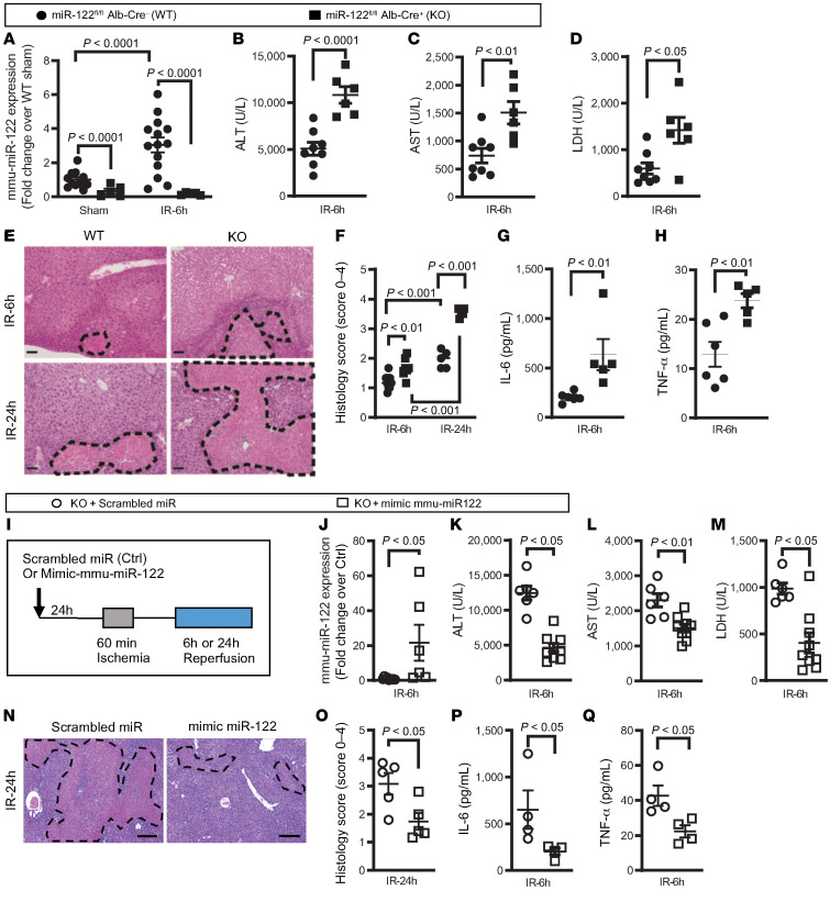
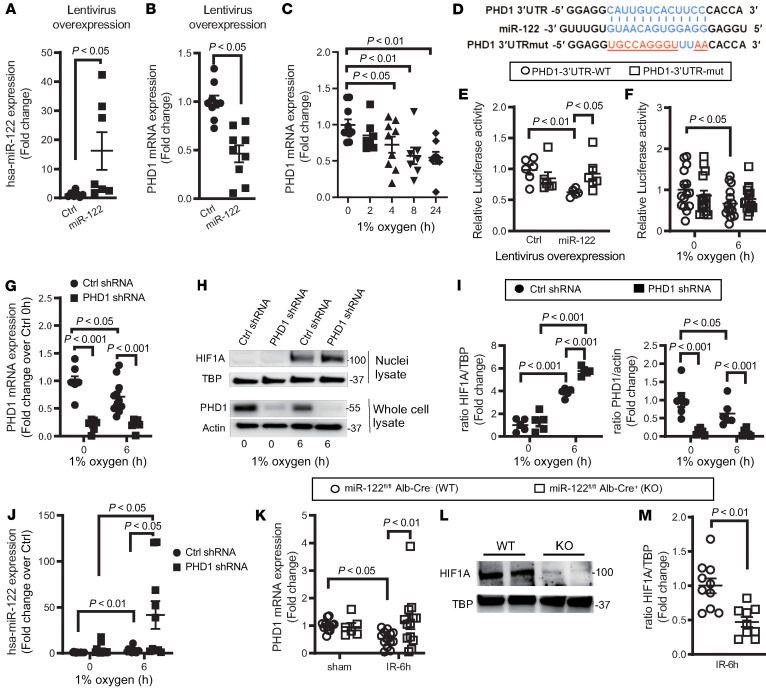
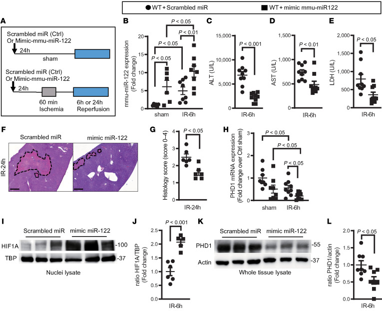
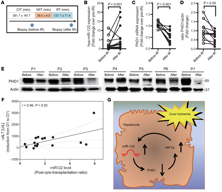
Hepatic ischemia and reperfusion (IR) injury contributes to the morbidity and mortality associated with liver transplantation. microRNAs (miRNAs) constitute a family of noncoding RNAs that regulate gene expression at the posttranslational level through the repression of specific target genes. Here, we hypothesized that miRNAs could be targeted to enhance hepatic ischemia tolerance. A miRNA screen in a murine model of hepatic IR injury pointed us toward the liver-specific miRNA miR122. Subsequent studies in mice with hepatocyte-specific deletion of miR122 (miR122loxP/loxP Alb-Cre+ mice) during hepatic ischemia and reperfusion revealed exacerbated liver injury. Transcriptional studies implicated hypoxia-inducible factor-1α (HIF1α) in the induction of miR122 and identified the oxygen-sensing prolyl hydroxylase domain 1 (PHD1) as a miR122 target. Further studies indicated that HIF1α-dependent induction of miR122 participated in a feed-forward pathway for liver protection via the enhancement of hepatic HIF responses through PHD1 repression. Moreover, pharmacologic studies utilizing nanoparticle-mediated miR122 overexpression demonstrated attenuated liver injury. Finally, proof-of-principle studies in patients undergoing orthotopic liver transplantation showed elevated miR122 levels in conjunction with the repression of PHD1 in post-ischemic liver biopsies. Taken together, the present findings provide molecular insight into the functional role of miR122 in enhancing hepatic ischemia tolerance and suggest the potential utility of pharmacologic interventions targeting miR122 to dampen hepatic injury during liver transplantation.

Keywords: Gastroenterology; Gene therapy; Hypoxia; Organ transplantation; Transplantation.


Conflict of interest statement

Conflict of interest: The authors have declared that no conflict of interest exists.

Figures

Figure 1
Figure 1. Identification of miR122 as a hypoxia-responsive miRNA during hepatic IR injury.
(A) Targeted miRNA array in WT liver after hepatic ischemia (60 minutes) and reperfusion (6 hours). The data shown indicate fold changes of the indicated miRNAs in mouse livers subjected to IR injury relative to sham control liver tissues. *P < 0.05 compared with values for the WT sham mice indicated by the dashed line, by 2-tailed, unpaired Student’s t test. n = 11/group. (B) Hepatic mmu-miR122 transcript levels following the indicated hepatic ischemia times and a 6-hour reperfusion. Statistical significance was determined by 1-way ANOVA. n = 9/group, except n = 10 in the 30-minute ischemia group. (C) Hepatic mmu-miR122 transcript levels after 60 minutes of liver ischemia and the indicated reperfusion times. Statistical significance was determined by 1-way ANOVA. n = 13, 9, 9, 12, and 5 for 0, 1, 3, 6, and 24 hours, respectively. (D) Hsa-miR122 transcript levels in cultured human hepatocytes (HepG2 cells) exposed to hypoxia (1% oxygen) for the indicated durations. Statistical significance was determined by 1-way ANOVA. n = 12, 12, 11, 11, and 11 for the 0-, 2-, 4-, 8-, and 24-hour groups, respectively). All data are shown as the mean ± SEM.
Figure 2
Figure 2. Role of HIF in miR122 induction.
(A) The promoter for miR122 includes 2 putative HIF-binding sites — hypoxia response elements (HREs). Maps indicate the promoter constructs generated for analysis of promoter activity. (B) ChIP analysis demonstrated binding of HIF1α to the putative HRE within the miR122 promoter (2-way ANOVA. n = 4/group from 4independent experiments). (C) Effective knockdown of HIF1A mRNA following lentivirus-mediated HIF1α shRNA delivery into HepG2 cells (2-tailed, unpaired Student’s t test. n = 8 and n = 7 for the control [Ctrl] and shRNA groups, respectively). (D and E) HIF1α shRNA abolished HIF1α induction in HepG2 cell nuclei following 6-hour hypoxia culturing in 1% oxygen (results are representative of 3 independent experiments). (GI) Experiments analogous to those depicted in D and E demonstrate preserved induction of miR122 following shRNA-mediated repression of HIF2α. (G) Two-tailed, unpaired Student’s t test; n = 8 and 11 for control and shRNA groups, respectively. (H and I) Results are representative of 4 independent experiments. (F and J) Time-dependent induction of miR122 in HepG2 cells exposed to hypoxia was abolished following HIF1α, but not HIF2α, knockdown compared with the same control shRNA group (2-way ANOVA, n = 4, and 6 at 0, 2, 4, 8 hours for the control group shared by F and J, and n = 6 for all shRNA groups, respectively, at 0, 2, 4, and 8 hours). (KN) In mice with hepatocyte-specific deletion of HIF1α (HIF1Afl/fl Alb-Cre+), HIF1A mRNA and protein were abolished, and induction of miR122 was abolished following ischemia and reperfusion (2-tailed, unpaired Student’s t test). (K) n = 5 and 8 in WT and KO groups, respectively. (M) n = 3/group. (N) n = 8 and 6 in WT and KO groups, respectively. All data are shown as the mean ± SEM.
Figure 3
Figure 3. Exacerbated liver IR injury in mice with hepatocyte-specific deletion of miR122 is reduced by reconstitution of the miR122 mimic.
Male miR122fl/fl Alb-Cre+ mice and littermate controls (miR122fl/fl Alb-Cre) were exposed to liver ischemia and reperfusion. (A) Lack of miR122 induction in the miR122fl/fl Alb-Cre+ mice after 60 minutes of ischemia and 6 hours of reperfusion. Two-way ANOVA. n = 11, 5 in the sham WT and KO groups, respectively; n = 13, 5 in the IR WT and KO groups, respectively. miR122fl/fl Alb-Cre+ mice showed increased hepatic injury, as determined by (B) ALT, (C) AST, and (D) LDH levels after 60 minutes of ischemia and 6 hours of reperfusion. Two-tailed Student’s t test. n = 8, 6 in the WT and KO groups, respectively. (E and F) Liver histology after 6 hours and 24 hours of reperfusion. Two-way ANOVA. n = 10, 7 for the 6-hour WT and KO groups, respectively; n = 5, 7 for the 24-hour WT and KO groups, respectively. Scale bars: 50 μm. (G and H) Protein levels of the liver inflammation markers IL-6 and TNF-α. Two-tailed, unpaired Student’s t test. n = 6, 5 in the WT and KO groups, respectively. (I) miR122fl/fl Alb-Cre+ mice were reconstituted with synthetic miRNA-122 or a scrambled miR control using nanoparticles 24 hours prior to 60 minutes of ischemia, followed by 6 or 24 hours of reperfusion. (J) Robust increases in miR122 levels were observed in the liver from mice reconstituted with a miR122 mimic compared with those reconstituted with a scrambled miR after ischemia and reperfusion. Two-tailed, unpaired Student’s t test. n = 9, 6 in the scrambled miR and miR122 groups, respectively. (KM) Compared with miR122fl/fl Alb-Cre+ mice that received scrambled miR treatment, mice reconstituted with synthetic miR122 show decreased levels of (K) ALT, (L) AST, and (M) LDH. n = 6, 9 in the scrambled miR and miR122 groups, respectively. (N and O) Histology images and improved histology scores. n = 5/group. Scale bars: 100 μm.(P and Q) Reduced levels of the proinflammatory cytokines IL-6 and TNF-α. n = 4/group. Two-tailed, unpaired Student’s t test. All data are shown as the mean ± SEM.
Figure 4
Figure 4. Identification of PHD1 as a miR122 target gene.
(A and B) Hsa-miR122 levels and PHD1 transcript levels in HepG2 cells with miR122 overexpression compared with control cells. Two-tailed, unpaired Student’s t test. n = 7/group (A); n = 9/group (B). (C) Repression of PHD1 transcripts in HepG2 cells following hypoxia exposure. One-way ANOVA. n = 10, 7, 9, 7, and 8 at 0, 2, 4, 8, and 24 hours, respectively. (D) Sequences used to generate 2 luciferase reporter plasmids, with 1 containing the WT 3′-UTR of PHD1, or a mutated version prohibiting binding of miR122 to the 3′-UTR. (E and F) Decreased luciferase activity caused by miR122 overexpression (n = 6/group) or following 24 hours of hypoxia (n = 15/group) in HepG2 cells transfected with the WT PHD1–3′-UTR. These responses were absent in cells transfected with the mutated PHD1–3′-UTR plasmid (PHD1-3′-UTR-mut), where specific binding of miR122 to the 3′-UTR does not occur. A 2-way ANOVA was performed. (G) Effective PHD1 knockdown following lentivirus-mediated PHD1 shRNA delivery into HepG2 cells. n = 8, 11 in CTL-shRNA groups at 0 hours and 6 hours, respectively; n = 6 in PHD1 shRNA groups at 0 hours and 6 hours. (HJ) HepG2 cells were cultured for 6 hours in hypoxic or normoxic conditions(0 hours). In PHD1-knockdown cells, hypoxia-induced HIF1α protein stabilization (n = 5/group) and miR122 augmentation were enhanced compared with control shRNA-transduced HepG2 cells. (J) n = 15, 12 in the control shRNA groups at 0 and 6 hours, respectively; n = 12, 10 in the PHD-1 shRNA groups at 0 and 6 hours, respectively. A 2-way ANOVA was performed. (K) PHD1 transcript expression was not suppressed in miR122f/f Alb-Cre+ mice after 60 minutes of ischemia and 6 hours of reperfusion. Two-way ANOVA. n = 13, 6 in the sham WT and KO groups, respectively; n = 13/group in the IR groups. (L and M) Lack of stabilization of HIF1α in miR122fl/fl Alb-Cre+ mice compared with littermate controls. Two-tailed, unpaired Student’s t test. n = 10, 9 in the WT and KO groups, respectively. All data are shown as the mean ± SEM.
Figure 5
Figure 5. Hepatic overexpression of miR122 provides liver protection during hepatic IR injury.
(A) Male C57Bl/6 WT mice were injected intravenously with a single dose of 20 μg synthetic mouse miR122 or scrambled miR formulated in NLE, 24 hours prior to 60 minutes of ischemia followed by 6 hours and 24 hours of reperfusion. (B) Treatment of exogenous miR122 increased miR122 levels in sham and IR livers. Two-way ANOVA. n = 6/group in the sham groups, n = 8/group in the IR groups. Mice treated with the miR122 mimetic show decreased levels of ALT (C), AST (D), and LDH (E) following 6 hours of reperfusion. Two-tailed, unpaired Student’s t test. n = 9/group. (F and G) Exogenous miR122 treatment improved histology scores following 24 hours of reperfusion. Two-tailed, unpaired Student’s t test. n = 6/group. (HL) Exogenous miR122 treatment (H) decreased hepatic PHD1 transcript levels (n = 8/group in the sham groups, n = 9/group in the IR groups), (I and J) augmented stabilization of HIF1α (n = 6, 5 in the scrambled miR and miR122 groups, respectively), and (K and L) repressed PHD1 protein levels (n = 8/group) after liver IR injury. Two-way ANOVA (H) and 2-tailed, unpaired Student’s t test (J and L).
Figure 6
Figure 6. MiR122 expression is increased in human hepatic IR injury.
(A) Two subsequent liver biopsies were obtained during cadaveric human liver transplantation — the first before IR and the second after warm ischemia and reperfusion after IR. Each liver biopsy served as its own control. (B and C) The miR122 (n = 16/group) and PHD1 transcript levels (n = 11/group) were measured by real-time RT-PCR. Two-tailed, paired Student’s t test. Data are shown as the mean ± SEM. (D and E) PHD1 protein levels in pre- and post-IR liver samples were measured by Western blot analysis and quantified. Representative blots are shown. Two-tailed, paired Student’s t test. n = 11/group. data are shown as the mean ± SEM. (F) Correlation between the ratio of miR122 levels (post/pre-transplantation ratio) and the reduction in ALT levels from postoperative days 1 to 7 (D1 to D7) as an indicator of liver recovery (n = 16 samples). Data are depicted as linear regression (black line) with a 95% CI (dashed lines). (G) Schematic summary of the main findings. HIF1α-dependent upregulation of miR122 and miR122-dependent repression of PHD1, which results in hepatic HIF1α stabilization, represent a feed-forward pathway for liver protection during IR injury.

References

    1. Eltzschig HK, Eckle T. Ischemia and reperfusion--from mechanism to translation. Nat Med. 2011;17(11):1391–1401. doi: 10.1038/nm.2507. - DOI - PMC - PubMed
    1. Eltzschig HK, Carmeliet P. Hypoxia and inflammation. N Engl J Med. 2011;364(7):656–665. doi: 10.1056/NEJMra0910283. - DOI - PMC - PubMed
    1. Hilmi I, et al. The impact of postreperfusion syndrome on short-term patient and liver allograft outcome in patients undergoing orthotopic liver transplantation. Liver Transpl. 2008;14(4):504–508. doi: 10.1002/lt.21381. - DOI - PubMed
    1. Cameron AM, et al. Optimal utilization of donor grafts with extended criteria: a single-center experience in over 1000 liver transplants. Ann Surg. 2006;243(6):748–753. doi: 10.1097/01.sla.0000219669.84192.b3. - DOI - PMC - PubMed
    1. Pirenne J, et al. Influence of ischemia-reperfusion injury on rejection after liver transplantation. Transplant Proc. 1997;29(1–2):366–367. - PubMed

Publication types