

MRI index lesion radiomics and machine learning for detection of extraprostatic extension of disease: a multicenter study
- PMID: 33792737
- PMCID: PMC8452573
- DOI: 10.1007/s00330-021-07856-3
MRI index lesion radiomics and machine learning for detection of extraprostatic extension of disease: a multicenter study
Abstract
Objectives: To build a machine learning (ML) model to detect extraprostatic extension (EPE) of prostate cancer (PCa), based on radiomics features extracted from prostate MRI index lesions.
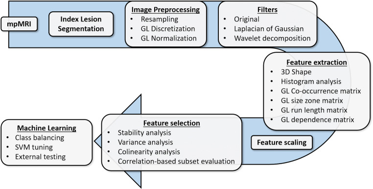
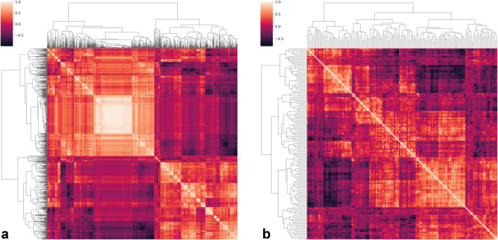
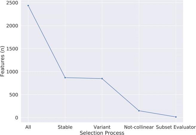
Methods: Consecutive MRI exams of patients undergoing radical prostatectomy for PCa were retrospectively collected from three institutions. Axial T2-weighted and apparent diffusion coefficient map images were annotated to obtain index lesion volumes of interest for radiomics feature extraction. Data from one institution was used for training, feature selection (using reproducibility, variance and pairwise correlation analyses, and a correlation-based subset evaluator), and tuning of a support vector machine (SVM) algorithm, with stratified 10-fold cross-validation. The model was tested on the two remaining institutions' data and compared with a baseline reference and expert radiologist assessment of EPE.
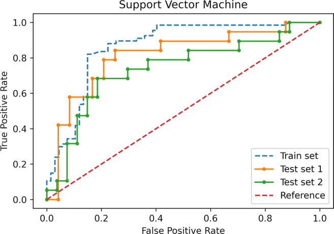
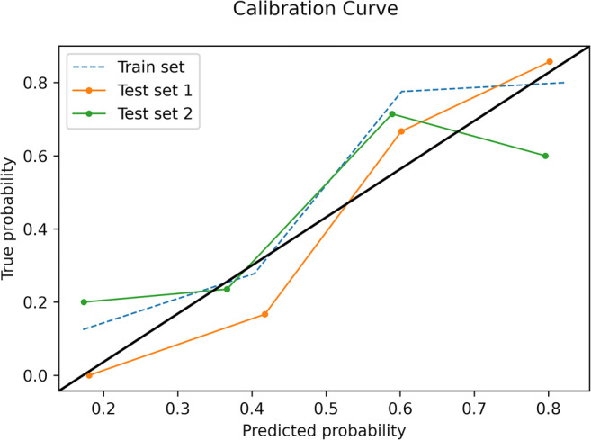
Results: In total, 193 patients were included. From an initial dataset of 2436 features, 2287 were excluded due to either poor stability, low variance, or high collinearity. Among the remaining, 14 features were used to train the ML model, which reached an overall accuracy of 83% in the training set. In the two external test sets, the SVM achieved an accuracy of 79% and 74% respectively, not statistically different from that of the radiologist (81-83%, p = 0.39-1) and outperforming the baseline reference (p = 0.001-0.02).
Conclusions: A ML model solely based on radiomics features demonstrated high accuracy for EPE detection and good generalizability in a multicenter setting. Paired to qualitative EPE assessment, this approach could aid radiologists in this challenging task.
Key points: • Predicting the presence of EPE in prostate cancer patients is a challenging task for radiologists. • A support vector machine algorithm achieved high diagnostic accuracy for EPE detection, with good generalizability when tested on multiple external datasets. • The performance of the algorithm was not significantly different from that of an experienced radiologist.
Keywords: Machine learning; Magnetic resonance imaging; Prostate cancer; Prostatectomy; Support vector machine.
© 2021. The Author(s).
Conflict of interest statement
The authors of this manuscript declare no relationships with any companies, whose products or services may be related to the subject matter of the article.
Figures

References
-
- (2019) EAU Guidelines. Edn. presented at the EAU Annual Congress Barcelona 2019. https://uroweb.org/guideline/prostate-cancer. Accessed 13 May 2019
Publication types
MeSH terms
LinkOut - more resources
Full Text Sources
Other Literature Sources
Medical
