. 2021 Apr 15;12(14):3509-3513.
doi: 10.1021/acs.jpclett.1c00223. Epub 2021 Apr 1.

A Proposed Mechanism for the Initial Myosin Binding Event on the Cardiac Thin Filament: A Metadynamics Study

Affiliations

A Proposed Mechanism for the Initial Myosin Binding Event on the Cardiac Thin Filament: A Metadynamics Study

Anthony P Baldo et al. J Phys Chem Lett. .

Abstract

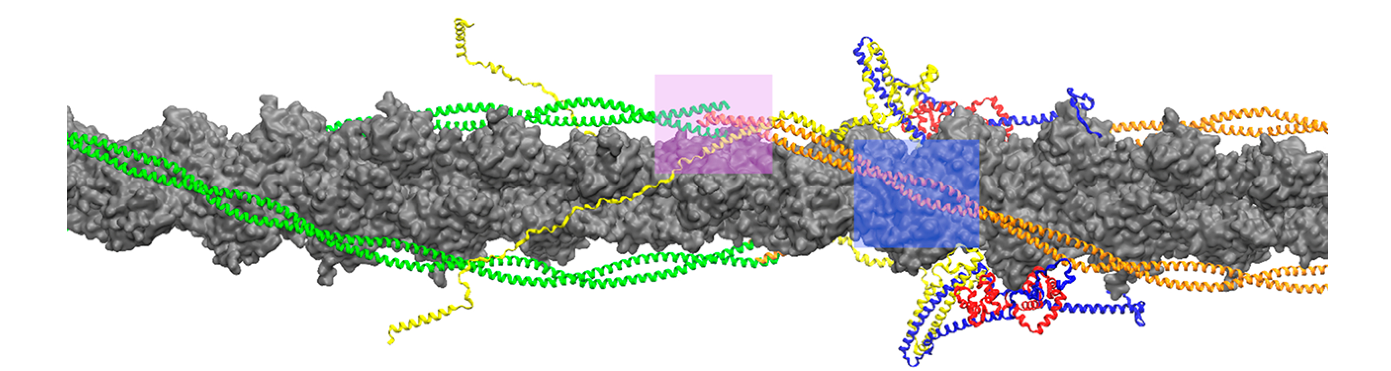
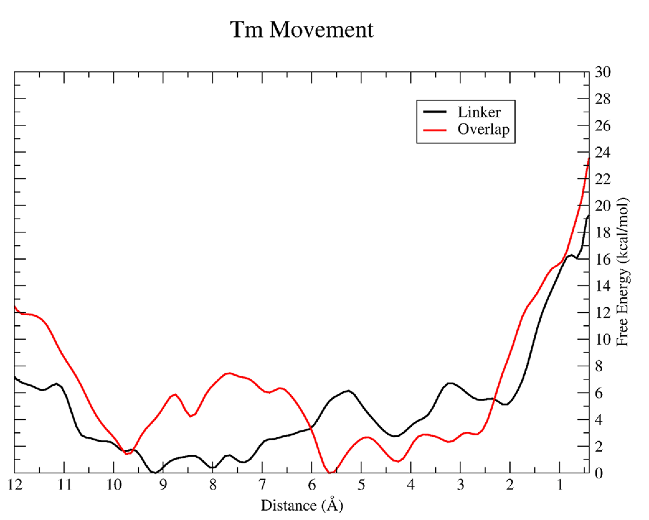
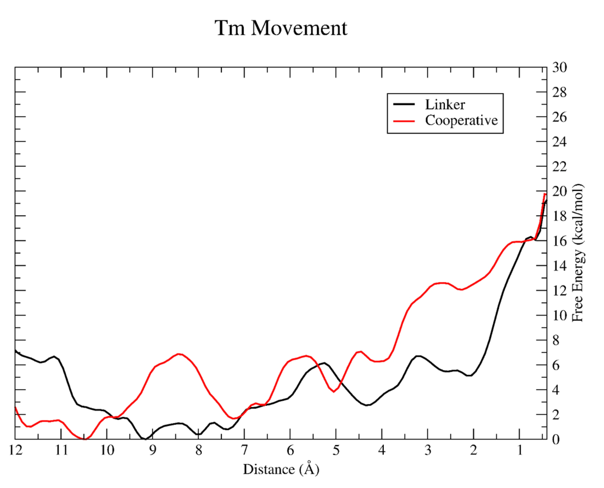
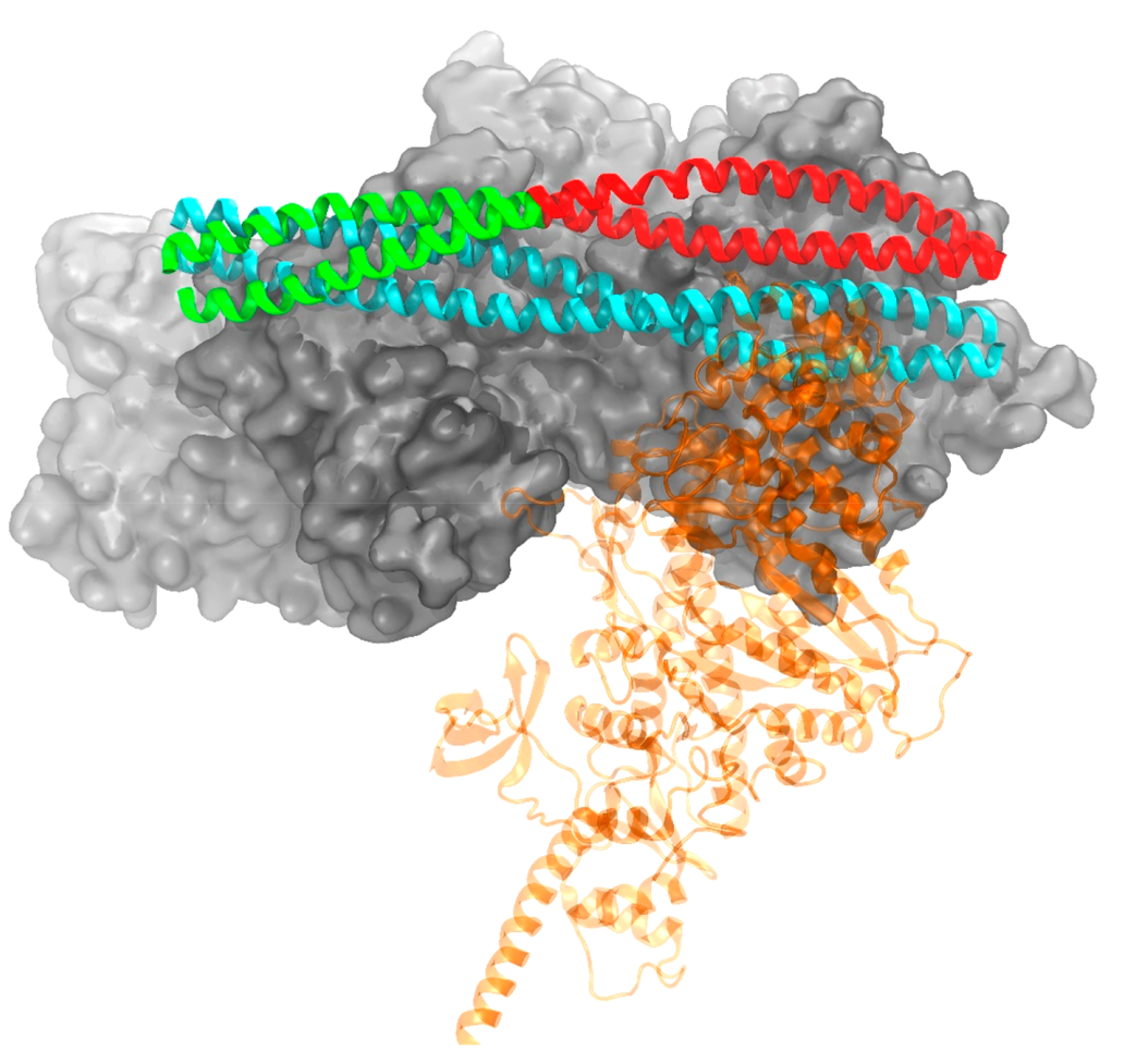
The movement of tropomyosin over filamentous actin regulates the cross-bridge cycle of the thick with thin filament of cardiac muscle by blocking and revealing myosin binding sites. Tropomyosin exists in three, distinct equilibrium states with one state blocking myosin-actin interactions (blocked position) and the remaining two allowing for weak (closed position) and strong myosin binding (open position). However, experimental information illuminating how myosin binds to the thin filament and influences tropomyosin's transition across the actin surface is lacking. Using metadynamics, we mimic the effect of a single myosin head binding by determining the work required to pull small segments of tropomyosin toward the open position in several distinct regions of the thin filament. We find differences in required work due to the influence of cardiac troponin T lead to preferential binding sites and determine the mechanism of further myosin head recruitment.


Conflict of interest statement

Notes

The authors declare no competing financial interest.

Figures

Figure 1.
Figure 1.
Organization of tropomyosin (orange/green), troponin complex (yellow, red, and blue) and actin (gray) in the cardiac thin filament. Overlap and linker regions where tropomyosin and troponin T (yellow) interact are highlighted in pink and blue, respectively.
Figure 2.
Figure 2.
Resulting PMF for tropomyosin segment pulling in the linker (black) and overlap (red). The CV defined in both simulations is the center of mass of a subset of α carbons from the Tm chain (linker: Tm residues 180 to 220; overlap: Tm residues 266 to 284 from the C-terminal chain and residues 1 to 35 from the N-terminal chain) shown here as a distance in angstroms. A CV value of zero denotes the final M-state. The free energy (ΔG) is given in units of kcal/mol.
Figure 3.
Figure 3.
Resulting PMF for tropomyosin pulling in the linker (black) and cooperative (red) simulation.
Figure 4.
Figure 4.
Conformations of tropomyosin in a blocked (cyan) position and at the end of a metadynamics simulation (green/red) mimicking a single myosin head (transparent for clarity) binding. The green portion of Tm illustrates the segment biased in the cooperative simulation, while red was biased in the linker simulation. Troponin not shown for clarity.

References

    1. Tobacman LS Thin Filament-Mediated Regulation of Cardiac Contraction. Annu. Rev. Physiol 1996, 58, 447–481. - PubMed
    1. Gordon AM; Homsher E; Regnier M Regulation of Contraction in Striated Muscle. Physiol. Rev 2000, 80, 853–924. - PubMed
    1. McKillop DF; Geeves MA Regulation of the Interaction between Actin and Myosin Subfragment 1: Evidence for Three States of the Thin Filament. Biophys. J 1993, 65, 693–701. - PMC - PubMed
    1. Li X; Tobacman LS; Mun JY; Craig R; Fischer S; Lehman W Tropomyosin Position on F-Actin Revealed by EM Reconstruction and Computational Chemistry. Biophys. J 2011, 100, 1005–1013. - PMC - PubMed
    1. Risi C; Eisner J; Belknap B; Heeley DH; White HD; Schröder GF; Galkin VE Ca2+-Induced Movem Tropomyosin on Native Cardiac Thin Filaments Revealed by Cryoelectron Microscopy. Proc. Natl. Acad. Sci. U. S. A 2017, 114, 6782–6787. - PMC - PubMed

LinkOut - more resources