. 2021 Apr 1;17(4):e1008889.
doi: 10.1371/journal.pcbi.1008889. eCollection 2021 Apr.

Building blocks and blueprints for bacterial autolysins

Affiliations

Building blocks and blueprints for bacterial autolysins

Spencer J Mitchell et al. PLoS Comput Biol. .

Abstract

Bacteria utilize a wide variety of endogenous cell wall hydrolases, or autolysins, to remodel their cell walls during processes including cell division, biofilm formation, and programmed death. We here systematically investigate the composition of these enzymes in order to gain insights into their associated biological processes, potential ways to disrupt them via chemotherapeutics, and strategies by which they might be leveraged as recombinant antibacterial biotherapies. To do so, we developed LEDGOs (lytic enzyme domains grouped by organism), a pipeline to create and analyze databases of autolytic enzyme sequences, constituent domain annotations, and architectural patterns of multi-domain enzymes that integrate peptidoglycan binding and degrading functions. We applied LEDGOs to eight pathogenic bacteria, gram negatives Acinetobacter baumannii, Klebsiella pneumoniae, Neisseria gonorrhoeae, and Pseudomonas aeruginosa; and gram positives Clostridioides difficile, Enterococcus faecium, Staphylococcus aureus, and Streptococcus pneumoniae. Our analysis of the autolytic enzyme repertoires of these pathogens reveals commonalities and differences in their key domain building blocks and architectures, including correlations and preferred orders among domains in multi-domain enzymes, repetitions of homologous binding domains with potentially complementarity recognition modalities, and sequence similarity patterns indicative of potential divergence of functional specificity among related domains. We have further identified a variety of unannotated sequence regions within the lytic enzymes that may themselves contain new domains with important functions.


Conflict of interest statement

I have read the journal’s policy and the authors of this manuscript have the following competing interests: KEG and CB-K are member-managers of Lyticon LLC. No other authors have a conflict of interest. Potential conflicts of interest for KEG and CB-K are under management at Dartmouth. The authors declare that the work presented here is free of any bias.

Figures

Fig 1
Fig 1. LEDGOs workflow.
For each organism of interest, the user provides as input a set of “seed” proteins, here based on GO terms indicative of peptidoglycan recognition and catalysis. The LEDGOs data collection pipeline then gathers organism-specific homologs of the seed proteins by repeated PSI-BLAST searches. The LEDGOs pipeline further annotates the catalytic and cell-wall binding domains within the collected sequences according to Pfam families, and catalogs the domain architectures of the proteins. Note that the identified homologs can extend (past the marked “||”) to include additional domains beyond those in the seeds. There can also be uncharacterized sequence regions (marked “?”) between the annotated domains. The domain sequences, annotations, and architectures within full enzymes are stored in the LEDGOs database. The LEDGOs data analysis tools then query this database to characterize and compare/contrast the lysin domain building blocks and architectures employed by the different organisms.
Fig 2
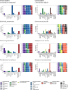
Fig 2. Domain usage frequency by organism.
Bar charts indicate percentages of representative proteins containing each of the domain types. Waffle plots indicate percentages of each domain type among the set of domains comprising the representative proteins, separately counting duplicates of a domain type within a protein. With the entire set of domain sequences totaling 100%, each block in a waffle indicates that 1% of the domains are of a given domain type. Only domain types that appear in at least 2% of some organism’s representative proteins are shown. Bars and waffle cells are ordered and colored by domain type as summarized in the legend.
Fig 3
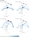
Fig 3. Common gram negative lytic enzyme architectures.
In each graph, the nodes indicate domains (along with the N terminus and C terminus), with size reflecting frequency within the organism’s clustered proteins and empty circles for domains with no representation in that species. The edges represent connections within a single protein, with edge shading and thickness representing relative frequency. Note that there are some self-edges (e.g., LysM loops back to itself), indicating a repeated domain. A path from Nterm through one or more domains to Cterm thus represents a protein, though not all such proteins have been observed in LEDGOs (see text).
Fig 4
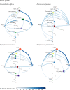
Fig 4. Common gram positive lytic enzyme architectures.
In each graph, the nodes indicate domains (along with the N terminus and C terminus), with size reflecting frequency within the organism’s clustered proteins and empty circles for domains with no representation in that species. The edges represent connections within a single protein, with edge shading and thickness representing relative frequency. Note that there are some self-edges (e.g., LysM loops back to itself), indicating a repeated domain. A path from Nterm through one or more domains to Cterm thus represents a protein, though not all such proteins have been observed in LEDGOs (see text).
Fig 5
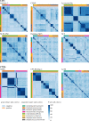
Fig 5. Domain sequence diversity.
In each heatmap, each row and column represent a single non-redundant domain sequence in the LEDGOs database, and the cell for a pair of sequences is colored to indicate sequence identity (darker blue, higher). Cells are grouped by organism and clustered within an organism based on sequence identity patterns, so that similar sequences within an organism appear together as “blocks” on the diagonal, and blocks of similar sequences across organisms as off-diagonal blocks.
Fig 6
Fig 6. Domain sequence diversity by architecture.
Heatmaps as in Fig 5, except limited to non-redundant domain sequences appearing in architectures with a frequency of at least 2% are shown, and with row/column colors indicating the organism and architecture rather than the organism and gram status.
Fig 7
Fig 7. Domain sequence diversity by repeat position.
Heatmaps as in Fig 5, except with the colors above the columns indicating the organism and architecture, and the colors beside the rows indicating the organism and repeat number. Thus cells are grouped by organism and repeat number, and clustered within those based on sequence identity patterns.
Fig 8
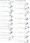
Fig 8. Regions of Unknown Function, RUFs.
Entries give each RUF’s number of sequences, median sequence length, median pairwise sequence identity, organism in which it appears, and architecture graph (as in Figs 3 and 4) constructed from all architectures occurring at least 5 times.

Similar articles

Cited by

References

    1. Rodríguez-Rubio L, Martínez B, Donovan DM, Rodríguez A, García P. Bacteriophage virion-associated peptidoglycan hydrolases: Potential new enzybiotics. Critical Reviews in Microbiology. Crit Rev Microbiol; 2013. pp. 427–434. 10.3109/1040841X.2012.723675 - DOI - PubMed
    1. Nelson DC, Schmelcher M, Rodriguez-Rubio L, Klumpp J, Pritchard DG, Dong S, et al.. Endolysins as Antimicrobials. Advances in Virus Research. Academic Press Inc.; 2012. pp. 299–365. 10.1016/B978-0-12-394438-2.00007-4 - DOI - PubMed
    1. Fischetti VA. Lysin therapy for Staphylococcus aureus and other bacterial pathogens. Current Topics in Microbiology and Immunology. Springer Verlag; 2017. pp. 529–540. 10.1007/82_2015_5005 - DOI - PubMed
    1. Wittekind M, Schuch R. Cell wall hydrolases and antibiotics: Exploiting synergy to create efficacious new antimicrobial treatments. Current Opinion in Microbiology. Elsevier Ltd; 2016. pp. 18–24. 10.1016/j.mib.2016.05.006 - DOI - PubMed
    1. Scanlon TC, Teneback CC, Gill A, Bement JL, Weiner JA, Lamppa JW, et al.. Enhanced Antimicrobial Activity of Engineered Human Lysozyme. ACS Chem Biol. 2010;5: 809–818. 10.1021/cb1001119 - DOI - PMC - PubMed

Publication types

MeSH terms

Substances

LinkOut - more resources