. 2021 Apr 1;16(4):e0248290.
doi: 10.1371/journal.pone.0248290. eCollection 2021.

CD40 signaling augments IL-10 expression and the tolerogenicity of IL-10-induced regulatory dendritic cells

Affiliations

CD40 signaling augments IL-10 expression and the tolerogenicity of IL-10-induced regulatory dendritic cells

Wojciech Dawicki et al. PLoS One. .

Abstract

CD40 expressed on stimulatory dendritic cells (DC) provides an important accessory signal for induction of effector T cell responses. It is also expressed at lower levels on regulatory DC (DCreg), but there is little evidence that CD40 signaling contributes to the tolerogenic activity of these cells. Indeed, CD40 silencing within DCreg has been reported to induce T cell tolerance in multiple disease models, suggesting that CD40 is superfluous to DC-induced tolerance. We critically assessed whether CD40 does have a role in tolerance induced by IL-10-differentiated DC (DC10) by using DC10 generating from the bone marrow of wild-type (w.t.) or CD40-/- donor mice, or IL-10-complemented CD40-/- DC10 to treat asthmatic mice. Wild-type DC10 ablated the OVA-asthma phenotype via induction of Foxp3+ Treg responses, but CD40-/- DC10 had no discernible effects on primary facets of the phenotype (e.g., IL-5, IL-9, IL-13 levels, IgE & IgG1 antibodies; p>0.05) and were ≤40% effective in reversal of others. Foxp3+ T cells from the lungs of CD40-/- DC10-treated mice expressed reduced levels of a panel of six Treg-specific activation markers relative to Treg from w.t. DC10-treated mice. Coculture with effector T cells from asthmatic mice induced a marked upregulation of cell surface CD40 on w.t. DC10. While untreated CD40-/- and w.t. DC10 secreted equally low levels of IL-10, stimulation of w.t. DC10 with anti-CD40 for 72 h increased their expression of IL-10 by ≈250%, with no parallel induction of IL-12. Complementing IL-10 expression in CD40-/- DC10 by IL-10 mRNA transfection fully restored the cells' abilities to suppress the asthma phenotype. In summary, CD40 signaling in DC10 contributes importantly to their expression of IL-10 and to a robust induction of tolerance, including activation of induced Treg.


Conflict of interest statement

The authors have declared that no competing interests exist.

Figures

Fig 1
Fig 1. CD40-/- DC10 have a reduced capacity to suppress T-cell proliferation relative to wild-type DC10.
DC10 were differentiated from the bone marrow of wild-type (w.t.) or CD40 knock-out mice as noted in the Methods section and added at the indicated DC10:T-cell ratios to 3-day cultures of OVA-pulsed stimulatory DC-LPS and Teff cells (3x10⁴ DC-LPS, 2x10⁵ T cells) from the lungs of OVA-asthmatic mice. T cell proliferation was determined using standard 3H-thymidine-uptake assays. The data presented are representative of one of 3 independent experiments. NS or ***, p>0.05 or ≤0.001, respectively versus the DC-LPS/T cell control, as determined using one-way ANOVA tests with Tukey’s post-hoc testing.
Fig 2
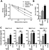
Fig 2. CD40 expression by DC10 is important in their ability to suppress the asthma phenotype.
C57BL/6 mice were injected i.p. with 200 μl of OVA-alum (2 μg OVA/mg alum) on days 0 and 14, and then exposed to 1% OVA aerosols for 20 min on days 28, 30 and 32. They were rested for two weeks and then given a single i.p. injection of 1x10⁶ w.t. or CD40-/- OVA-presenting DC10. Control mice included saline-treated asthmatic (Sal.) and healthy normal animals. Four weeks later all mice were again challenged with aerosolized OVA and their asthma phenotype was assessed. (A) Airway hyperresponsiveness was determined by head-out plethysmography 24h post allergen exposure. At 48h post allergen exposure mice were sacrificed, and (B) BAL eosinophils were enumerated using Wright’s stained cytocentrifuge preparations, while (C) BALF Th2 cytokines were determined by ELISA. Bars represent mean with SEM from 9–15 mice from three independent experiments. NS, *, ** or ***, p>0.05, ≤0.05, 0.01 or 0.001, respectively versus CD40-/- DC treatment (AHR) or versus the saline control (all other parameters), as determined using one-way ANOVA tests with Tukey’s post-hoc testing.
Fig 3
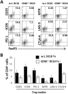
Fig 3. CD40 is important for the induction of Treg in DC10-treated asthmatic mice.
Lungs were removed from asthmatic mice treated three weeks earlier with DC10 generated from wild-type (w.t.) and CD40-/- mice, as in Fig 2, and then enzymatically dispersed as in the Materials and Methods. Lung CD4+ cells were magnetically sorted, stained for the indicated Treg markers and marker expression was analyzed by flow cytometry. (A) Representative scatter plots from one experiment of three undertaken depicting the expression of the indicated markers on the Foxp3+/marker-positive CD4+ cells. The quadrant boundaries are based on isotype staining, with the percentage of cells in the upper–right quadrant. (B). Graphic presentation of the mean +/- SD of the proportion of the CD4+ cells that stained with each marker. This experiment was repeated three times.
Fig 4
Fig 4. Interaction with cognate T cells leads to upregulation of cell surface CD40 on DC10.
DC10 were generated as in Fig 1 and (A) stained with anti-CD40 antibody in the absence (cell surface) or presence (intracellular) of a permeabilizing agent and analyzed by flow cytometry. (B) OVA-pulsed DC10 (thick line) or DC10 without OVA (thin line, 48 and 72 hr panels) were co-cultured with OVA-specific CD4+ cells from D011.10 mice for the indicated times and then their cell surface CD40 was evaluated by flow cytometry. Shaded histograms represent isotype staining. The data presented depict the results of one experiment that is representative of 3–4 independent experiments with duplicate analysis of each sample.
Fig 5
Fig 5. CD40 signaling increases IL-10, but not IL-12 production by DC10.
CD40-/- or w.t. DC10 were stimulated with plate-bound hamster IgG1 anti-mouse CD40 or irrelevant hamster IgG1 control antibodies (Ctrl) for two days and expression of (A) IL-10 mRNA and (B, left panel) IL-12p35 mRNA were determined by qRT-PCR and ELISA. (B, right panel) LPS-matured immunostimulatory DC (DC-LPS) were also stimulated with anti-CD40 or irrelevant control antibodies for 48 h, and their expression of IL-12p70 was assessed by ELISA. Bars represent mean with SEM from duplicate or triplicate repetitions from one of three independent experiments. NS or ***, p>0.05 or ≤0.001, respectively versus the w.t. or saline control, as appropriate. For the left hand panel in Fig 5A, we used a one-way ANOVA test with Tukey’s post-hoc testing, while students T tests were used for all other panels.
Fig 6
Fig 6. Complementing IL-10 expression in CD40-deficient DC10 restores their ability to suppress the asthma phenotype.
Mice were rendered asthmatic as described in Fig 2 and treated with 1x10⁶ w.t. or with CD40-/- DC10 that had been transfected with medium- (SHAM) or IL-10 mRNA-containing (IL-10 RNA) liposomes. Control groups included saline-treated asthmatic mice and normal mice. Four weeks after treatment, mice were challenged again with aerosolized OVA and (A) the asthmatic phenotype assessed by measuring AHR, (B) BAL eosinophils, (C) OVA-specific plasma IgG1 and IgE and (D) BALF cytokines. Bars represent mean with SEM from 6–10 mice from two independent experiments. NS, * or **, p>0.05, ≥0.05 or 0.01, respectively versus CD40-/- DC10 treatment (AHR) or versus the saline control (all other parameters), as determined using one-way ANOVA tests with Tukey’s post-hoc testing.

References

    1. Koch F, Stanzl U, Jennewein P, Janke K, Heufler C, Kampgen E, et al.. High level IL-12 production by murine dendritic cells: upregulation via MHC class II and CD40 molecules and downregulation by IL-4 and IL-10. J Exp Med. 1996;184(2):741–6. 10.1084/jem.184.2.741 - DOI - PMC - PubMed
    1. Maldonado-Lopez R, Moser M. Dendritic cell subsets and the regulation of Th1/Th2 responses. Semin Immunol. 2001;13(5):275–82. 10.1006/smim.2001.0323 - DOI - PubMed
    1. Akbari O, DeKruyff RH, Umetsu DT. Pulmonary dendritic cells producing IL-10 mediate tolerance induced by respiratory exposure to antigen. Nat Immunol. 2001;2(8):725–31. 10.1038/90667 - DOI - PubMed
    1. Gordon JR, Ma Y, Churchman L, Gordon SA, Dawicki W. Regulatory dendritic cells for immunotherapy in immunologic diseases. Front Immunol. 2014;5:7. 10.3389/fimmu.2014.00007 - DOI - PMC - PubMed
    1. Lu M, Dawicki W, Zhang X, Huang H, Nayyar A, Gordon JR. Therapeutic induction of tolerance by IL-10-differentiated dendritic cells in a mouse model of house dust mite-asthma. Allergy. 2011;66(5):612–20. 10.1111/j.1398-9995.2010.02526.x - DOI - PubMed

Publication types

MeSH terms

Grants and funding